

Editorial
doi: 10.1093/cvr/cvt058.
Epub 2013 Mar 14.
Imaging T-tubules: dynamic membrane structures for deep functions
- PMID: 23504549
- PMCID: PMC3633161
- DOI: 10.1093/cvr/cvt058
Item in Clipboard
Editorial
Imaging T-tubules: dynamic membrane structures for deep functions
Cardiovasc Res.
.
No abstract available
Figures
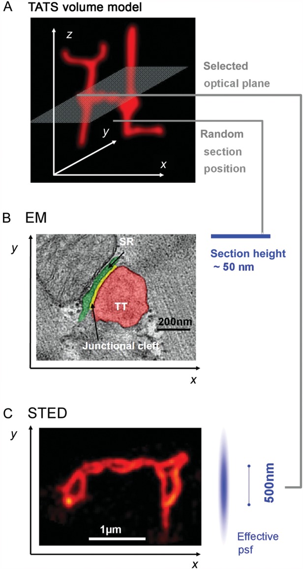
The cardiac cell membrane exerts deep intracellular functions through TT invaginations, forming a continuous three-dimensional TATS network composed of transversal and longitudinal components. (A) Minimal three-dimensional TATS volume model composed of 2 transversal TTs continuously connected with longitudinal TT elements; example imaging/sectioning planes in (B) and C) are indicated. (B) Individual TT–jSR contacts were investigated in control and HF samples in end-stage DCM/ICM by Zhang et al. Increased spatial separation between Z-lines and jSR junctions (green) was shown among other observations in human HF samples. While ultrathin sectioning allows for precise EM measurements of TT–jSR membrane architectures, sectioning occurs randomly relative to the complex three-dimensional membrane structures. (C) STED image of deep intracellular TT membrane structures specifically labelled with di-8-ANEPPS showing abnormally enlarged TT cross-sections and TATS network remodelling 4 weeks post-MI in mouse hearts. Optical planes can be directed to specific objects (e.g. transverse-axial TATS intersections) in the focal plane based on image contrast. Image analysis based on identified objects can be semi-automated for quantification. The effective psf indicates a lateral resolution of ∼55 nm and a z resolution of ∼500 nm, the latter limiting 3D resolution similar to confocal imaging here; imaging by z stacks allows for targeted three-dimensional sampling. The Field of view can be adapted to the TATS network dimensions or individual TT objects as needed. B and C reproduced with permission.
Comment on
-
Emerging mechanisms of T-tubule remodelling in heart failure.Cardiovasc Res. 2013 May 1;98(2):204-15. doi: 10.1093/cvr/cvt020. Epub 2013 Feb 7. Cardiovasc Res. 2013. PMID: 23393229 Free PMC article. Review.
-
Ultrastructural uncoupling between T-tubules and sarcoplasmic reticulum in human heart failure.Cardiovasc Res. 2013 May 1;98(2):269-76. doi: 10.1093/cvr/cvt030. Epub 2013 Feb 11. Cardiovasc Res. 2013. PMID: 23405000
References
-
- Kohl T, Westphal V, Hell SW, Lehnart SE. Super-resolution microscopy in heart—cardiac nanoscopy. J Mol Cell Cardiol. 2013 - PubMed
-
- Forssmann WG, Girardier L. A study of the T system in rat heart. J Cell Biol. 1970;44:1–19. doi:10.1083/jcb.44.1.1. - DOI - PMC - PubMed
-
- Fawcett DW, McNutt NS. The ultrastructure of the cat myocardium. I. Ventricular papillary muscle. J Cell Biol. 1969;42:1–45. doi:10.1083/jcb.42.1.1. - DOI - PMC - PubMed
-
- Wagner E, Lauterbach MA, Kohl T, Westphal V, Williams GS, Steinbrecher JH, et al. Stimulated emission depletion live-cell super-resolution imaging shows proliferative remodeling of T-tubule membrane structures after myocardial infarction. Circ Res. 2012;111:402–414. doi:10.1161/CIRCRESAHA.112.274530. - DOI - PMC - PubMed
-
- Scriven DRL, Asghari P, Moore EDW. Microarchitecture of the dyad. Cardiovasc Res. 2013;98:169–176. - PubMed
Publication types
MeSH terms
LinkOut - more resources
Full Text Sources
Other Literature Sources
Medical
