. 2013 Sep 1;521(13):2927-46.
doi: 10.1002/cne.23323.

Somatodendritic targeting of M5 muscarinic receptor in the rat ventral tegmental area: implications for mesolimbic dopamine transmission

Affiliations

Somatodendritic targeting of M5 muscarinic receptor in the rat ventral tegmental area: implications for mesolimbic dopamine transmission

Miguel Garzón et al. J Comp Neurol. .

Abstract

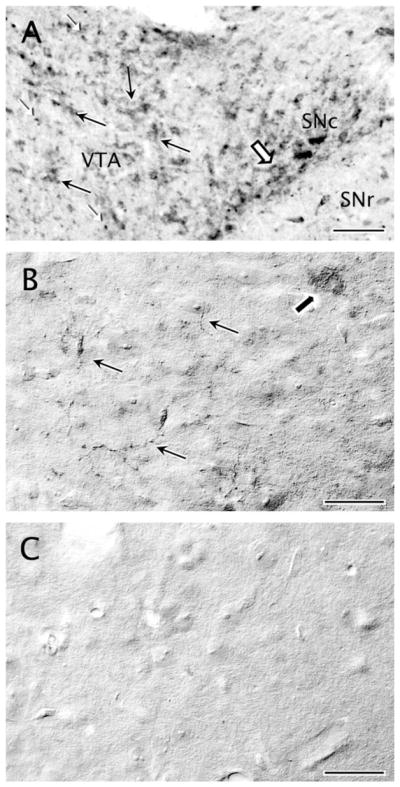
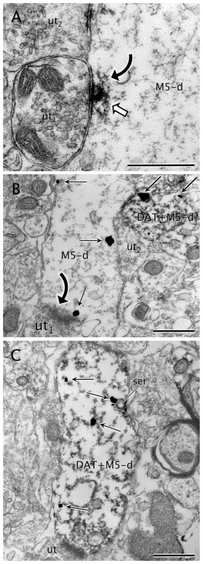
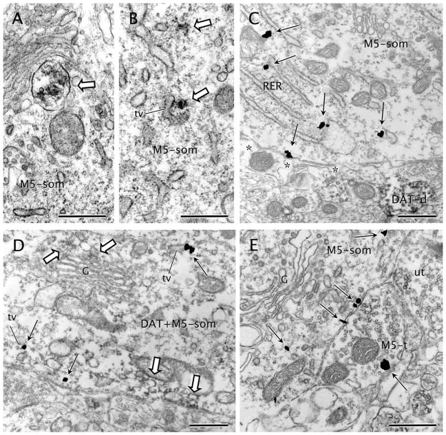
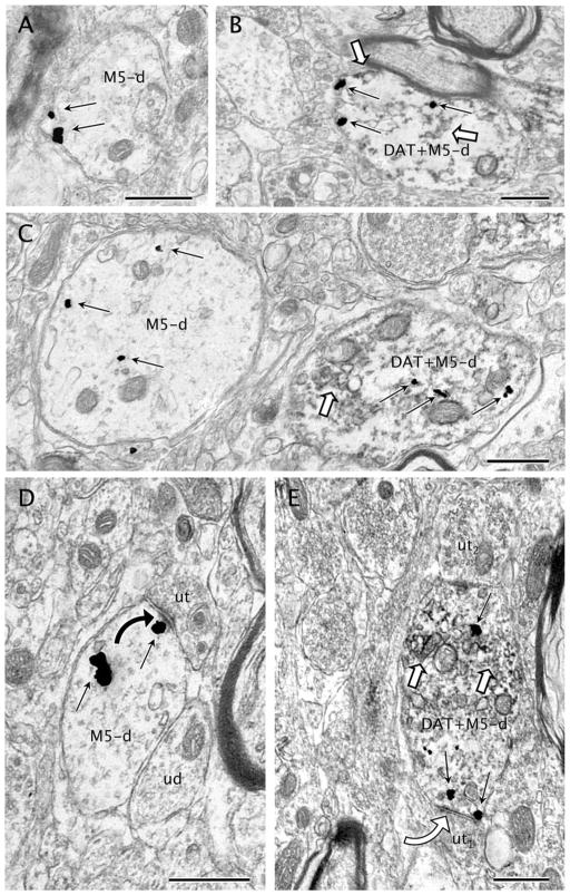
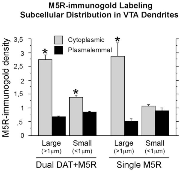
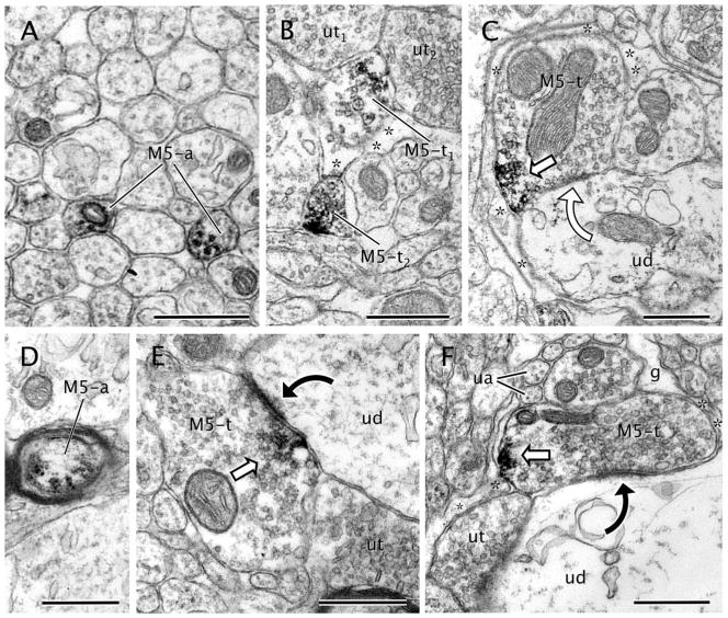
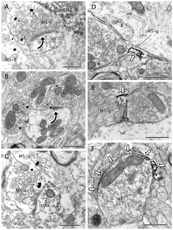
Muscarinic modulation of mesolimbic dopaminergic neurons in the ventral tegmental area (VTA) plays an important role in reward, potentially mediated through the M5 muscarinic acetylcholine receptor (M5R). However, the key sites for M5R-mediated control of dopamine neurons within this region are still unknown. To address this question we examined the electron microscopic immunocytochemical localization of antipeptide antisera against M5R and the plasmalemmal dopamine transporter (DAT) in single sections through the rat VTA. M5R was located mainly to VTA somatodendritic profiles (71%; n = 627), at least one-third (33.2%; n = 208) of which also contained DAT. The M5R immunoreactivity was distributed along cytoplasmic tubulovesicular endomembrane systems in somata and large dendrites, but was more often located at plasmalemmal sites in small dendrites, the majority of which did not express DAT. The M5R-immunoreactive dendrites received a balanced input from unlabeled terminals forming either asymmetric or symmetric synapses. Compared with dendrites, M5R was less often seen in axon terminals, comprising only 10.8% (n = 102) of the total M5R-labeled profiles. These terminals were usually presynaptic to unlabeled dendrites, suggesting that M5R activation can indirectly modulate non-DAT-containing dendrites through presynaptic mechanisms. Our results provide the first ultrastructural evidence that in the VTA, M5R has a subcellular location conducive to major involvement in postsynaptic signaling in many dendrites, only some of which express DAT. These findings suggest that cognitive and rewarding effects ascribed to muscarinic activation in the VTA can primarily be credited to M5R activation at postsynaptic plasma membranes distinct from dopamine transport.

Keywords: acetylcholine; motivation; reinforcement; reward; ultrastructure.


Conflict of interest statement

CONFLICT OF INTEREST STATEMENT

The authors declare that they have no conflict of interest.

Figures

Figure 1
Figure 1
Specific M5 immunoreactivity is expressed in the ventral tegmental area (VTA). A: Distribution of M5 immunoreactivity in the neuropil of a wild-type rat midbrain section at −5.60 mm AP level from Bregma, as indicated in the Paxinos and Watson atlas (1986). Moderate immunoreaction is observed in profiles resembling neuronal somata and continuous dendrites in the VTA (black arrows) and in the adjacent substantia nigra compacta (SN; block arrow). A denser immunoreaction product is seen throughout the neuropil as punctate deposit in thin neuritic processes (white arrowheads). B: Distribution of M5 immunoreactivity in the neuropil of a wild-type mouse VTA. Light M5 immunolabeling is observed in the VTA, detected diffusely in some neuronal somata (block arrow) and also in thin varicose punctate processes that may be axons, as was the case for rats (A). C: Similar immunohistochemical processing in tissue sections collected at the same AP level as in B of an M5 null mouse shows no detectable M5 immunoreactivity. Scale bar = 25 μm in A–C.
Figure 2
Figure 2
M5 labeling in DAT and non-DAT labeled dendrites. A: Immunoperoxidase reaction product for M5 (white block arrow) is seen within a dendrite (M5-d). The labeling is located beneath an apparently symmetric synapse (curved arrow) from an unlabeled axon terminal (ut1). M5-immunoperoxidase is also evident near the plasma membrane of M5-d in a zone near an apposition with another unlabeled axon terminal (ut2). B: An M5-labeled dendrite (M5-d) showing M5-immunogold particles (straight black arrows) localized to cytoplasmic areas near the plasma membrane. Note that one M5-immunogold particle is definitely attached to the postsynaptic density on an asymmetric synapse (curved black arrow) from an unlabeled axon terminal (ut). The M5-d also contacts a dendrite (DAT+M5-d) that contains both DAT-immunoperoxidase and M5-immunogold. An unlabeled axon terminal (ut) apposes both the single M5-d and the dual DAT+M5-d. C: M5-immunogold particles (straight black arrows) are seen within the cytoplasm of a dendrite (M5+DAT-d) also showing intense immunoreactivity for DAT. Note that one of the M5-immunogold particles is located on tubulovesicles of smooth endoplasmic reticulum (ser). This dendrite receives a synapse from an unlabeled terminal (ut). Scale bar = 0.5 μm in A–C.
Figure 3
Figure 3
M5 distribution in neuronal somata. A: M5-immunoperoxidase (white block arrow) is seen in an endomembrane body within a soma (M5-som). B: M5-immunoperoxidase precipitate (white block arrows) is localized to cytoplasm and cytoplasmic tubulovesicles (tv) in a neuronal soma (M5-som). C: An M5-immunoreactive soma (M5-som) shows prominent M5-immunogold particles (black straight arrows) on membranes of endoplasmic reticulum cisterns (RER) and on the plasma membrane. A small dendrite intensely immunoreactive for DAT (DAT-d) is seen in the adjoining neuropil. Asterisks, astrocytic coverage. D: Moderate DAT-immunoperoxidase deposits (white block arrows) are present on the cytoplasmic surface of the plasma membrane or near to endomembranes of the Golgi complex (G) in a soma showing also M5-immunogold particles (black straight arrows) mainly in tubulovesicles (tv) of the endoplasmic reticulum. E: An M5-immunolabeled soma (M5-som) shows widespread immunogold labeling (black straight arrows) on or near portions of the plasma membrane apposing an axon terminal (M5-t). The terminal exhibits one plasmalemmal M5-immunogold particle and apposes an adjacent unlabeled terminal (ut). Scale bar = 0.5 μm in A–E.
Figure 4
Figure 4
M5-immunogold labeling in dendrites with or without DAT. A: M5-immunogold particles (black straight arrows) are seen on the plasma membrane of a small dendrite devoid of DAT. B: A dually labeled small dendrite (DAT+M5-d) contains M5-immunogold particles (black straight arrows) mostly localized on the plasma membrane as well as immunoperoxidase reaction product for DAT (white block arrows). C: M5-immunogold particles (black straight arrows) are localized within the cytoplasm of a transversally sectioned large dendrite (M5-d) without expression of DAT. The dendrite is near another large dendrite (DAT+M5) that shows DAT-immunoperoxidase (white block arrow) and M5-immunogold. D: An unlabeled axon terminal (ut) makes an asymmetric synapse (black curved arrow) with an M5-immunogold (black straight arrows) labeled dendrite (M5-d). One of the M5-immunogold particles is located near the postsynaptic density of the established synapse. The plasmalemmal surface of M5-d is also in contact with an adjoining unlabeled dendrite (ud). E: A dual-labeled dendrite (DAT+M5-d) showing DAT-immunoperoxidase (white block arrows) and M5-immunogold particles (black straight arrows), one of which is localized on the postsynaptic density of an incoming symmetric synapse (white curved arrow) from an unlabeled terminal (ut1). Another unlabeled axon terminal (ut2) also provides synaptic input to DAT+M5-d in a distant opposite portion of M5-d plasma membrane. Scale bar = 0.5 μm in A–E.
Figure 5
Figure 5
Bar graph summarizing the mean ± SE immunogold densities (number of gold particles per profile) in M5R-single and dopamine transporter (DAT)+M5R-dual large (>1 μm) or small (<1 μm) dendrites within the VTA. Mean densities were calculated based on the numbers obtained from 627 VTA dendrites (215 large and 412 small) taken from ultrathin sections from 14 Vibratome sections in four rats (2,288 total immunolabeled profiles) processed for dual labeling. *, P < 0.05, Fisher’s test for cytoplasmic versus plasmalemmal subcellular localization.
Figure 6
Figure 6
M5-immunolabeling in axons and axon terminals. A: M5-immunoperoxidase precipitate is seen within the cytoplasm of transversally sectioned small unmyelinated axons (M5-a) that traverse the neuropil forming bundles with many other unlabeled axons. B: Axon terminal showing a patch of immunoperoxidase labeling for M5 (M5-t1) in small synaptic vesicles contacts two unlabeled axon terminals (ut1,2). A more conspicuous distribution of M5-immunoperoxidase is seen on the plasma membrane of another axon terminal (M5-t2). Asterisks indicate surrounding astrocytic processes. C: M5-immunoperoxidase is localized on portions of the plasma membrane in an axon terminal (M5-t). The terminal is covered by an astrocytic process (asterisk) and located near a contact with an unlabeled dendrite (ud). D: M5-immunoperoxidase (white block arrows) is observed on the plasma membrane and within the cytoplasm of a myelinated axon (M5-a). E: Axon terminal (M5-t) showing M5 immunoreactivity (white block arrow) on endomembranes and small synaptic vesicles localized near or apposed to the plasma membrane. The M5-t forms an asymmetric synapse (black curved arrow) onto an unlabeled dendrite (ud). F: A dense patch of M5-immunoperoxidase (white block arrow) is observed on the plasma membrane of an axon terminal (M5-t). This terminal shows extensive glial coverage (asterisks), including the zone adjacent to the M5-peroxidase reaction product. The M5-t makes an asymmetric synapse (black curved arrow) with a large unlabeled dendrite (ud). g, glia; ua, unlabeled small unmyelinated axon. Scale bar = 0.5 μm in A–F.
Figure 7
Figure 7
M5-immunogold labeling in axon terminals and glial processes. A: Axon terminal showing immunogold particles (black straight arrows) for M5 (M5-t) makes an asymmetric synapse (black curved arrow) with an unlabeled dendrite (ud). The terminal also apposes an intense DAT-immunoperoxidase labeled dendrite (DAT-d) and an M5-immunogold labeled dendrite (M5-d). B: M5-immunogold particles (black straight arrows) are localized to small synaptic vesicles (ssv) within an M5-immunolabeled terminal (M5-t1). The M5-immunogold is also seen on the presynaptic side of an asymmetric synapse (black curved arrow) made with an unlabeled dendrite (ud). This dendrite contacts another M5-immunoreactive terminal (M5-t2) showing both plasmalemmal and cytoplasmic M5-immunogold particles (black straight arrows). C: M5-immunogold particles (black straight arrows) are seen in an axon terminal (M5-t) making contact with a dendrite containing DAT-immunoperoxidase (DAT-d). The M5-t is wrapped by an astrocytic expansion showing M5-immunogold particles (M5-g) and an unlabeled astrocytic profile (asterisks). D: M5-immunoperoxidase reaction product (white block arrow) neatly rims the plasma membrane of an astrocytic process (M5-g). The labeled process is immersed among other unlabeled profiles (ut, unlabeled terminal; ud, unlabeled dendrite) within the neuropil. E: M5-immunoperoxidase is prevalently distributed on the inner surface of the plasma membrane (white block arrow) in an astrocytic profile (M5-g). A gap junction (white block arrow) is seen between the M5-g and an adjacent astrocytic process (asterisks). M5-g also apposes an unlabeled terminal (ut) and an unlabeled dendrite (ud). F: An astrocytic leaflet extension (M5-g) containing M5 immunolabeling (white block arrows) wraps partially around the surface of an unlabeled axon terminal (ut). Scale bar = 0.5 μm in A–F.

Similar articles

Cited by

References

    1. Achour L, Labbé-Jullié C, Scott MGH, Marullo S. An escort for GPCRs: implications for regulation of receptor density at the cell surface. Trends Pharmacol Sci. 2007;29:528–533. - PubMed
    1. Alderson HL, Latimer MP, Blaha CD, Phillips AG, Winn P. An examination of d-amphetamine self-administration in pedunculopontine tegmental nucleus-lesioned rats. Neuroscience. 2004;125:349–358. - PubMed
    1. Araque A, Martín ED, Perea G, Arellano JI, Buño W. Synaptically released acetylcholine evokes Ca2+ elevations in astrocytes in hippocampal slices. J Neurosci. 2002;22:2443–2450. - PMC - PubMed
    1. Augelli-Szafran CE, Blankley CJ, Jaen JC, Moreland DW, Nelson CB, Penvose-Yi JR, Schwarz RD, Thomas AJ. Identification and characterization of m1 selective muscarinic receptor antagonists1. J Med Chem. 1999;42:356–363. - PubMed
    1. Basile AS, Fedorova I, Zapata A, Liu X, Shippenberg T, Duttaroy A, Yamada M, Wess J. Deletion of the M5 muscarinic acetylcholine receptor attenuates morphine reinforcement and withdrawal but not morphine analgesia. Proc Natl Acad Sci U S A. 2002;99:11452–11457. - PMC - PubMed

Publication types

MeSH terms

Substances