. 2013 May;5(5):751-61.
doi: 10.1002/emmm.201302466. Epub 2013 Apr 15.

Disruption of SMIM1 causes the Vel- blood type

Affiliations

Disruption of SMIM1 causes the Vel- blood type

Bryan A Ballif et al. EMBO Mol Med. 2013 May.

Abstract

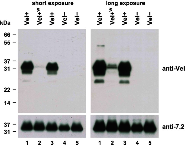
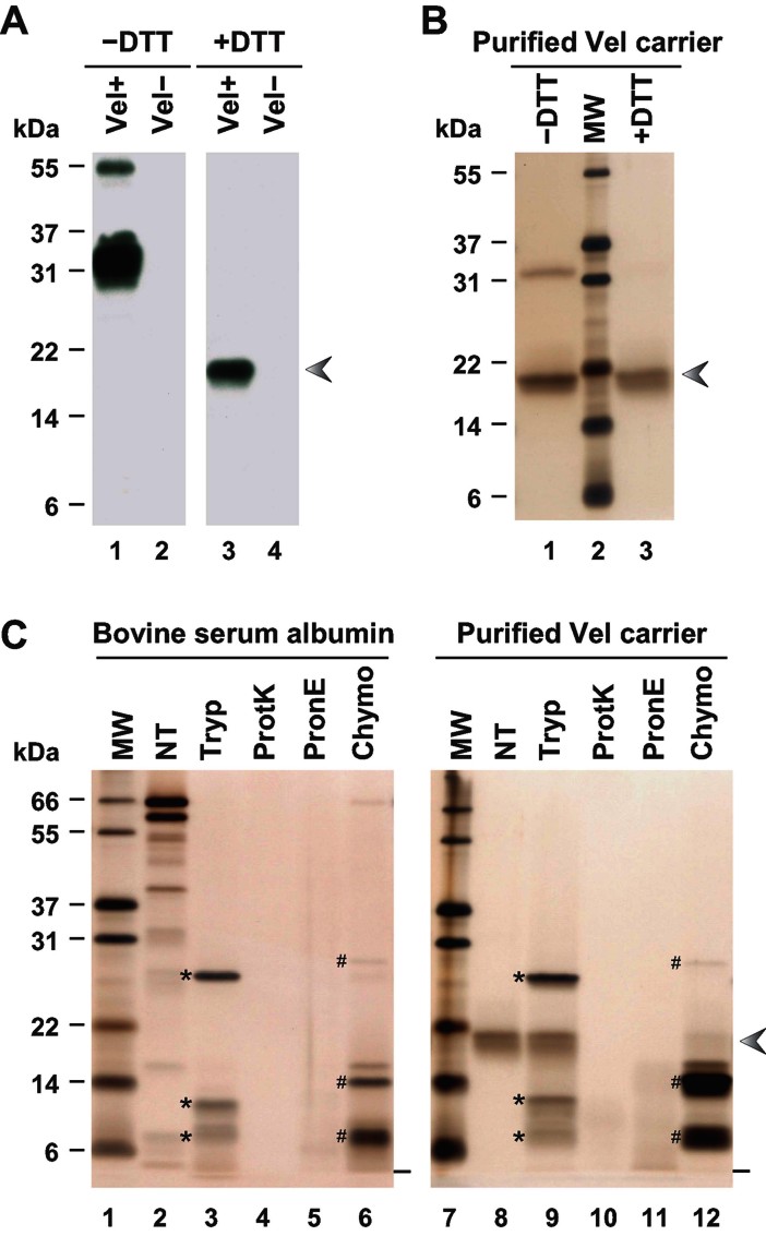
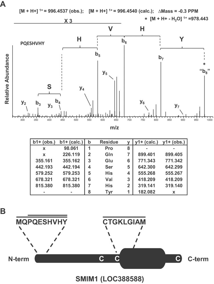
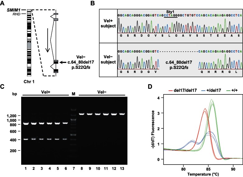
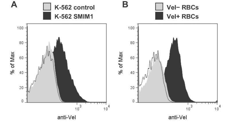
Here, we report the biochemical and genetic basis of the Vel blood group antigen, which has been a vexing mystery for decades, especially as anti-Vel regularly causes severe haemolytic transfusion reactions. The protein carrying the Vel blood group antigen was biochemically purified from red blood cell membranes. Mass spectrometry-based de novo peptide sequencing identified this protein to be small integral membrane protein 1 (SMIM1), a previously uncharacterized single-pass membrane protein. Expression of SMIM1 cDNA in Vel- cultured cells generated anti-Vel cell surface reactivity, confirming that SMIM1 encoded the Vel blood group antigen. A cohort of 70 Vel- individuals was found to be uniformly homozygous for a 17 nucleotide deletion in the coding sequence of SMIM1. The genetic homogeneity of the Vel- blood type, likely having a common origin, facilitated the development of two highly specific DNA-based tests for rapid Vel genotyping, which can be easily integrated into blood group genotyping platforms. These results answer a 60-year-old riddle and provide tools of immediate assistance to all clinicians involved in the care of Vel- patients.


Figures

Figure 1
Figure 1
Anti-Vel Western blot of non-reduced RBC membrane extracts identifies a 32 kDa band present only in Vel+ individuals. RBC membrane extracts prepared from two Vel+ (lanes 1 and 3), one Vel+w (lane 2), and two Vel− (lanes 4 and 5) subjects were resolved by non-reducing SDS–PAGE, and probed with an anti-Vel (top panels). A long exposure time was required to detect the 32 kDa reactive band in the Vel+w subject (top right panel), who later appeared to be heterozygous for the 17 nt deletion in SMIM1. The same blot was reprobed with anti-7.2b (bottom panels).
Figure 2
Figure 2
Purification and protease sensitivity of the 18 kDa reduced form of the Vel antigen carrier. A. RBC membrane extracts prepared from a Vel+ subject (lanes 1 and 3) and a Vel− subject (lanes 2 and 4) were resolved by SDS–PAGE under non-reducing (lanes 1 and 2) or reducing conditions (lanes 3 and 4), and probed with an anti-Vel. B. The 18 kDa material purified from a Vel+ RBC membrane extract was analyzed by SDS–PAGE, under non-reducing (lane 1) or reducing conditions (lane 3), and detected by silver staining. C. Bovine serum albumin (lanes 2–6) and the 18 kDa band purified from a Vel+ RBC membrane extract (lanes 8–12) were treated with trypsin (Tryp), proteinase K (ProtK), pronase E (PronE) or chymotrypsin (Chymo), or were left untreated (NT), and analyzed by reducing SDS–PAGE followed by silver staining. The electrophoretic migration fronts are indicated by dashes, trypsin and its autoproteolytic fragments by asterisks, and chymotrypsin and its autoproteolytic fragments by number signs. The arrowhead indicates the migration of the Vel antigen carrier under reducing conditions.
Figure 3
Figure 3
Mass spectrometry-based identification of the Vel antigen carrier as SMIM1. A. The top panel shows the high resolution MS/MS spectrum acquired in the orbitrap mass spectrometer corresponding to the SMIM1 peptide PQESHVHY that was annotated following de novo peptide sequencing as described in the Methods Section. The bottom panel provides the corresponding calculated (calc.) and observed (obs.) m/z values of the singly charged y- and b-type ions. B. Schematic representation of the SMIM1 protein showing the predicted transmembrane domain, the peptides that were identified by mass spectrometry, and the three cysteine residues potentially involved in dimer formation.
Figure 4
Figure 4
Identification and screening of the 17 nt deletion in SMIM1 that is responsible for the Vel− phenotype. A. Schematic representation of the SMIM1 gene showing its location in chromosome 1 (ISCN 550 band ideogram) and its organization in four exons (black represents coding regions, and grey represents untranslated regions). B. Detail of Sanger sequencing of SMIM1 in a Vel+ subject (top) and a Vel− subject (bottom) showing the homozygous deletion of 17 nt in the SMIM1 coding sequence (c.64_80del17; pS22Qfs) that is responsible for the Vel− blood type. The StyI restriction site on which is based the RFLP analysis is underlined. C. RFLP analysis of the 17 nt deletion in SMIM1 in six random Vel+ subjects (lanes 1–6) and in six random Vel− subjects (lanes 8–13). D. HRM analysis of the 17 nt deletion in SMIM1 in three homozygous (red curves), two heterozygous (blue curves) and three wildtype (green curves) individuals, performed in duplicate by two different operators.
Figure 5
Figure 5
Cell-surface expression of the Vel antigen in SMIM1-transfected K-562 cells compared with RBCs. A. Flow cytometry analysis of native K-562 cells stably transfected with a SMIM1 expression construct (black profile) or with the corresponding empty vector (grey profile) that were labelled with an anti-Vel. The dashed profile corresponds to K-562 SMIM1 cells incubated with only the secondary antibody. B. Flow cytometry analysis of native RBCs taken from a Vel+ subject (black profile) or a Vel− subject (grey profile) labelled with an anti-Vel. The dashed profile corresponds to Vel+ RBCs incubated with only the secondary antibody.

References

    1. Alfred BM, Stout TD, Lee M, Birkbeck J, Petrakis NL. Blood groups, phosphoglucomutase, and cerumen types of the Anaham (Chilcotin) Indians. Am J Phys Anthropol. 1970;32:329–337. - PubMed
    1. Allen FH, Jr, Corcoran PA. Blood groups of the Penobscot Indians. Am J Phys Anthropol. 1960;18:109–114. - PubMed
    1. Altschul SF, Gish W, Miller W, Myers EW, Lipman DJ. Basic local alignment search tool. J Mol Biol. 1990;215:403–410. - PubMed
    1. Ballif BA, Cao Z, Schwartz D, Carraway KL, III, Gygi SP. Identification of 14-3-3epsilon substrates from embryonic murine brain. J Proteome Res. 2006;5:2372–2379. - PubMed
    1. Ballif BA, Carey GR, Sunyaev SR, Gygi SP. Large-scale identification and evolution indexing of tyrosine phosphorylation sites from murine brain. J Proteome Res. 2008;7:311–318. - PubMed

Publication types