

Novel FOXF1 mutations in sporadic and familial cases of alveolar capillary dysplasia with misaligned pulmonary veins imply a role for its DNA binding domain
- PMID: 23505205
- PMCID: PMC3663886
- DOI: 10.1002/humu.22313
Novel FOXF1 mutations in sporadic and familial cases of alveolar capillary dysplasia with misaligned pulmonary veins imply a role for its DNA binding domain
Abstract
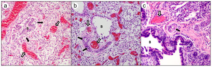
Alveolar capillary dysplasia with misalignment of pulmonary veins (ACD/MPV) is a rare and lethal developmental disorder of the lung defined by a constellation of characteristic histopathological features. Nonpulmonary anomalies involving organs of gastrointestinal, cardiovascular, and genitourinary systems have been identified in approximately 80% of patients with ACD/MPV. We have collected DNA and pathological samples from more than 90 infants with ACD/MPV and their family members. Since the publication of our initial report of four point mutations and 10 deletions, we have identified an additional 38 novel nonsynonymous mutations of FOXF1 (nine nonsense, seven frameshift, one inframe deletion, 20 missense, and one no stop). This report represents an up to date list of all known FOXF1 mutations to the best of our knowledge. Majority of the cases are sporadic. We report four familial cases of which three show maternal inheritance, consistent with paternal imprinting of the gene. Twenty five mutations (60%) are located within the putative DNA-binding domain, indicating its plausible role in FOXF1 function. Five mutations map to the second exon. We identified two additional genic and eight genomic deletions upstream to FOXF1. These results corroborate and extend our previous observations and further establish involvement of FOXF1 in ACD/MPV and lung organogenesis.
© 2013 Wiley Periodicals, Inc.
Conflict of interest statement
The authors declare no conflict of interest.
Figures


References
-
- Ahmed S, Ackerman V, Faught P, Langston C. Profound hypoxemia and pulmonary hypertension in a 7-month-old infant: late presentation of alveolar capillary dysplasia. Pediatr Crit Care Med. 2008;9:e43–6. - PubMed
-
- Al-Hathlol K, Phillips S, Seshia MK, Casiro O, Alvaro RE, Rigatto H. Alveolar capillary dysplasia. Report of a case of prolonged life without extracorporeal membrane oxygenation (ECMO) and review of the literature. Early Hum Dev. 2000;57:85–94. - PubMed
-
- Astorga J, Carlsson P. Hedgehog induction of murine vasculogenesis is mediated by Foxf1 and Bmp4. Development. 2007;134:3753–61. - PubMed
-
- Bhuvanagiri M, Schlitter AM, Hentze MW, Kulozik AE. NMD: RNA biology meets human genetic medicine. Biochem J. 2010;430:365–77. - PubMed
Publication types
MeSH terms
Substances
Grants and funding
- R01 HL086324/HL/NHLBI NIH HHS/United States
- R01 HL065174/HL/NHLBI NIH HHS/United States
- R01 HL101975/HL/NHLBI NIH HHS/United States
- UL1 TR001108/TR/NCATS NIH HHS/United States
- R01 HL116358/HL/NHLBI NIH HHS/United States
- R01 HL097195/HL/NHLBI NIH HHS/United States
- R01 HL105447/HL/NHLBI NIH HHS/United States
- 1R01HL101975-01/HL/NHLBI NIH HHS/United States
- L40 GM102895/GM/NIGMS NIH HHS/United States
- T32 GM008638/GM/NIGMS NIH HHS/United States
- K08 HL105891/HL/NHLBI NIH HHS/United States
- K08 HL130666/HL/NHLBI NIH HHS/United States
- K12 HD068371/HD/NICHD NIH HHS/United States
- L40 AI096442/AI/NIAID NIH HHS/United States
- L40 HL098005/HL/NHLBI NIH HHS/United States
LinkOut - more resources
Full Text Sources
Other Literature Sources
Medical
Molecular Biology Databases
