
Acyl-lipid metabolism

Yonghua Li-Beisson et al. Arabidopsis Book. 2013.

Abstract

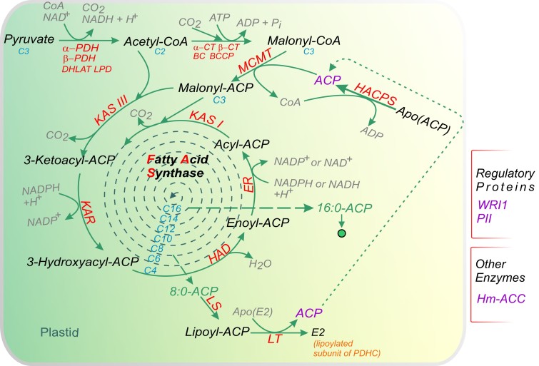
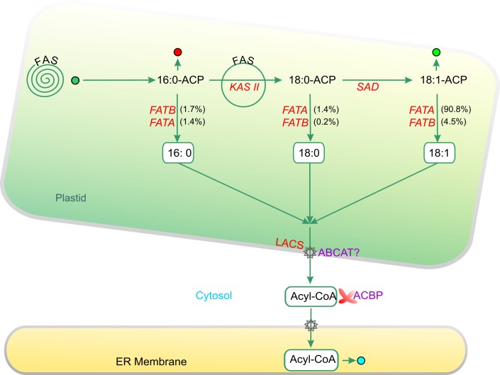
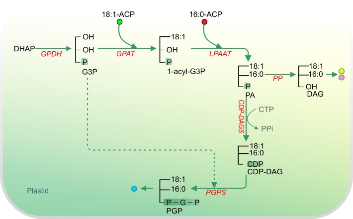
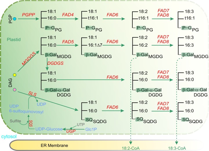
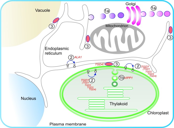
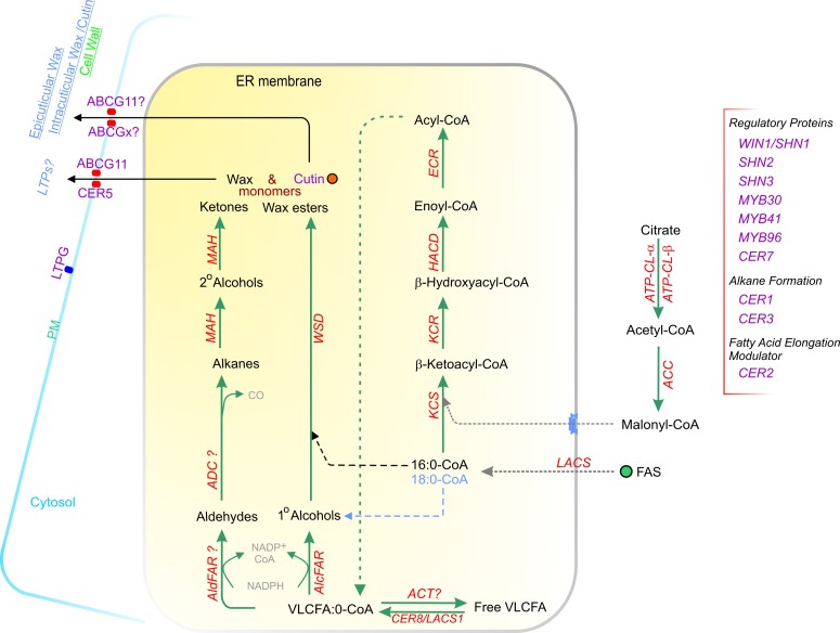
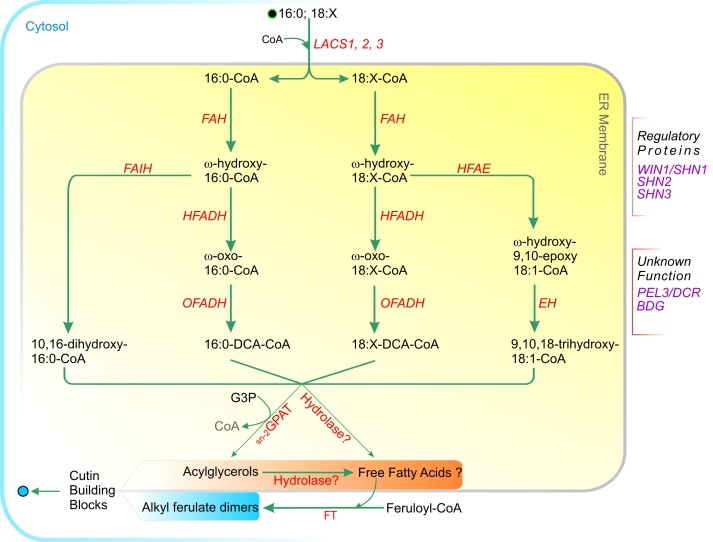
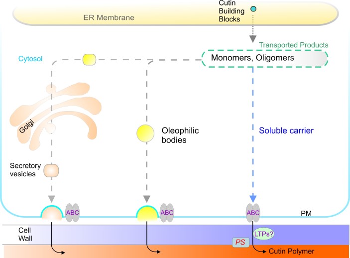
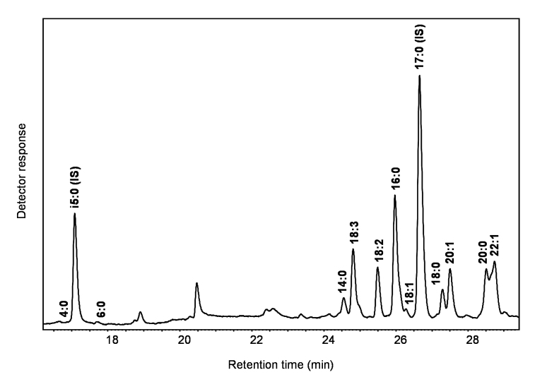
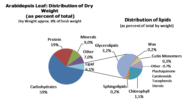
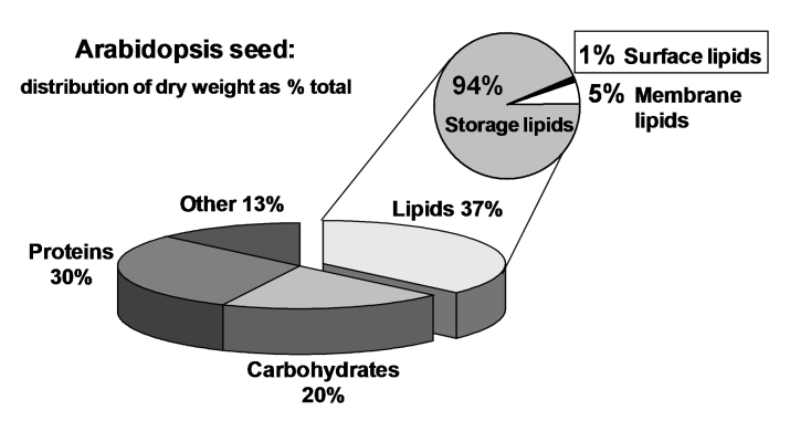
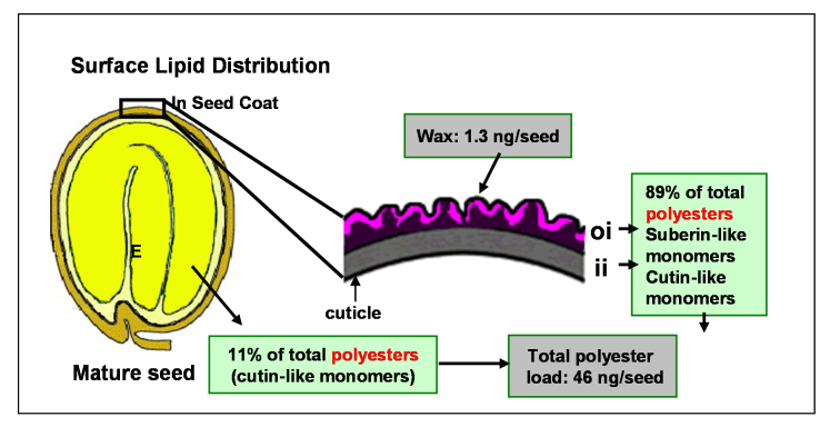
Acyl lipids in Arabidopsis and all other plants have a myriad of diverse functions. These include providing the core diffusion barrier of the membranes that separates cells and subcellular organelles. This function alone involves more than 10 membrane lipid classes, including the phospholipids, galactolipids, and sphingolipids, and within each class the variations in acyl chain composition expand the number of structures to several hundred possible molecular species. Acyl lipids in the form of triacylglycerol account for 35% of the weight of Arabidopsis seeds and represent their major form of carbon and energy storage. A layer of cutin and cuticular waxes that restricts the loss of water and provides protection from invasions by pathogens and other stresses covers the entire aerial surface of Arabidopsis. Similar functions are provided by suberin and its associated waxes that are localized in roots, seed coats, and abscission zones and are produced in response to wounding. This chapter focuses on the metabolic pathways that are associated with the biosynthesis and degradation of the acyl lipids mentioned above. These pathways, enzymes, and genes are also presented in detail in an associated website (ARALIP: http://aralip.plantbiology.msu.edu/). Protocols and methods used for analysis of Arabidopsis lipids are provided. Finally, a detailed summary of the composition of Arabidopsis lipids is provided in three figures and 15 tables.


Figures

Figure 1.
Figure 1.
Fatty Acid Synthesis and Export. (A) De Novo Fatty Acid Synthesis in Plastids of Arabidopsis thaliana. The plastidial pyruvate dehydrogenase complex generates acetyl-coenzyme A that is used as a building block for fatty acid production. Fatty acids are grown by sequential condensation of two-carbon units by enzymes of the fatty acid synthase complex. During each cycle, four reactions occur: condensation, reduction, dehydration, and reduction. Acyl carrier protein is a cofactor in all reactions. Synthesis of a C16 fatty acid requires that the cycle be repeated seven times. During the first turn of the cycle, the condensation reaction is catalyzed by ketoacyl-ACP synthase (KAS) III. For the next six turns of the cycle, the condensation reaction is catalyzed by isoform I of KAS. Finally, KAS II is used during the conversion of 16:0 to 18:0. Abbreviations: ACC, acetyl-CoA carboxylase; ACP, acyl carrier protein; BC, biotin carboxylase; BCCP, biotin carboxyl carrier protein; CT, carboxyltransferase; DHLAT, dihydrolipoamide acetyltransferase; ENR, enoyl-ACP reductase; HACPS, holo-ACP synthase; HAD, hydroxyacyl-ACP dehydrase; KAR, ketoacyl-ACP reductase; KAS, ketoacyl-ACP synthase; LPD, dihydrolipoamide dehydrogenase; LS, lipoate synthase; LT, lipoyltransferase; MCMT, malonyl-CoA: ACP malonyltransferase; PDH, pyruvate dehydrogenase; PDHC, pyruvate dehydrogenase complex. For additional details on genes involved in these reactions, please see http://aralip.plantbiology.msu.edu/pathways/fatty_acid_synthesis.
continued
continued
(B) Fatty Acid Elongation, Desaturation, and Export From Plastid. C16:0 fatty acids produced by the pathways shown in Figure 1A can enter three possible reactions. First, they can be elongated by an additional cycle of fatty acid synthesis. In these cases, KAS II is used during the conversion of 16:0 to 18:0. Alternatively, C16:0 can enter the prokaryotic glycerolipid pathway as shown in Figure 2. Finally, 16:0-ACP can be hydrolyzed by FATB thioesterase to release free fatty acids that are exported from the plastid. Most 18:0-ACP produced by elongation is desaturated by the stearoyl-ACP desaturase.The resulting 18:1-ACP can either enter the prokaryotic glycerolipid pathway (Figure 2) or be hydrolyzed by FATA for export from the plastid. Abbreviations: ABCAT, ABC acyl transporter; ACBP, acyl-CoA binding protein; ACP, acyl carrier protein; FAS, fatty acid synthase; FATA (B), fatty acyl thioesterase A (B); KAS, ketoacyl-ACP synthase; LACS, long-chain acylCoA synthetase; SAD, stearoyl-ACP desaturase. For additional details on genes involved in these reactions, please see http://aralip.plantbiology.msu.edu/pathways/fatty_acid_elongation_desaturation_export_from_plastid.
Figure 2.
Figure 2.
Prokaryotic Galactolipid, Sulfolipid, and Phospholipid Synthesis. (A) The pool of diacylglycerol (DAG) backbones for the so-called prokaryotic lipid synthesis is generated exclusively and entirely inside the plastid: Dihydroxyacetonephosphate (DHAP) is reduced to glycerol 3-phosphate (G3P), which is then first acylated at the sn-1 position with an activated fatty acid (18:1 ACP) by glycerol-3-phosphate acyltransferase (GPAT) to lysophosphatidic acid (LPA). LPA in turn is then acylated at the sn-2 position with 16:0 by the lysophosphatidic acid acyltransferase (LPAAT) to phosphatidic acid (PA). The specificity of this particular acyltransferase for C16 fatty acids distinguishes the prokaryotic from the eukaryotic lipids. PA is either dephosphorylated by PA phosphatase (PP) to DAG, which serves as precursor for galactolipid and sulfolipid biosynthesis (see diagram B), or a CDP-DAG synthase (CDP-DAGS) uses PA to synthesize activated CDP-DAG, which—together with G3P—is required for phosphatidylglycerol phosphate (PGP) synthesis, the precursor of phosphatidylglycerol (PG). Abbreviations: CDP-DAGS, CDP-DAG synthase; DAG, diacylglycerol; DHAP, dihydroxyacetonephosphate; G3P, glycerol 3-phosphate; GPAT, glycerol-3-phosphate acyltransferase; LPA, lysophosphatidic acid; LPAAT, lysophosphatidic acid acyltransferase; PA, phosphatidic acid; PP, PA phosphatase; PGP, phosphatidylglycerol phosphate; PG, phosphatidylglycerol. For additional details on genes involved in these reactions, please see http://aralip.plantbiology.msu.edu/pathways/prokaryotic_galactolipid_sulfolipid_phospholipid_synthesis
continued
continued
(B) PGP is dephosphorylated by the PGP phosphatase (PGPP) to phosphatidylglycerol (PG), which is subjected to several desaturation steps. The relevant fatty acid desaturases (FADs) insert cis (or transΔ3 for 16:0 at the sn-2 position of PG) double bonds at specific sites in the acyl groups at the sn-1 or sn-2 position. The monogalactosyldiacylglycerol transferase (MGDGS) transfers a galactose moiety from UDP-galactose to DAG, thus generating monogalactosyldiacylglycerol. A small proportion of this MGDG is subsequently glycosylated by the also UDP-galactose-dependent digalactoslydiacylglycerol synthase (DGDGS) to digalactosyldiacylglycerol (DGDG) carrying two galactose molecules in its headgroup. Both MGDG and DGDG acyl chains are also characterized by a high degree of desaturation introduced by the various FAD (fatty acid desaturase) enzymes. The first step of sulfoquinovosyldiacylglycerol (SQDG) synthesis is performed by the UDP-glucose pyrophosphorylase (UGP), which catalyzes the formation of UDP-glucose from glucose-1-phosphate and UTP (UGP3Glc1P). UDP-Sulfoquinovose Synthase (SQS) then condenses UDP-Glucose with sulfite to generate UDP 6-sulfoquinovosyl, which is then transferred by the sulfolipid synthase (SLS) on to DAG to serve as sugar donor for the headgroup. Again, the acyl chains in SQDG are desaturated by the various FAD enzymes. Abbreviations: FAD, fatty acid desaturases; MGDGS, monogalactosyldiacylglycerol transferase; PG, phosphatidylglycerol; PGPP, PGP phosphatase; SLS, sulfolipid synthase SQDG, sulfoquinovosyldiacylglycerol; SQS, UDP-sulfoquinovose synthase. For additional details on genes involved in these reactions, please see http://aralip.plantbiology.msu.edu/pathways/prokaryotic_galactolipid_sulfolipid_phospholipid_synthesis_2
Figure 3.
Figure 3.
Eukaryotic Galactolipid and Sulfolipid Synthesis. The eukaryotic galactolipid and sulfolipid pathway differs from its prokaryotic version with regard to the DAG backbone, which is derived from eukaryotic PC and is characterized by a C18 acyl chain in the sn-2 position as well as C16:0 at the sn-1 position in some cases. The exact route and identity of the transported lipid moiety or moieties from the ER is still unknown, yet several possibilities have been discussed: DAG precursors are transported into the plastid involving TGD1, a permease-like protein of the inner chloroplast envelope. Other possibilities involve transport of PC to the chloroplast, where it is then either dephosphorylated by a nonspecific phospholipase C (nsPLC) to DAG or first hydrolyzed to PA by phospholipase D (PLDζ) and then dephosphorylated to DAG by PA phosphatase (PP). Instead of a direct transport, lysophosphatidylcholine (LPC), generated by the phospholipase A2 (PLA2), has also been suggested as a possible intermediate for PC transport into the chloroplast, where acylation by the lysophosphatidylcholine acyltransferase (LPCAT) would revert it to PC. Starting from DAG, the prokaryotic and eukaryotic pathways share the same activities, but they are sometimes encoded by an extra/different set of genes (see text). Note that when a C16:0 acyl chain is present at the sn-1 position, it is not desaturated. Abbreviations: DAG, diacylglycerol; FAD, fatty acid desaturase; LPCAT, lysophosphatidylcholine acyltransferase; nsPLC, nonspecific phospholipase C; PC, phospholipid choline; PLDζ, phospholipase D; PA, phosphatidic acid; PP, PA phosphatase; PP, phosphatidate phosphatase; PLA2, phospholipase A2; TGD1, permease-like protein of inner chloroplast envelope. For additional details on genes involved in these reactions, please see http://aralip.plantbiology.msu.edu/pathways/eukaryotic_galactolipid_sulfolipid_synthesis
Figure 4.
Figure 4.
Eukaryotic Phospholipid Synthesis and Editing. G3P produced by GPDH is converted to PA by sequential acylation reactions regulated by GPAT and LPAAT. PA is converted to CDP-DAG, from which Pl is produced by PIS. Alternatively, the PA is hydrolyzed to DAG by PP. DAGs are combined with CDP-choline and CDP-ethanolamine to produce PC and PE, respectively; the enzyme responsible for these reactions (AAPTs) may have dual substrate specificity. Phosphoethanolamine produced by EK is an important intermediate for PE and PC biosyntheses. Cytosolic CKs also provide phosphocholine for PC biosynthesis from free choline, which may be recovered from PC by PLDs or derived from other tissues by phloem translocation. Phosphoethanlamine is converted to PE via CDP-ethanolamine, whereas the same substrate is methylated to phosphocholine by PEAMT and then converted to PC via CDP-choline. PE is a substrate for PS biosynthesis by BE-PSS, whereas PS is converted to PE by PSD. PC is the major substrate for desaturation and acyl editing. Acyl editing involves a dynamic exchange of fatty acids predominatly between the sn-2 (but also sn-1 ) position of PC and acyl-CoA pools, which may be regulated by PLA2 and LPCAT. PLMT catalyzes a putative pathway to PC via methylation of methyl-PE. In addition to the above pathways, the conversion of PGP to PG by PGP phosphatase (PGPP) is not shown. Abbreviations: AAPT, aminoalchoholphosphotransferase; BE-PSS, base-exchange-type phosphatydylserine synthase; CCT, CTP:phosphorylcholine cytidylyltransferase; CDP-DAGS, CDP-diacylglycerol synthetase; CK, choline kinase; DAG, diacylglycerol; DAG-CPT, CDP-choline:diacylglycerol cholinephosphotransferase; DAG-EPT, CDP-ethanolamine:diacylglycerol cholinephosphotransferase; DHAP: dihydroxyacetone phosphate; EK, ethanolamine kinase; FAD2, oleate desaturase; FAD3, linoleate desaturase; G3P, glycerol 3-phosphate; GIc 6-P, glucose 6-phosphate; GPAT, glycerol-3-phosphate acyltransferase; GPDH, glycerol-phosphate dehydrogenase; lno 3-P, inositol 3-phosphate; LACS, long chain acyl-CoA synthetase; LPA, lysophosphatidic acid; LPAAT, lysophosphatidic acid acyltransferase; LPC, lysophosphatidylcholine; LPCAT: lysophosphatidylcholine acyltransferase; LPL, lysophospholipid; lysophospholipid acyltransferase; MIPS, myo-inositol-3-phosphate synthase; PA, phasphatidic acid; PC, phosphatidylcholine; PE, phosphatidylethanolamine; PEAMT, phosphoethanolamine N-methyltransferase; PECT, CTP:phosphorylethanolamine cytidyltransferase; PG, phosphatidylglycerol; PGP, phosphatidyglycerophosphate; PGPS, phosphatidylglycerophosphate synthase; Pl, phosphatidylinositol; PIS, phosphatidylinositol synthase; PLA2, PLMT, N-methylphospholipid methyltransferase; phospholipase A2 (Cytosolic); PLD, phospholipase D; PP, phosphatidate phosphatase; PS, phosphatidylserine; PSD, phosphatidylserine decarboxylase. For additional details on genes involved in these reactions, please see http://aralip.plantbiology.msu.edu/pathways/eukaryotic_phospholipid_synthesis_editing
Figure 5.
Figure 5.
Sphingolipid Biosynthesis in Arabidopsis. (A) Biosynthesis of Sphingobases and Ceramides in the ER. Palmitoyl-CoA for the serine-palmitoyl transferase (SPT) reaction and ceramide synthesis is captured by SPT at the cytosolic face of the ER membrane. Very long chain fatty acids (VLCFA) also generated by elongation at the cytosolic face of the ER are incorporated into ceramides by ceramide synthase (CS). Phosphorylation of sphingobases by long-chain base kinase (LCBK) may facilitate their increased solubilization in the cytosol. Abbreviations: DSD, dihydrosphinganine Δ4-desaturase; FA2H, fatty acid 2-hydroxylase; KSR, 3-ketosphinganine reductase; LCBPP, long-chain base phosphate phosphatase; SBH, sphingoid base hydroxylase; SLD, sphingolipid Δ8-desaturase; STP, sphingosine transfer protein. For additional details on genes involved in these reactions, please see http://aralip.plantbiology.msu.edu/pathways/sphingolipid_biosynthesis
continued
continued
(B) Fate of Ceramide Within the Cell. Synthesis of ceramide in the ER results in two distinct pools of ceramide. One pool is glycosylated by glucosylceramide synthase (GCS). The other pool is transported to the Golgi apparatus, where it received a phosphorylinositol headgroup from phosphatidylinositol through the action of inositolphosphorylceramide synthase (IPCS). Both complex sphingolipids end up in the plasma membrane where they are eventually turned over by hydrolysis. Abbreviations: CERK, ceramide kinase; CES, ceramidase; DPL1, dihydrosphingosine phosphate lyase; GCG, glucosylceramide glucosidase; GIPCS, glycosylinositolphosphoylceramide synthase; LCBK, long-chain base kinase; PLC, phospholipase C; STP, sphingosine transfer protein. For additional details on genes involved in these reactions, please see http://aralip.plantbiology.msu.edu/pathways/sphingolipid_biosynthesis_2
Figure 6.
Figure 6.
Mitochondrial Fatty Acid Synthesis. (A) Mitochondrial Fatty Acid and Lipoic Acid Synthesis. In mitochondria, fatty acids are synthesized by type II fatty acid synthase located in the matrix using malonate as a precursor. Octanoic acid, which is a major fatty acid synthesized in mitochondria, is used for biosynthesis of lipoic acid that is bound to the H protein of the GDC complex and the E2 subunits of PDH, KGDH, and BCKADH complexes as a cofactor. Abbreviations: BCKADH, branched chain α-keto acid dehydrogenase; ENR, enoyl-ACP reductase; GDC, glycine decarboxylase; HAD, 3-hydroxyacyl-ACP dehydrase; HACPS, holo-ACP synthase; KAR, 3-ketoacylACP reductase; KAS, 3-ketoacyl-ACP synthase; KGDH, α-ketoglutarate dehydrogenase; LS, lipoic acid synthase; LT, lipoyltransferase; MAS, Malonyl-ACP synthetase; MCAMT, malonyl-CoA: ACP malonyltransferase; MCS, malonyl-CoA synthase; PDH, pyruvate dehydrogenase. For additional details on genes involved in these reactions, please see http://aralip.plantbiology.msu.edu/pathways/mitochondrial_fatty_acid_lipoic_acid_synthesis
continued
continued
(B) Arabidopsis mitochondria can channel 3-hydroxy-14:0-ACP into lipopolysaccharide via a pathway homologous to the E. coli Lipid A pathway at least as far as Lipid IVA . Lipid IVA is further metabolized by a homolog to LpxK, which in E. coli incorporates 3-deoxy-D-manno-octulosonic acid (Kdo) moieties, but products of this enzyme have not yet been detected in Arabidopsis. Abbreviations: LpxA, UDP-N-acetylglucosamine acyltransferase; LpxB, Lipid-A-disaccharide synthase; LpxC, UDP-3-O-acyl N-acetylglucosamine deacetylase; LpxD, UDP-3-0-(3-hydroxymyristoyl)glucosamine N-acyltransferase; LpxH, UDP-2,3-diacylglucosamine pyrophosphatase; LpxK, tetraacyldisaccharide 4'-kinase; KdtA, 3-deoxy-D-manno-octulosonic acid (Kdo) transferase. For additional details on genes involved in these reactions, please see http://aralip.plantbiology.msu.edu/pathways/mitochondrial_lipopolysaccharide_synthesis
continued
continued
(C) Mitochondrial Phospholipid Synthesis. Although mitochondrial membranes contain phosphatidylcholine (PC), phosphatidylethanolamine (PE), phosphatidylinositol (Pl), and cardiolipin (CL) as major phospholipids, PC, PE, and Pl are mainly synthesized in the ER and transported to mitochondria. Phosphatidylserine (PS) synthesized in the ER is also transported to mitochondria and is used for biosynthesis of PE by PS decarboxylase. It has been suggested that CL is synthesized de novo in mitochondria from glycerol 3-phosphate and acyl-ACP. However, a part of PG and CDP-DAG synthesized in the ER might be transported to mitochondria and used for biosynthesis of CL. Abbreviations: ALCAT, acyl-CoA:monolysocardiolipin acyltransferase; CDP-DAG, CDP-diacylglycerol; CDP-DAGS, CDP-diacylglycerol synthase; CLD, cardiolipin deacylase; CLS, cardiolipin synthase; DAG3P, diacylglycerol 3-phosphate; G3P, glycerol-3-phosphate; GPAT, glycerol-3-phosphate acyltransferase; LPAAT, lysophosphatidic acid acyltransferase; PGP, phosphatidylglycerolphosphate; PGPP, phosphatidylglycerol-phosphate phosphatase; PGPS, phosphatidylglycerol-phosphate synthase; PSD, phosphatidylserine decarboxylase; TAZ, Taffazzin (cardiolipin transacylase); VLCFA, very long chain fatty acid. For additional details on genes involved in these reactions, please see http://aralip.plantbiology.msu.edu/pathways/mitochondrial_phospholipid_synthesis
Figure 7.
Figure 7.
Triacylglycerol Synthesis in Arabidopsis. Newly synthesized acyl-CoAs (shown in green) are exported from the plastid to the endoplasmic reticulum (ER) membrane, where they join a larger acyl-CoA pool. A series of reactions adds the fatty acids to a glycerol backbone to form triacylglycerol (TAG). TAG molecules coalesce to form oil droplets that bud out of the ER membrane. Additional reactions integrate the synthesis of TAG with that of phosphatidylcholine (PC), an important membrane lipid. Fatty acids can be further desaturated when they are part of the PC pool. Abbreviations: CCT, choline-phosphate cytidylyltransferase; CK, choline kinase; DAG, diacylglycerol; DAG-CPT, diacylglycerol cholinephosphotransferase; DAGTA, diacylglycerol transacylase; DGAT, acyl-CoA: diacylglycerol acyltransferase; DHAP, dihydroxyacetone phosphate; FAD2, oleate desaturase; FAD3, linoleate desaturase; G3P, glycerol-3-phosphate; GPAT, glycerol-3-phosphate acyltransferase; GPDH, glycerol-3-phosphate dehydrogenase; LPA, 2-lysophosphatidic acid; LPAAT, 2-lysophosphatidic acid acyltransferase; LPC, 2-lysophosphatidylcholine; LPCAT, 2-lysophosphatidylcholine acyltransferase; MAG, monoacylglycerol; MAGAT, monoacylglycerol acyltransferase; PA, phosphatidic acid; PDAT, phospholipid:diacylglycerol acyltransferase; PDCT, phosphatidylcholine:diacylglycerol cholinephosphotransferase; PLA2, phospholipase A2; PP, phosphatidate phosphatase. For additional details on genes involved in these reactions, please see http://aralip.plantbiology.msu.edu/pathways/triacylglycerol_biosynthesis
Figure 8.
Figure 8.
Summary of Intracellular Lipid Transport Processes in Arabidopsis. Process 1a. A vesicular mechanism is proposed for trafficking of membrane proteins and certain lipids between cellular organelles in the secretory pathways. Process 1b. The vesicular transfer of lipids from the inner chloroplast envelope to the thylakoids. Process 2. Polar lipid flipping across the ER and outer and inner chloroplast envelope membranes. Process 3. Lipid transfer through membrane contact sites between ER and plastids, mitochondria, plasma membrane, or vacuoles. Abbreviations: ALA1, aminophospholipid ATPase; TGD1, permease-like protein of inner chloroplast envelope; TGD2, phosphatidic acid-binding protein; TGD3, ATPase; TGD4, protein involved in lipid transport; VIPP1, vesicle-inducing protein in plastids. For additional details on genes involved in these reactions, please see http://aralip.plantbiology.msu.edu/pathways/lipid_trafficking
Figure 9.
Figure 9.
Fatty Acid Elongation and Cuticular Wax Biosynthesis Pathways for the formation in epidermal cells of very long chain fatty acyl-CoAs (VLCFAs) and cuticular waxes. Analogous FA elongation pathways occur in all cells to produce VLCFA for sphingolipids, and in seeds for elongation of 18:1 to 20:1 and 2:1. Saturated C16 and C18 fatty acids, produced in the plastid (FAS), are esterifed to coenzyme A (CoA) by LACS. 16:0- and 18:0-CoA esters are elongated by reiteratively adding a two-carbon unit to the carboxy terminal to generate VLCFA wax precursors between 20 and 34 carbons in length. The endoplasmic reticulum (ER)-associated fatty acid elongase consists of four enzymatic activities (KCS, KCR, HACD, and ECR) that work sequentially to elongate the fully saturated acyl-CoA chains. Once the VLCFAs are synthesized, they are converted to cuticular waxes by the coordinated activities of a number of enzymes. A proportion of the elongated acyl-CoAs are hydrolyzed by a thioesterase to release free fatty acids. Conversely, free VLCFAs can be reactivated into CoA esters by LACS1. However, most of the elongated fatty acyl-CoAs enter one of two wax biosynthetic pathways: an alkane-forming pathway (also known as the decarbonylation pathway) that produces aldehydes, alkanes, secondary alcohols, and ketones, and a primary alcohol forming pathway (also known as the acyl reduction pathway) that produces primary alcohols and wax esters. The waxes are transported to the plasma membrane (PM) by an unknown mechanism, and transport across the PM is facilitated by ABC transporters (CER5 and ABCG). Lipid transfer proteins (LTPs) may be involved in transport of waxes across the cell wall in order to reach the final destination at the cuticle. The elongase, decarbonylation, and acyl reduction pathways are localized to the ER or ER membrane, however the location of the LACS enzymes has yet to be established. Abbreviations: ABCG, ATP-binding cassette transporter G subfamily; ACC, acetyl-CoA carboxylase (Cytosolic; Homomeric); ACT, acyl-CoA thioesterase; ADC, aldehyde decarbonylase; AIcFAR, alcohol-forming fatty acyl-CoA reductase; AIdFAR, aldehyde-forming fatty acyl-CoA reductase; ATP-CL-α(β), ATP citrate lyase α(β) subunit; CER5, ECERIFERUM 5; ECR, enoyl-CoA reductase; HACD, hydroxyacyl-CoA dehydrase; KCS, ketoacyl-CoA synthase; KCR, ketoacyl-CoA reductase; LACS, long-chain acyl-CoA synthetase; LTP, lipid transfer protein; LTPG, GPl-anchored lipid transfer protein; MAH, midchain alkane hydroxylase; MAO, midchain alkanol oxidase; VLCFA, very long-chain fatty acid; WSD, bifunctional wax ester synthase/diacylglycerol acyltransferase. For additional details on genes involved in these reactions, please see http://aralip.plantbiology.msu.edu/pathways/fatty_acid_elongation_wax_biosynthesis
Figure 10.
Figure 10.
Proposed Pathways for the Synthesis of the Cutin Polyester. (A) This biosynthetic scheme represents one possible scenario for the synthesis of cutin building blocks (possibly acylglycerols and free fatty acids) and their assembly. It is assumed that the sequential order of reaction on acyl chains is activation to coenzyme-A by LACS, oxidation by P450s, and esterification to glycerol-3-phosphate by GPATs.Abbreviations: EH, epoxide hydrolase; FAH, Fatty acyl ω-hydroxylase; FAIH, Fatty acyl in-chain hydroxylase; FT, Feruloyl Transferase; G3P, glycerol-3-phosphate; GPAT, glycerol 3-phosphate acyltransferase; HFADH, ù-hydroxy fatty acyl dehydrogenase; HFAE, ω-hydroxy fatty acyl epoxygenase; LACS, long-chain acyl-coA synthetase; LTP, lipid transfer proteins; OFADH, ω-oxo fatty acyl dehydrogenase; PS, polyester synthase. For additional details on genes involved in these reactions, please see http://aralip.plantbiology.msu.edu/pathways/cutin_synthesis_transport
continued
continued
(B) The site of polymerization is now known as extracellular, but, the nature of the molecules transported across the cytoplasm and cell wall remain unknown (adapted from Pollard et al., 2008). Abbreviations: EH, epoxide hydrolase; FAH, Fatty acyl ω-hydroxylase; FAIH, Fatty acyl in-chain hydroxylase; FT, Feruloyl Transferase; G3P, glycerol-3-phosphate; GPAT, glycerol 3-phosphate acyltransferase; HFADH, ù-hydroxy fatty acyl dehydrogenase; HFAE, ω-hydroxy fatty acyl epoxygenase; LACS, long-chain acyl-coA synthetase; LTP, lipid transfer proteins; OFADH, ω-oxo fatty acyl dehydrogenase; PS, polyester synthase. For additional details on genes involved in these reactions, please see http://aralip.plantbiology.msu.edu/pathways/cutin_synthesis_transport_2
Figure 11.
Figure 11.
Proposed Pathways for the Synthesis of Suberin Aliphatic Polyester. This biosynthetic scheme represents one possible scenario for the synthesis of suberin building blocks (i.e. acyl glycerols), where we assume that acyl oxidation occurs after elongation and before esterification to glycerol (or G3P). So far, the order of such reactions remains unclear. Likewise, subcellular compartments where these pathways may take place are unknown, but fatty acid-modification and acyl glycerol synthesis steps are believed to take place in the endoplasmic reticulum (ER) membranes. Transport mechanisms required to transfer monomers or oligomers/polymers to the site of polyester assembly remain hypothetical (Beisson et al., 2012). One ABC transporter has been associated with both cutin and suberin synthesis (ABCG11; Panikashvili et al., 2010); a membrane-bound LTP (LTPG1) have been associated with cuticular wax synthesis (DeBono et al., 2009), and homologous proteins could be involved in suberin assembly. Polyester synthase(s), required to link monomers to produce high molecular weight suberin polyesters, as well as polymerization site(s) remain unknown. However, two extracellular proteins, BDG (Kurdyukov et al., 2006) and CD1 (Yeats et al., 2012), belonging to the αβ-hydrolase superfamily and the GDSL lipase/hydrolase family respectively, have been proposed as a cutin synthases. (A) Synthesis of aliphatic suberin monomers. Abbreviations: ABC, ATP binding cassette transporter; FAR, alcohol-forming fatty acyl-CoA reductase; ASFT, aliphatic suberin feruloyl transferase; BDG, BODYGUARD; CD1, cutin deficient 1; FAH, Fatty acyl ω-hydroxylase; GDSL, family of serine lipase/esterase enzymes; G3P, glycerol-3-phosphate; sn-2 GPAT, glycerol 3-phosphate acyltransferase; HFADH, ω-hydroxy fatty acyl dehydrogenase; LACS, long chain acylcoA synthase; LTP, lipid transport protein; LTPG, glycosylphosphatidylinositol (GPI)-anchored protein; OFADH, ω-oxo fatty acyl dehydrogenase; ω-hydroxy-FA, ω-hydroxy fatty acid. (B) Synthesis of aromatic suberin monomers for the aliphatic domain. Abbreviations: 4CL, 4-coumarate ligase; ASFT, aliphatic suberin feruloyl transferase; C3H, Coumarate 3-hydroxylase; C4H, Cinnamate 4-hydroxylase; CCoAOMT, caffeoyl coA O-methyltransferase; MAG, monoacylglycerol. (C) Potential transport mechanisms involved in the assembly of suberin polyesters. Abbreviations: ABC, ATP binding cassette transporter; ABCG1/DSO, a member of the ABCG subgroup of the ABC transport family; BDG, BODYGUARD; CCoAOMT, caffeoyl coA O-methyltransferase; GDSL, family of serine lipase/ esterase enzymes; LTP, lipid transport protein; LTPG, glycosylphosphatidylinositol (GPI)-anchored protein; PS, polyester synthase. For additional details on genes involved in these reactions, please see http://aralip.plantbiology.msu.edu/pathways/suberin_synthesis_transport
continued
continued
(B) Synthesis of aromatic suberin monomers for the aliphatic domain. Abbreviations: 4CL, 4-coumarate ligase; ASFT, aliphatic suberin feruloyl transferase; C3H, Coumarate 3-hydroxylase; C4H, Cinnamate 4-hydroxylase; CCoAOMT, caffeoyl coA O-methyltransferase; MAG, monoacylglycerol. For additional details on genes involved in these reactions, please see http://aralip.plantbiology.msu.edu/pathways/suberin_synthesis_transport_2
continued
continued
(C) Potential transport mechanisms involved in the assembly of suberin polyesters. Abbreviations: ABC, ATP binding cassette transporter; BDG, BODYGUARD; DCR, DEFECTIVE IN CUTICULAR RIDGES; CCoAOMT, caffeoyl coA O-methyltransferase; FAH, Fatty acyl ω-hydroxylase; G3P, glycerol LTP, lipid transport protein; PS, polyester synthase For additional details on genes involved in these reactions, please see http://aralip.plantbiology.msu.edu/pathways/suberin_synthesis_transport_3
Figure 12.
Figure 12.
Triacylglycerol and Fatty Acid Degradation. Triacylglycerols are first degraded by cellular lipases to release free fatty acids from glycerol. Then reactions involved in the β-oxidation of straight-chain fatty acids occur in the peroxisome (adapted from Goepfert and Poirier, 2007). “2x cycles” indicates that the molecule undergoes two successive rounds through the β-oxidation cycle. CTS is an ABC transporter involved in the import of β-oxidation substrates to the peroxisome. (A) Examples of the involvement of auxiliary activities in the degradation of oleic acid (C18:1Δ9c/s). (B) The core β-oxidation cycle. (C) Example of the involvement of the monofunctional type 2 enoyl-CoA hydratase (ECH2) in the degradation of fatty acids with a cis double bond on an even-numbered carbon (C18:1Δ6cis, petroselinic acid). Abbreviations: ACT, acyl-CoA thioesterase; ACX, acyl-CoA oxidase; DCH, D3,5,D2,4-dienoyl-CoA isomerase; lsom, D3,D2-enoyl-CoA isomerase; KAT, 3-ketothiolase; LACS, long chain acyl-CoA synthetase; MAGL, monoacylglycerol lipase; MFP, multifunctional protein containing a 2E-enoyl-CoA hydratase (ECH) and a 3S-hydroxyacyl-CoA dehydrogenase (HACDH); Red, 2,4-dienoyl-CoA reductase; TAGL, triacylglycerol lipase. For additional details on genes involved in these reactions, please see http://aralip.plantbiology.msu.edu/pathways/triacylglycerol_fatty_acid_degradation
Figure 13.
Figure 13.
Separation of the Methyl Ester Derivatives of Fatty Acids From Arabidopsis Leaf.A capillary fused silica column coated with (50% Cyanopropyl)-methylpolysiloxane (DB-23; J&W Scientific) was temperature-programmed from 140°C to 260°C at 10°C min-1 with helium as carrier gas. One µL of sample was injected in a 270°C inlet with a 30:1 split ratio. Methyl ester derivatives were detected using a FID at 270°C. (Courtesy of Dr. Imad Ajjawi, Michigan State University, Michigan)
Figure 14.
Figure 14.
TLC Image Examples and Flow Chart for Analysis of Radiolabeled Glycerolipids From [14C]acetate and [14C]glycerol Labeled Soybean Embryos. Examples of the major TLC systems used during analysis of radiolabeled glycerolipid classes, molecular species, and FA composition. Molecular species (e.g. SSS, SM, etc.) are represented as a combination of two or three FA, total saturates, S; monoenes (18:1), M; dienes (18:2), D; trienes (18:3), T. (A) Neutral lipid class TLC, pictured 6 min [14C]acetate labeling. (B) Polar lipid class TLC, pictured 6 min [14C]acetate labeling. (C) TAG molecular species AgNO3-TLC, pictured 30 min [14C]glycerol labeling. (D) Molecular species separation of PC (after phospholipase C digestion) and DAG as 3-acetyl-1,2-diacyl-glycerols, pictured 6 min [14C]acetate labeled PC. (E) FAME AgNO3-TLC, pictured FAMEs from different 6 min [14C]acetate labeled PC molecular species separated in D. See (Bates et al., 2009) for labeling experiment details and TLC protocols. Abbreviations: DAG, diacylglycerol; FFA, free fatty acid; PG, phosphaglycerol; Pl, phosphainostol; PE, phosphoethanolamine; PC, phosphacholine; PLC, phosphalipase C; PS, phosphaserine; PUFA, polyunsaturated fatty acid; TAG, triacylglycerol;
Figure 15.
Figure 15.
GC-FlD Analysis of Fatty Acid Methyl Esters Derived From Neutral Lipids Isolated From Arabidopsis Col-0 Seed. Neutral lipids were isolated from wild type Col-0 seed and transmethylated and the subsequent fatty acid methyl esters separated on a J+W DB-23 (50% cyanopropyl) methylpolysiloxane 30 m column and detected using a flame ionization detector. The column temperature was initially held at 150 °C for 3 min, then increased to 240 °C at rate of 10 °C min-1, and then held at 240°C for 10 min. Tripentadecanoin (15:0) and triheptadecanoin (C17:0) were used as seed lipid extraction and transmethylation internal standards, respectively. IS = internal standard (Courtesy of Dr. Timothy Durrett, Michigan State University)
Figure 16.
Figure 16.
TAG Profile of Col-0 Dry Seed. The total ion current (A) is used for the data-dependent selection of parent ions (inset in (B)), which are subjected to MS2 fragmentation analysis. Up to three daughter diacylglycerol (DAG) fragments are generated per triacylglycerol (TAG); in this example, two are generated because there are only two unique constituent fatty acids. The neutral losses for the observed DAG fragments (after correction for adducts) can be used for a constrained calculation of fatty acid identity (B). The correct stoichometric combination of these fatty acids to give the parent ion is used to calculate the TAG molecular formula.
Figure 17.
Figure 17.
High-Performance Liquid Chromatography Trace of Acyl-CoA From a Col-0 Seedling Extract.The extract was made as described from 30 seedlings (grown on media plates) harvested at 5 days after imbibition. This developmental stage encompasses both rapid storage lipid breakdown and de novo lipid biosynthesis associated with seedling establishment. Peak areas and heights are directly proportional to the absolute amounts of the indicated acyl-CoAs. lS = internal standard.
Figure 18.
Figure 18.
Wax Crystals of Arabidopsis Stems Viewed by Confocal Microscopy and Cryo-Scanning Electron Microscopy. (A) Nile Red and confocal microscopy of the Arabidopsis stem epidermis surface with wax crystal structures. (B) CryoSEM of Arabidopsis stem epidermis surface with wax crystal structures (closed arrowhead).Bars = 8 µm (A) and 5 µm (B).
Figure 19.
Figure 19.
Arabidopsis Hypocotyl Cells Damaged During Handling Prior to Live Imaging. (A) Hypocotyl cells stained with FM4-64 are vesiculated after crushing (arrowheads). (B) Yellow fluorescent protein tagged glycosylphosphatidyl-inositol (GPI)anchored lipid transfer protein (YFP-LTPG) is plasma membrane localized in undamaged cells. In contrast, due to damage during sample dissection, cells along the incision (dotted line) contain numerous large vesicles. Bars = 14 µm.
Figure 20.
Figure 20.
Arabidopsis Stem Epidermal Cells Stained With FM4-64. Prolonged exposure to FM4-64 stains endocytic compartments (arrowheads) in addition to the plasma membrane. Bar = 9 µm.
Figure 21.
Figure 21.
Relative Distribution of Lipids and Other Components of Arabidopsis Leaf. Data adapted from Browse and Somerville (1994). (Prepared by John Ohlrogge)
Figure 22.
Figure 22.
Relative Distribution of Lipids and Other Components of Arabidopsis Seeds. Relative contribution of storage lipids and proteins were obtained from Y.H. Li et al., 2006. Percentage of membrane glycerolipids relative to total lipids is from Ohlrogge and Browse, 1995. Content of surface lipids is from Molina et al. (2006) and Beisson et al. (2007). (Prepared by Isabel Molina)
Figure 23.
Figure 23.
Distribution of Extracellular Lipids in Mature Seeds. Values of total polyester monomers and distribution between seed coat and embryo (inferred from B. napus data) are from Molina et al. (2006). Distribution of polyester monomers in inner integument (ii) and outer integument (oi) summarize results from Molina et al. (2008). Surface wax load was reported by Beisson et al. (2007). (Prepared by Isabel Molina)

References

    1. Aarts M.G.M., Keijzer C.J., Stiekema W.J., Pereira A. Molecular characterization of the CER1 gene of arabidopsis involved in epicuticular wax biosynthesis and pollen fertility. Plant Cell. 1995;7:2115–2127. - PMC - PubMed
    1. Acosta I.F., Farmer E.E. The Arabidopsis Book. Rockville, MD: American Society of Plant Biologists; 2010. Jasmonates. - PMC - PubMed
    1. Adham A.R., Zolman B.K., Millius A., Bartel B. Mutations in Arabidopsis acyl-CoA oxidase genes reveal distinct and overlapping roles in beta-oxidation. Plant J. 2005;41:859–874. - PubMed
    1. Agrawal V.P., Kolattukudy P.E. Biochemistry of suberization: Omega-hydroxyacid oxidation in enzyme preparations from suberizing potato tuber disks. Plant Physiol. 1977;59:667–672. - PMC - PubMed
    1. Agrawal V.P., Kolattukudy P.E. Purification and characterization of a wound-induced omega-hydroxyfatty acid:NADP oxidoreductase from potato tuber disks (Solanum tuberosum L.). Arch. Biochem. Biophys. 1978;191:452–465. - PubMed