. 2013;9(3):e1003307.
doi: 10.1371/journal.pgen.1003307. Epub 2013 Mar 7.

Distinct molecular strategies for Hox-mediated limb suppression in Drosophila: from cooperativity to dispensability/antagonism in TALE partnership

Affiliations

Distinct molecular strategies for Hox-mediated limb suppression in Drosophila: from cooperativity to dispensability/antagonism in TALE partnership

Nagraj Sambrani et al. PLoS Genet. 2013.

Abstract

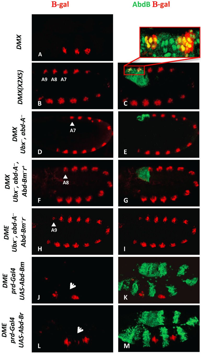
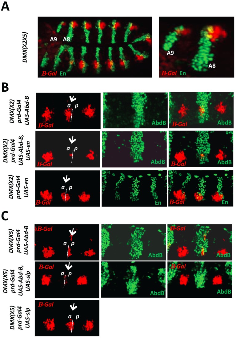
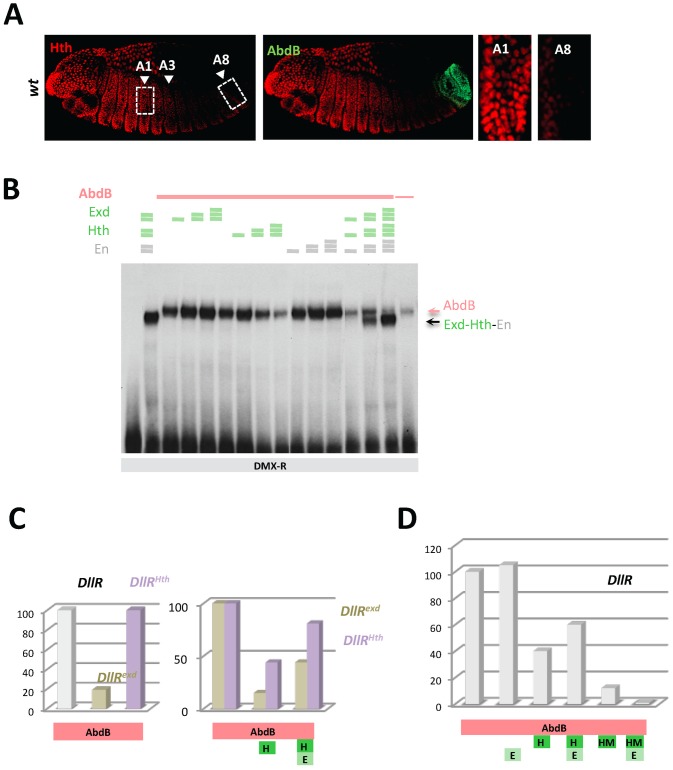
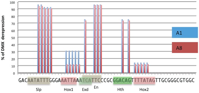
The emergence following gene duplication of a large repertoire of Hox paralogue proteins underlies the importance taken by Hox proteins in controlling animal body plans in development and evolution. Sequence divergence of paralogous proteins accounts for functional specialization, promoting axial morphological diversification in bilaterian animals. Yet functionally specialized paralogous Hox proteins also continue performing ancient common functions. In this study, we investigate how highly divergent Hox proteins perform an identical function. This was achieved by comparing in Drosophila the mode of limb suppression by the central (Ultrabithorax and AbdominalA) and posterior class (AbdominalB) Hox proteins. Results highlight that Hox-mediated limb suppression relies on distinct modes of DNA binding and a distinct use of TALE cofactors. Control of common functions by divergent Hox proteins, at least in the case studied, relies on evolving novel molecular properties. Thus, changes in protein sequences not only provide the driving force for functional specialization of Hox paralogue proteins, but also provide means to perform common ancient functions in distinct ways.


Conflict of interest statement

The authors have declared that no competing interests exist.

Figures

Figure 1
Figure 1. AbdB m and r isoforms repress Dll in the posterior abdominal segments A8 and A9.
(A) Embryo stained for β-gal driven by the DMX enhancer (red) showing restricted thoracic activity. (B–C) DMX(X2X5) embryos co-stained for β-gal (red) and AbdB (green). This modified DMX enhancer is derepressed in all abdominal segments, including A8 and A9. Co-localization of β-gal and AbdB in A8 and A9 cells normally subject to enhancer activity repression is highlighted in the magnified view. (D–E) Embryo lacking Ubx and AbdA function (Df(109)) shows DMX abdominal de-repression (red) up to A7 (arrowhead) and remains repressed in A8 and A9 segments where AbdB is expressed at high levels (green). (F–G) Embryo lacking Ubx, AbdA and the AbdBm isoform, but retaining the AbdB r isoform, shows derepression of DMX (red) till A8 (arrowhead). (H–I) Embryos lacking Ubx, AbdA and the AbdB m and r isoforms (Df P9) show DMX de-repression (red) in all abdominal segments, including in A9 (arrowhead). (J–K) prd-Gal4 driven ectopic expression of the AbdBm isoform represses DMX activity (arrow in T2). (L–M) prd-Gal4 driven ectopic expression of the AbdBr isoform represses DMX activity (arrow in T2).
Figure 2
Figure 2. AbdB-mediated repression of DMX requires the activity of posterior and anterior compartment specific cofactors En and Slp.
A) Embryo co-stained for β-gal driven by DMX(X2X5) (red) and En (green). Magnification of the posterior abdominal segments A8 and A9 highlights derepression of DMX(X2X5) in anterior (En negative) and posterior (En positive) compartment cells. B) Thoracic segments of embryos bearing the DMX(X2) reporter and expressing ectopically AbdB, En or AbdB and En, in every other segments driven by the prd-GAL4 driver. The repressive potential on DMX(X2) is evaluated in the thoracic T2 segment (arrow). Upper panels: In embryos ectopically expressing AbdB, repression only occurs in posterior compartment cells (p). Middle panels: In embryos ectopically expressing AbdB and En, repression occurs in both posterior (p) and anterior compartment cells (a). Lower panels: In embryos ectopically expressing En, only weak and compartment non-specific repression is observed. C) Thoracic segments of embryo bearing the DMX(X5) reporter and expressing ectopically AbdB, Slp or AbdB and Slp, in every other segments driven by the prd-GAL4 driver. The repressive potential on DMX(X5) is evaluated in the thoracic T2 segment (arrow). Upper panels: In embryos ectopically expressing AbdB, repression occurs in anterior compartment cells (a). Middle panels: In embryos ectopically expressing AbdB and Slp, repression occurs in both anterior (a) as well as in posterior compartment cells (p). Lower panels: In embryos ectopically expressing Slp, only weak and compartment non specific repression is observed.
Figure 3
Figure 3. Dispensability of Exd/Hth cofactors for AbdB-mediated DMX posterior repression in posterior segments.
A) Embryos co-stained for Hth (red) and AbdB (green). Arrowheads point to segments A1, A3 and A8. wild type embryo, decrease of Hth expression is seen from segment A3 reaching almost undetectable levels in A8. B) EMSA of DMX-R (containing binding sites Slp,Hox1, Exd, En, Hth and Hox2) with AbdB and increasing amounts of Exd, Hth, En, or En and with combined increasing amounts of Exd, Hth and En. The amount of AbdB remains constant whenever present, except in the last lane (depicted by a thin pink line) where 1/3 of this quantity was used. Exd-Hth-En/DNA and AbdB/DNA complexes are highlighted by arrows. C) Quantification of AbdB binding in EMSA to DIIR (containing binding sites Hox1, Exd, En, and Hth) mutated in the Exd (DIIRexd) or Hth (DIIRHth) binding sites in the presence of AbdB alone, with Exd or Exd and Hth (see Figure S4). Note that mutation of the Exd binding site affect the formation of AbdB/DNA complexes. For the ease of comparison, AbdB binding to DIIRexd and DIIRHth have been arbitrarily set to 100%, allowing assessing the effect of Hth and Hth/Exd inhibitory effects independently off the effect of binding site mutations on AbdB/DNA complex assembly. D) Quantification of AbdB binding in EMSA to DIIR with various combinations of AbdB, Exd, Hth and truncated HM (HD less) form of Hth (See Figure S4). Note that the Exd-mediated release of inhibitory effect seen for full length Hth is lost with the truncated Hth HM protein.
Figure 4
Figure 4. DMX-R cis sequence requirements for repression by Ubx/AbdA and AbdB.
Schematic representation of cis sequence requirements for DMX repressive activity in A1 (Ubx/AbdA- mediated, blue) and A8 (AbdB-mediated, red) segments. 100% derepresion was defined by the level of abdominal DMX derepression in embryos fully deficient for Ubx, AbdA and AbdB m and r isoforms (Df P9). Cis sequence requirement was evaluated by quantifying the levels of derepression of 18 mutated forms of DMX-R (see Figures S5 and S6). These scanning mutations (altering simultaneously two to 5 nucleotide positions) cover 42 of the 57 nucleotide positions of the DMX-R element. Sequence is annotated according to transcription factor binding site (Slp, Exd, Hth, En and Hox) allocation from .
Figure 5
Figure 5. AbdB represses Hth expression.
A) Embryos co-stained for Hth (red) and AbdB (green). Arrowheads highlight segments A3 and A8. Upper panels: embryo lacking AbdB m isoform (AbdBm3), displaying posterior derepression till A8 (compare to wild type embryo (Figure 3A)). Derepression does not spread to A9, where the AbdBr isoform is still expressed. Lower panels: embryo lacking both AbdB m and r isoforms show extension of Hth derepression to segment A9. Right panels are magnifications of A3 and A8/9 segments. B) Embryos bearing the DME reporter co-stained for β-gal (white), Hth (red) and AbdB (right panels, green) or Ubx (left panels, green). Ectopic AbdB expression was driven in every other segments by prd-Gal4. AbdB, but not Ubx ectopic expression, strongly represses Hth expression. Magnifications of thoracic T2 segments are shown.
Figure 6
Figure 6. Protein sequence requirements for AbdB-mediated DME repression.
Sequence conservation in the AbdB HD (shown only for the N-terminal arm and helix 3) and HD flanking regions HX/LR and C-ter region. Sequences upstream and downstream of these regions display progressively weaker conservation. Web logo was obtained using sequences the following AbdB sequences (Drosophila (AAA84402), Tribolium (AAF36721.1), Anophela (XM311628), Sacculine (AAQ49317.1), Folsomia (AAK52499.1) and human (BCO10023)). The Drosophila melanogaster sequence is shown below the web logo. The position and the nature of the mutations generated are represented below the web logo. Effects of the mutations on the repressive activity of AbdB on DME are displayed in a box plot representation. While mutations in the HX/LR region have little effect on AbdB repressive activity, mutations within the HD, including the N-terminal arm, helix 3 and the Cter alter to different extent AbdB repressive potential. Illustration of data is given in Figure S10.
Figure 7
Figure 7. Requirement of protein domains in AbdB/Ubx chimeric proteins for DME repression.
Left part of the figure depicts wild type and mutated variants of Ubx (blue), including mutations (indicated by crosses) in the HX, and UbdA (UA) domains as previously described and in helix 3 (Q50 to K50) . AbdB protein sequences, including or not the QR domain, either with a wild type or mutated helix 3, is represented in red. Effects of the mutations on the repressive activity of AbdB on DME are displayed in a box plot representation. Switching the Ubx HD by that of AbdB endows the chimera with a posterior AbdB like dependent mode of DME repression. Illustration of data is given in Figure S11.
Figure 8
Figure 8. Models for distinct Hox cofactor partnership for Dll repression.
(A) Model for repression of Dll by Ubx/AbdA in anterior abdominal segments A1-7. Repression relies on the assembly of a Hox/Exd/Hth protein complex . DNA binding by Hox proteins is not essential (depicted by a dashed delineated pink zone of contact between the Hox protein and the DNA), as supported by the limited loss of repressive activity of a DNA binding deficient Ubx protein (Figure 7), and by the limited dereprepression associated to mutation in Hox binding sites. The non-essential character of Hox DNA binding may result from acting in a context of a multiprotein complex containing two additional DNA binding proteins (Exd and Hth). (B) Model for repression of Dll by AbdB in posterior abdominal segments in A8-9. AbdB represses Exd and Hth, and consequently act without the aid of Exd and Hth to repress Dll. This difference likely imposes a strict requirement for AbdB DNA binding for efficient repression.

Similar articles

Cited by

References

    1. Pearson JC, Lemons D, McGinnis W (2005) Modulating Hox gene functions during animal body patterning. Nat Rev Genet 6: 893–904. - PubMed
    1. Mann RS, Lelli KM, Joshi R (2009) Hox specificity unique roles for cofactors and collaborators. Curr Top Dev Biol 88: 63–101. - PMC - PubMed
    1. Merabet S, Hudry B, Saadaoui M, Graba Y (2009) Classification of sequence signatures: a guide to Hox protein function. Bioessays 31: 500–511. - PubMed
    1. Gehring WJ, Kloter U, Suga H (2009) Evolution of the Hox gene complex from an evolutionary ground state. Curr Top Dev Biol 88: 35–61. - PubMed
    1. Schubert FR, Nieselt-Struwe K, Gruss P (1993) The Antennapedia-type homeobox genes have evolved from three precursors separated early in metazoan evolution. Proc Natl Acad Sci Usa 90: 143–147. - PMC - PubMed

Publication types

MeSH terms

LinkOut - more resources