

Pemphigus vulgaris autoantibody profiling by proteomic technique
- PMID: 23505434
- PMCID: PMC3591405
- DOI: 10.1371/journal.pone.0057587
Pemphigus vulgaris autoantibody profiling by proteomic technique
Abstract
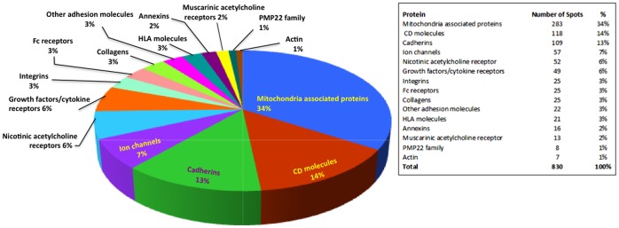
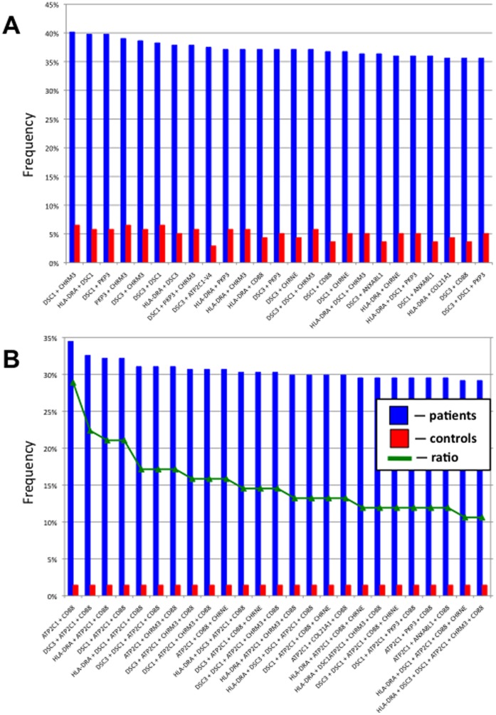
Pemphigus vulgaris (PV) is a mucocutaneous blistering disease characterized by IgG autoantibodies against the stratified squamous epithelium. Current understanding of PV pathophysiology does not explain the mechanism of acantholysis in patients lacking desmoglein antibodies, which justifies a search for novel targets of pemphigus autoimmunity. We tested 264 pemphigus and 138 normal control sera on the multiplexed protein array platform containing 701 human genes encompassing many known keratinocyte cell-surface molecules and members of protein families targeted by organ-non-specific PV antibodies. The top 10 antigens recognized by the majority of test patients' sera were proteins encoded by the DSC1, DSC3, ATP2C1, PKP3, CHRM3, COL21A1, ANXA8L1, CD88 and CHRNE genes. The most common combinations of target antigens included at least one of the adhesion molecules DSC1, DSC3 or PKP3 and/or the acetylcholine receptor CHRM3 or CHRNE with or without the MHC class II antigen DRA. To identify the PV antibodies most specific to the disease process, we sorted the data based on the ratio of patient to control frequencies of antigen recognition. The frequency of antigen recognition by patients that exceeded that of control by 10 and more times were the molecules encoded by the CD33, GP1BA, CHRND, SLC36A4, CD1B, CD32, CDH8, CDH9, PMP22 and HLA-E genes as well as mitochondrial proteins encoded by the NDUFS1, CYB5B, SOD2, PDHA1 and FH genes. The highest specificity to PV showed combinations of autoantibodies to the calcium pump encoded by ATP2C1 with C5a receptor plus DSC1 or DSC3 or HLA-DRA. The results identified new targets of pemphigus autoimmunity. Novel autoantibody signatures may help explain individual variations in disease severity and treatment response, and serve as sensitive and specific biomarkers for new diagnostic assays in PV patients.
Conflict of interest statement
Figures
References
-
- Beutner EH, Lever WF, Witebsky E, Jordon R, Chertock B (1965) Autoantibodies in Pemphigus Vulgaris: Response to an Intercellular Substance of Epidermis. JAMA 192: 682–688. - PubMed
-
- Anderson HJ, Newcomer VD, Landau JW, Rosenthal LH (1970) Pemphigus and other diseases. Results of indirect intercellular immunofluorescence. Arch Dermatol 101: 538–546. - PubMed
-
- Pisanti S, Sharav Y, Kaufman E, Posner LN (1974) Pemphigus vulgaris: incidence in Jews of different ethnic groups, according to age, sex, and initial lesion. Oral Surg Oral Med Oral Pathol 38: 382–387. - PubMed
-
- Chams-Davatchi C, Valikhani M, Daneshpazhooh M, Esmaili N, Balighi K, et al. (2005) Pemphigus: analysis of 1209 cases. International Journal of Dermatology 44: 470–476. - PubMed
-
- Carson PJ, Hameed A, Ahmed AR (1996) Influence of treatment on the clinical course of pemphigus vulgaris. Journal of the American Academy of Dermatology 34: 645–652. - PubMed
MeSH terms
Substances
LinkOut - more resources
Full Text Sources
Other Literature Sources
Medical
Research Materials
Miscellaneous
