. 2013;8(3):e57644.
doi: 10.1371/journal.pone.0057644. Epub 2013 Mar 7.

Crystal structures of the catalytic domain of human soluble guanylate cyclase

Affiliations

Crystal structures of the catalytic domain of human soluble guanylate cyclase

Charles K Allerston et al. PLoS One. 2013.

Abstract

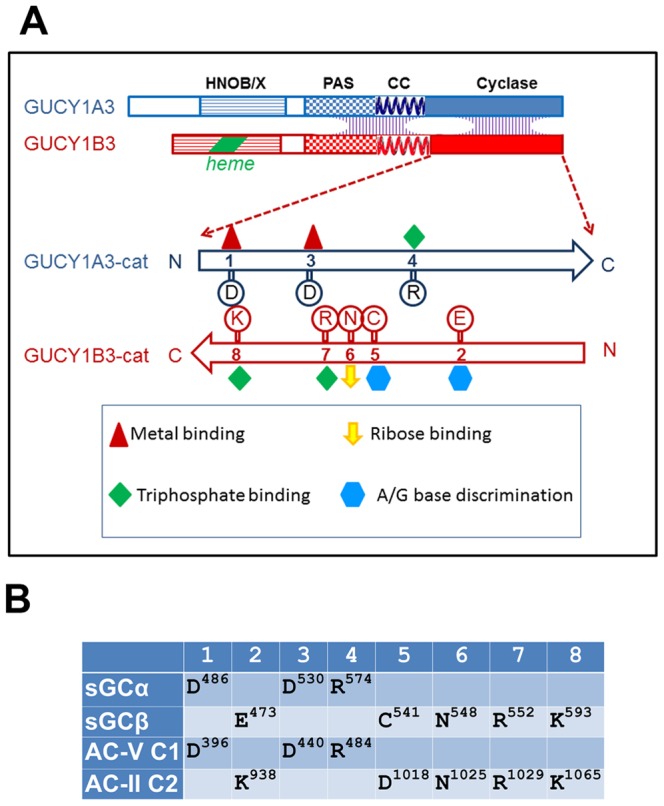
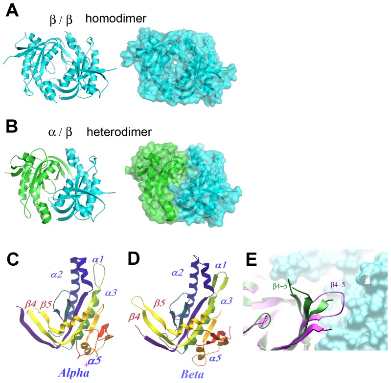
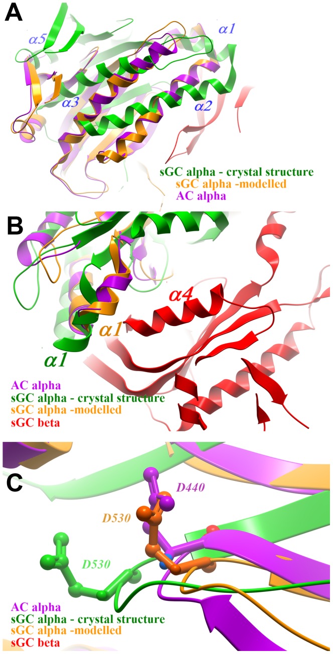
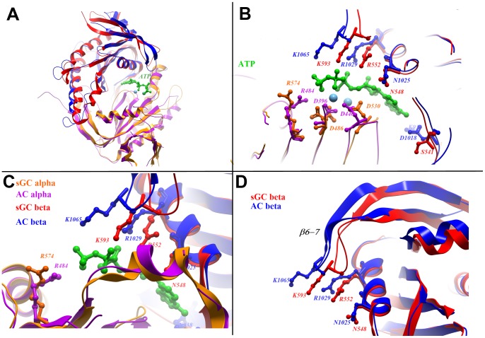
Soluble guanylate cyclase (sGC) catalyses the synthesis of cyclic GMP in response to nitric oxide. The enzyme is a heterodimer of homologous α and β subunits, each of which is composed of multiple domains. We present here crystal structures of a heterodimer of the catalytic domains of the α and β subunits, as well as an inactive homodimer of β subunits. This first structure of a metazoan, heteromeric cyclase provides several observations. First, the structures resemble known structures of adenylate cyclases and other guanylate cyclases in overall fold and in the arrangement of conserved active-site residues, which are contributed by both subunits at the interface. Second, the subunit interaction surface is promiscuous, allowing both homodimeric and heteromeric association; the preference of the full-length enzyme for heterodimer formation must derive from the combined contribution of other interaction interfaces. Third, the heterodimeric structure is in an inactive conformation, but can be superposed onto an active conformation of adenylate cyclase by a structural transition involving a 26° rigid-body rotation of the α subunit. In the modelled active conformation, most active site residues in the subunit interface are precisely aligned with those of adenylate cyclase. Finally, the modelled active conformation also reveals a cavity related to the active site by pseudo-symmetry. The pseudosymmetric site lacks key active site residues, but may bind allosteric regulators in a manner analogous to the binding of forskolin to adenylate cyclase. This indicates the possibility of developing a new class of small-molecule modulators of guanylate cyclase activity targeting the catalytic domain.


Conflict of interest statement

Competing Interests: The authors have the following interests. This study was funded by The SGC which is a charity funded by a public-private partnership that includes commercial entities (GlaxoSmithKline, Lilly Canada, the Novartis Research Foundation, Pfizer, Takeda, and AbbVie). There are no patents, products in development or marketed products to declare. This does not alter the authors' adherence to all the PLOS ONE policies on sharing data and materials, as detailed online in the guide for authors.

Figures

Figure 1
Figure 1. Structural organization of guanylate cyclase.
A. Schematic depiction of domain organization of human guanylate cyclase. The native enzyme is a heterodimer of α and β subunits (encoded by GUCY1A3 and GUCY1B3, respectively), with interaction interfaces spread across the PAS, CC and cyclase catalytic domains. The catalytic domains associate in a head to tail orientation; conserved residues involved in substrate binding and catalysis are distributed between the two subunits. B. A list of conserved active-site residues in guanylate cyclase (sGCα and β) and in the chimeric adenylate cyclase (AC-V C1 domain and AC-II C2 domain). A full alignment of adenylate and guanylate cyclases is shown in Supporting Fig. S1.
Figure 2
Figure 2. Overview of crystal structures.
A. Homodimer of sGCβ catalytic domain (PDB ID: 2WZ1). B. Heterodimer of sGCα (green) and sGCβ (cyan) catalytic domains (PDB ID: 3UVJ). C. Architecture of the sGCα catalytic domain. D. Architecture of the sGCβ catalytic domain. E. The β4–5 loop in AC-C1 (purple) and sGCα (green); the surface of the β/C2 subunit is shown in cyan.
Figure 3
Figure 3. Structural transitions in sGC activation.
A. sGCα in the crystal structure (green) compared with the same subunit (orange) modelled by alignment with the C1 domain of adelylate cyclase (purple). The rigid-body transition involves a 26° rotation, seen in the relative angles of the corresponding α helices. B. Detail: the change in position of the α1 helix (sGCα), bringing it closer to helix α4 (sGCβ). C. Detail: shift in position of the β6–7 loop, which brings a catalyitic residue D530 closer to the position of the corresponding residue in AC(D440).
Figure 4
Figure 4. Active site residues on sGC in the modelled active conformation.
The sGCα and sGCβ were separately aligned with AC domains C1 and C2, respectively. A. Overall view (the colour scheme is described in panel C). C. Active site residues surrounding an ATP analogue in the AC structure. C, D. Detailed views.
Figure 5
Figure 5. A possible allosteric site.
A. Forskolin in its binding pocket in adenylate cyclase. B. The same region in sGC in the crystal structure: the cavity is collapsed, with no space for small-molecule binding. C. sGC in the modelled active conformation: a cavity opens up; although forskolin does not fit, other small molecules may occupy the site.

References

    1. Poulos TL (2006) Soluble guanylate cyclase. Curr Opin Struct Biol 16: 736–743. - PubMed
    1. Mergia E, Russwurm M, Zoidl G, Koesling D (2003) Major occurrence of the new alpha2beta1 isoform of NO-sensitive guanylyl cyclase in brain. Cell Signal 15: 189–195. - PubMed
    1. Russwurm M, Koesling D (2002) Isoforms of NO-sensitive guanylyl cyclase. Mol Cell Biochem 230: 159–164. - PubMed
    1. Mergia E, Koesling D, Friebe A (2009) Genetic mouse models of the NO receptor ‘soluble’ guanylyl cyclases. Handb Exp Pharmacol 33–46. - PubMed
    1. Friebe A, Mergia E, Dangel O, Lange A, Koesling D (2007) Fatal gastrointestinal obstruction and hypertension in mice lacking nitric oxide-sensitive guanylyl cyclase. Proc Natl Acad Sci U S A 104: 7699–7704. - PMC - PubMed

Publication types

MeSH terms