. 2013;8(3):e58578.
doi: 10.1371/journal.pone.0058578. Epub 2013 Mar 7.

A unifying mechanism for cancer cell death through ion channel activation by HAMLET

Affiliations

A unifying mechanism for cancer cell death through ion channel activation by HAMLET

Petter Storm et al. PLoS One. 2013.

Abstract

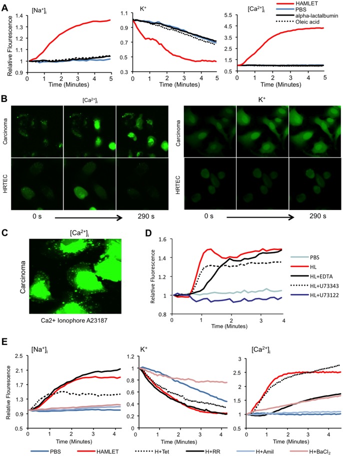
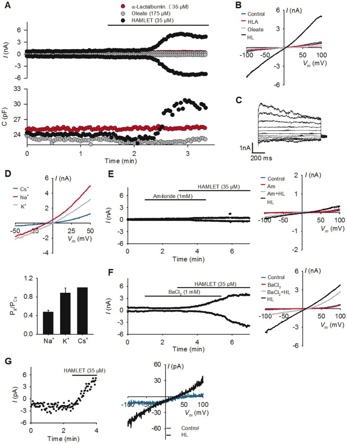
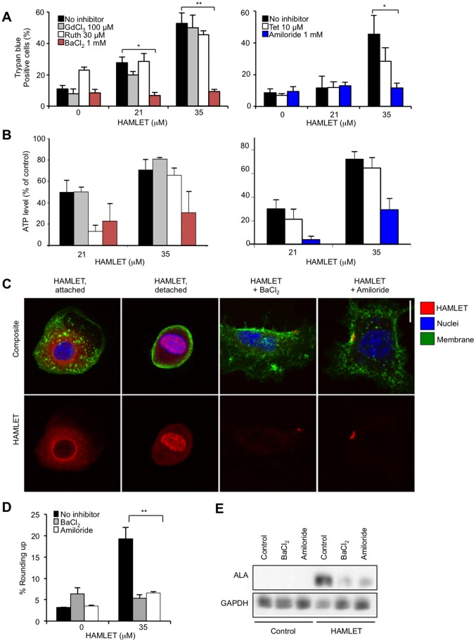
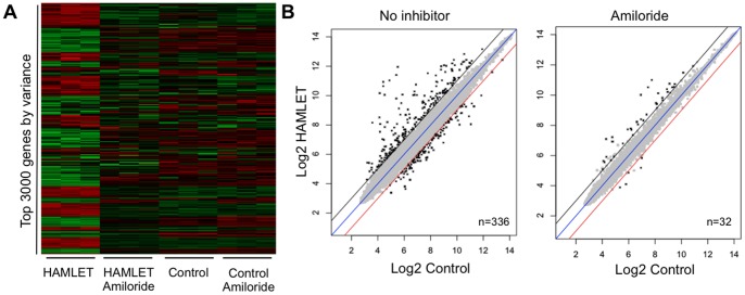
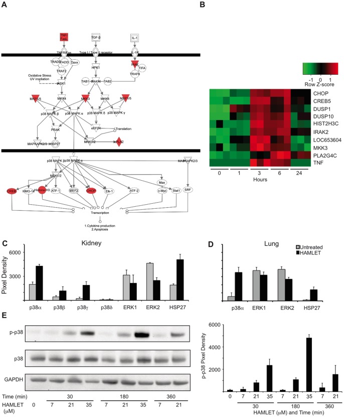
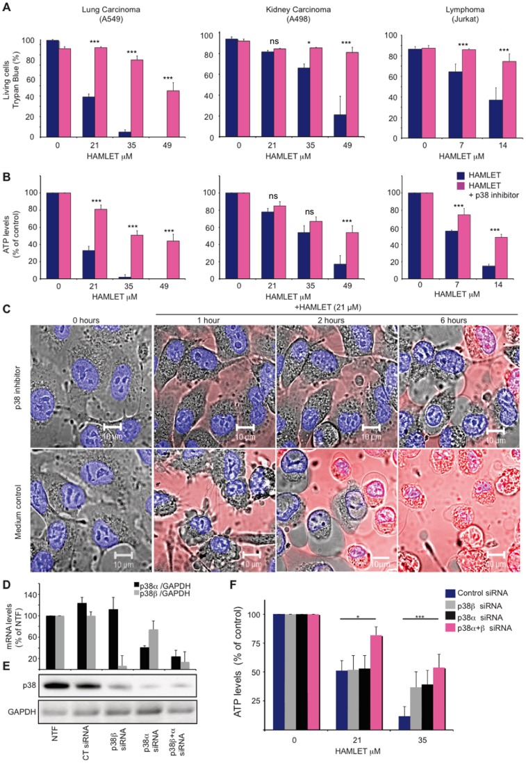
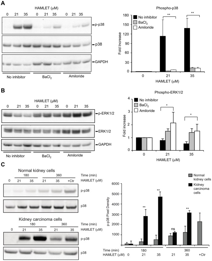
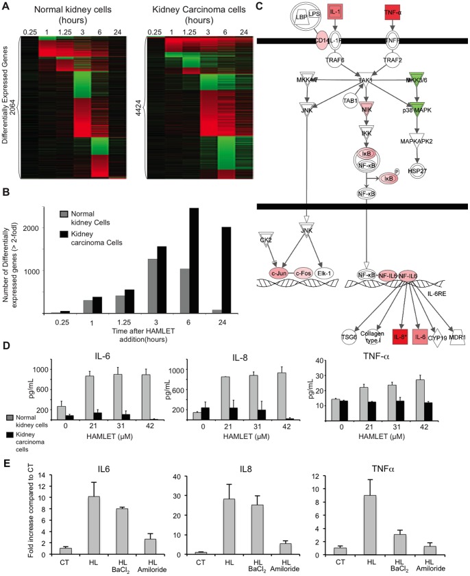
Ion channels and ion fluxes control many aspects of tissue homeostasis. During oncogenic transformation, critical ion channel functions may be perturbed but conserved tumor specific ion fluxes remain to be defined. Here we used the tumoricidal protein-lipid complex HAMLET as a probe to identify ion fluxes involved in tumor cell death. We show that HAMLET activates a non-selective cation current, which reached a magnitude of 2.74±0.88 nA within 1.43±0.13 min from HAMLET application. Rapid ion fluxes were essential for HAMLET-induced carcinoma cell death as inhibitors (amiloride, BaCl2), preventing the changes in free cellular Na(+) and K(+) concentrations also prevented essential steps accompanying carcinoma cell death, including changes in morphology, uptake, global transcription, and MAP kinase activation. Through global transcriptional analysis and phosphorylation arrays, a strong ion flux dependent p38 MAPK response was detected and inhibition of p38 signaling delayed HAMLET-induced death. Healthy, differentiated cells were resistant to HAMLET challenge, which was accompanied by innate immunity rather than p38-activation. The results suggest, for the first time, a unifying mechanism for the initiation of HAMLET's broad and rapid lethal effect on tumor cells. These findings are particularly significant in view of HAMLET's documented therapeutic efficacy in human studies and animal models. The results also suggest that HAMLET offers a two-tiered therapeutic approach, killing cancer cells while stimulating an innate immune response in surrounding healthy tissues.


Conflict of interest statement

Competing Interests: HAMLET patents are held by HAMLET Pharma - a commercially inactive company. The studies described in this manuscript were not supported by commercial sources/partnerships. The authors have filed a patent containing information relevant to this paper. The application number is WO 2012/069836. The authors hereby confirm that this patent does not alter the authors' adherence to all the PLOS ONE policies on sharing data and materials, as detailed online in the guide for authors.

Figures

Figure 1
Figure 1. HAMLET alters intracellular ion concentrations in tumor cells.
(A) Estimates of the relative free, intracellular concentrations of Na+, K+, and Ca2+ ([Na+]i, [K+]i, and [Ca2+]I, respectively) were obtained in A549 cells (K+ and Ca2+) and Jurkat cells (Na+) by fluorescence spectrometry using CoroNa Green, FluxOR and Fluo-4. Rapid ion fluxes were detected and these effects were HAMLET specific, as α-lactalbumin, oleic acid or PBS had no effect. (Means of four experiments, p<0.05 (Students t-test at t = 2 minutes) (B) Difference in HAMLET-induced ion fluxes between tumor cells and healthy cells. Ca2+ and K+ fluxes are visualized by real time confocal imaging of A549 lung carcinoma cells and HRTEC healthy, differentiated kidney cells, loaded with the Ca2+ and K+ fluorophores and treated with HAMLET (35 µM) for up to 290 seconds. Representative figures from three experiments are shown. (C) Calcium ionophore (A23187, 1 µM) response in A549 lung carcinoma cells loaded with Fluo-4. Representative figure from three experiments. (D) Inhibition of intracellular calcium release from ER reduces Ca2+ fluxes. A549 cells were pretreated with a PLC-inhibitor (10 µM, U73122), its inactive analogue (10 µM, U73343) or EDTA (1 mM) for 30 minutes and treated with HAMLET as indicated. Means of three experiments. (E) Amiloride (Amil, 1 mM) inhibited Na+ and Ca2+ fluxes in A549 cells (Ca2+) and Jurkat cells (Na+) induced by HAMLET (marked as H in figure). BaCl2 (1 mM) inhibited the Na+, K+ and Ca2+ fluxes. Cells were pre-loaded with the respective fluorophores, pretreated with inhibitors (30 minutes) and challenged with HAMLET (35 µM) for up to 5 minutes. Effects of amiloride on K+ fluxes could not be determined due to auto fluorescence. Ruthenium red (RR, 30 µM) reduced Ca2+ fluxes while tetranidrine (Tet, 10 µM) had no significant effect.
Figure 2
Figure 2. Whole cell currents activated by HAMLET in lung carcinoma cells.
HAMLET-induced currents were recorded in A549 cells, by whole cell patch clamp analysis, with the exception of data in G, which were obtained in inside-out patches. (A) Top panel: Currents were measured using the described ramp protocol and currents measured at +100 mV and −100 mV are depicted as a function of time for three representative and independent experiments stimulated with α-lactalbumin, oleate or HAMLET, the presence of which in the superfusate is indicated by the top line. Lower panel: Cell capacitance as a function of time. Same experiments as in top panel. (B) Same experiments as in A, showing the current- voltage relationship of cells following the indicated stimulation. The control trace represents the current-voltage relationship before exposure to HAMLET (n = 7). (C) Using a 1 s protocol stepping from −100 mV to +100 mV in 20 mV increments, time dependent characteristics of the HAMLET activated current were investigated. Note the clear time dependent inactivation at strongly depolarized potentials. n = 3. (D) Selectivity profile of HAMLET activated currents. Top panel: Reversal potentials of HAMLET activated currents where estimated from individual experiments as described in Materials and Methods. Lower panel: Permeability ratios for Na+ and K+ relative to Cs+, calculated as described. n = 3–5 (E) Amiloride inhibits HAMLET activated currents. Left panel: Current magnitude measured at −100 mV and +100 mV. The presence of amiloride (1 mM) and HAMLET in the superfusate is indicated by the top lines. Compare the current with that in the absence of amiloride in (A). Right panel: Current-Voltage relationship from same experiment as in the left panel. n = 4. (F) BaCl2 partially blocks the inward current. Left panel: Current magnitude measured at −100 mV and +100 mV. The presence of BaCl2 (1 mM) and HAMLET in the superfusate is indicated by the top lines. Right panel: Current-Voltage relationship from the same experiment as in left panel. Note the strong inhibition of the inward current compared to the outward current. n = 3. (G) HAMLET stimulates current activation in inside-out patches. Left Panel: Currents in inside-out patches were followed at the holding potential of the described ramp protocol and plotted as a function of time. The presence of HAMLET in the perfusion solution is indicated above. Right panel: Current voltage relationship measured in inside-out patches in the presence and absence of HAMLET. Same experiments as in the left panel. n = 3.
Figure 3
Figure 3. Ion channel inhibitors rescue carcinoma cells from death and block HAMLET uptake and morphologic change.
(A, B) Viability of lung carcinoma cells after exposure to HAMLET (21 or 35 µM, 3 h), quantified by trypan blue exclusion and ATP levels. Amiloride or BaCl2 inhibited the tumoricidal effect of HAMLET but Ruthenium Red and tetrandrine showed no effect (means+SEMs, two to three experiments per condition). (C) Internalization of Alexa-568 fluor labeled HAMLET by tumor cells (35 µM, 1 hour), visualized by high magnification (x63) confocal microscopy. HAMLET was localized to peri-nuclear and nuclear regions in tumor cells. Amiloride or BaCl2 inhibited internalization, leaving HAMLET associated with the cell surface. HAMLET was labeled red (Alexa-568), nuclei blue (Draq5) and membranes were labeled green (WGA). Scale bar = 10 µM. (D) BaCl2 and amiloride prevented changes in carcinoma cells morphology in response to HAMLET (Mean of two images in one experiment+SEMs). Rounding up signifies the shift from an adherent, extended to a rounded morphology. (E) Western blot of cell lysates confirming the reduction in cell-associated HAMLET by amiloride and BaCl2 (35 µM, 1 hour, detected with anti-α-lactalbumin antibodies, ALA). GAPDH was used as a loading control.
Figure 4
Figure 4. The transcriptomic response to HAMLET requires functional ion channels.
Genome wide expression analysis in lung carcinoma cells stimulated with HAMLET in the presence or absence of Amiloride. (A) Heatmap of the normalized expression of top 3000 genes sorted according to variance across all conditions. Amiloride markedly reduced the global transcriptional response to HAMLET but had little effect on control cells. (B) Scatter plots of normalized expression values for all genes present on the array. The number of differentially expressed genes (log2 fold change >1 and FDR-adjusted p-value <0.05, marked in black) was greatly reduced by amiloride.
Figure 5
Figure 5. HAMLET activates the p38 MAPK signaling pathway.
(A) Transcriptional changes were identified in HAMLET-treated A549 lung carcinoma cells. Three hundred sixty-seven genes showed a minimum log 2-fold change of 1.2 compared to PBS-treated control cells, with a Benjamini-Hochberg adjusted p-value <0.05. Ten genes in the p38 pathway were upregulated (red) three hours after HAMLET treatment (21 µM) of lung carcinoma cells (A549), as marked in the canonical pathway. (B) Heat map (triplicate for each time period) and log2 ratios of differentially expressed genes in the p38 MAPK pathway, 1, 3, 6 and 24 hours after HAMLET exposure. (D, E) Increased phosphorylation of p38α, β, γ or HSP27 and reduced ERK1/2 phosphorylation in kidney (C) and lung (D) carcinoma cells exposed to HAMLET (35 µM, 30 minutes). Membranes with phospho-specific antibodies were probed with protein lysates from HAMLET- or PBS-treated (control) carcinoma cells. Protein phosphorylation was quantified using ImageJ. Data are mean ± SEM of 3 experiments. Full array images are given in Figure S6. (E) Dose- and time-dependent p38 MAPK phosphorylation in response to HAMLET. Lung carcinoma cells were treated with HAMLET and compared to PBS-treated negative controls and whole cell lysates were probed with antibodies specific for phosphorylated p38 MAPK (Thr180/Tyr182). Membranes were stripped and reprobed with total p38 and GAPDH antibody as a loading control. For effects on p38 MAPK signaling in kidney carcinoma cells, see Figure S6.
Figure 6
Figure 6. p38 inhibition rescues carcinoma and lymphoma cells from death in response to HAMLET.
(A, B) p38 MAPK inhibition (SB202190, 20 µM, 30 minutes pre-incubation) rescued carcinoma (A549 and A498) and T-cell lymphoma (Jurkat) cells from death in response to HAMLET (7–49 µM, 3 h). Viability was quantified by Trypan blue exclusion (A) or as ATP levels (B). Data are means±SEMs for 3 independent experiments. The p38 inhibitor alone had no significant effect on cell death (leftmost bars in each graph). (C) Real-time images of cell morphology after HAMLET exposure, showing that p38 inhibition prevents morphological changes in carcinoma cells (nuclear condensation, rounding up and blebbing). Alexa-Fluor 568-labeled HAMLET is red and the nuclei are blue (Hoechst 33342). (D–E) A549 lung carcinoma cells were transfected using siRNA against p38α and/or p38β or non-targeting siRNA and compared to non-transfected controls (NTF). Relative p38α and p38βmRNA levels are shown (p38/GAPDH, in % of non-transfected cells) as means+SEMs for four independent experiments. Knockdown was also confirmed on the protein level by western blotting against total p38 MAPK (representative blot shown, *p<0.05, ***p<0.001.). GAPDH was used as a loading control. (F) The cytotoxic effect of HAMLET was quantified 48 hours after transfection as a reduction in ATP levels. Data are means+SEM for four independent experiments. *p<0.05, ***p<0.001. For effects of the p38 inhibitor Birb0796, see Figure S6.
Figure 7
Figure 7. Ion channel inhibitors reduce protein phosphorylation.
Amiloride or BaCl2 reduced phosphorylation of targets in the p38 signaling Lung carcinoma cells were exposed to HAMLET (21 and 35 µM) for 1 hour, protein lysates were blotted, incubated with antibodies as noted in the figure and quantified using ImageJ (Representative blot,+SEMs of 2–3 independent experiments). (A) Amiloride or BaCl2 (1 mM, 30 minutes pretreatment) inhibited HAMLET-induced p38 MAPK phosphorylation (B) Amiloride and BaCl2 reverse the suppression of p-ERK1/2 phosphorylation by HAMLET. (C) Difference in phosphorylation of p38 MAPK between normal differentiated cells (HRTEC) and kidney carcinoma cells, visualized by phospho-specific antibodies.
Figure 8
Figure 8. Innate immune response to HAMLET in normal, differentiated cells.
(A) The transcriptional response to HAMLET is qualitatively different in normal cells (RPTEC), as shown by the heat map of genes with a log2 fold change >2 at any time point. (B) The number of differentially expressed genes (log2 fold change ≥ 2) was reduced compared to carcinoma cells. (C) Seven innate immunity-related genes are upregulated in normal cells, 75 minutes after HAMLET treatment. Two genes, p38 MAPK and MKK3/6, are downregulated. (D) Confirmation of the innate immune response to HAMLET. Elevated TNF, IL-8 and IL-6 levels in supernatants of normal, differentiated cells, but not in carcinoma cells treated with HAMLET (21–42 µM, 6 h). Data are means ± SEMs of triplicates from 3 independent experiments. (E) The innate immune response of healthy cells (IL-6, IL-8 and TNF-α) was inhibited by amiloride and TNF-α expression by BaCl2. qRT-PCR quantification of cytokine mRNA-levels in normal, differentiated cells exposed to HAMLET (35 µM, 1 hour).

Similar articles

Cited by

References

    1. Martinac B (2004) Mechanosensitive ion channels: molecules of mechanotransduction. Journal of cell science 117: 2449–2460. - PubMed
    1. Nilius B, Owsianik G, Voets T, Peters JA (2007) Transient receptor potential cation channels in disease. Physiol Rev 87: 165–217. - PubMed
    1. Pedersen SF, Nilius B (2007) Transient receptor potential channels in mechanosensing and cell volume regulation. Methods Enzymol 428: 183–207. - PubMed
    1. Holzer P (2009) Acid-sensitive ion channels and receptors. Handb Exp Pharmacol: 283–332. - PMC - PubMed
    1. Arcangeli A (2011) Ion channels and transporters in cancer. 3. Ion channels in the tumor cell-microenvironment cross talk. Am J Physiol Cell Physiol 301: C762–771. - PubMed

Publication types

MeSH terms