. 2013;8(3):e58601.
doi: 10.1371/journal.pone.0058601. Epub 2013 Mar 7.

MUC1* ligand, NM23-H1, is a novel growth factor that maintains human stem cells in a more naïve state

Affiliations

MUC1* ligand, NM23-H1, is a novel growth factor that maintains human stem cells in a more naïve state

Benoit J Smagghe et al. PLoS One. 2013.

Abstract

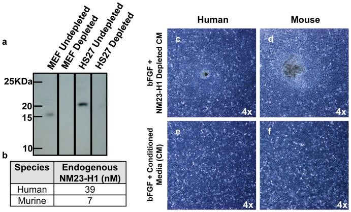
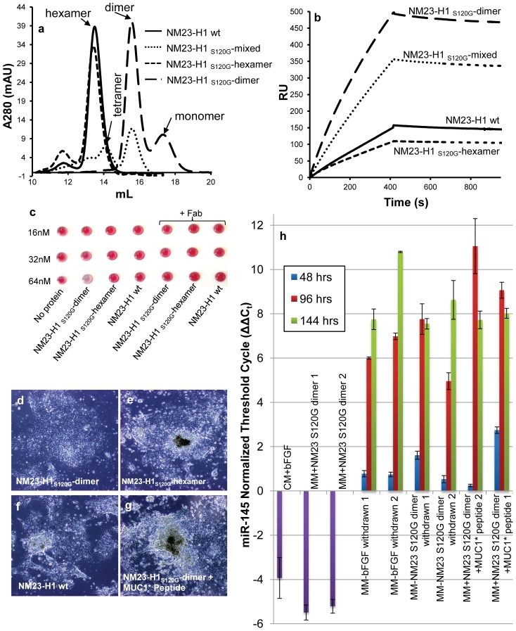
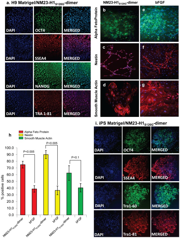
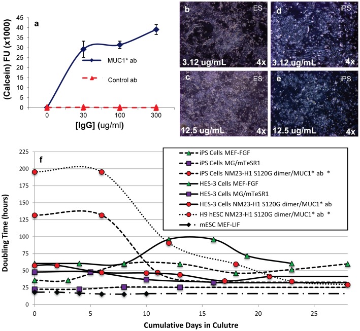
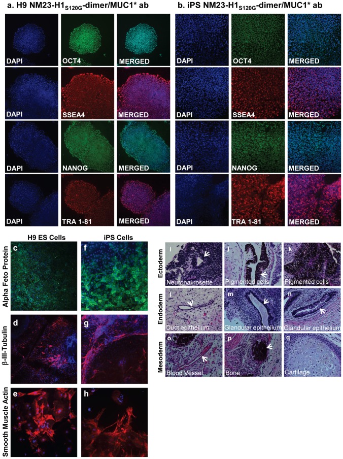
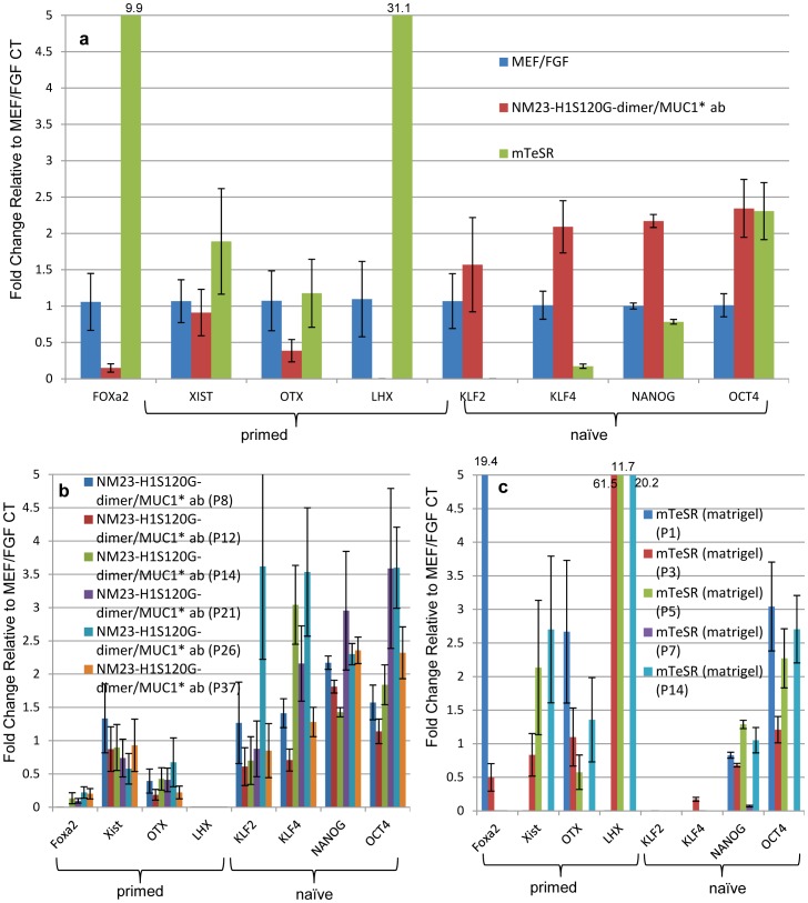
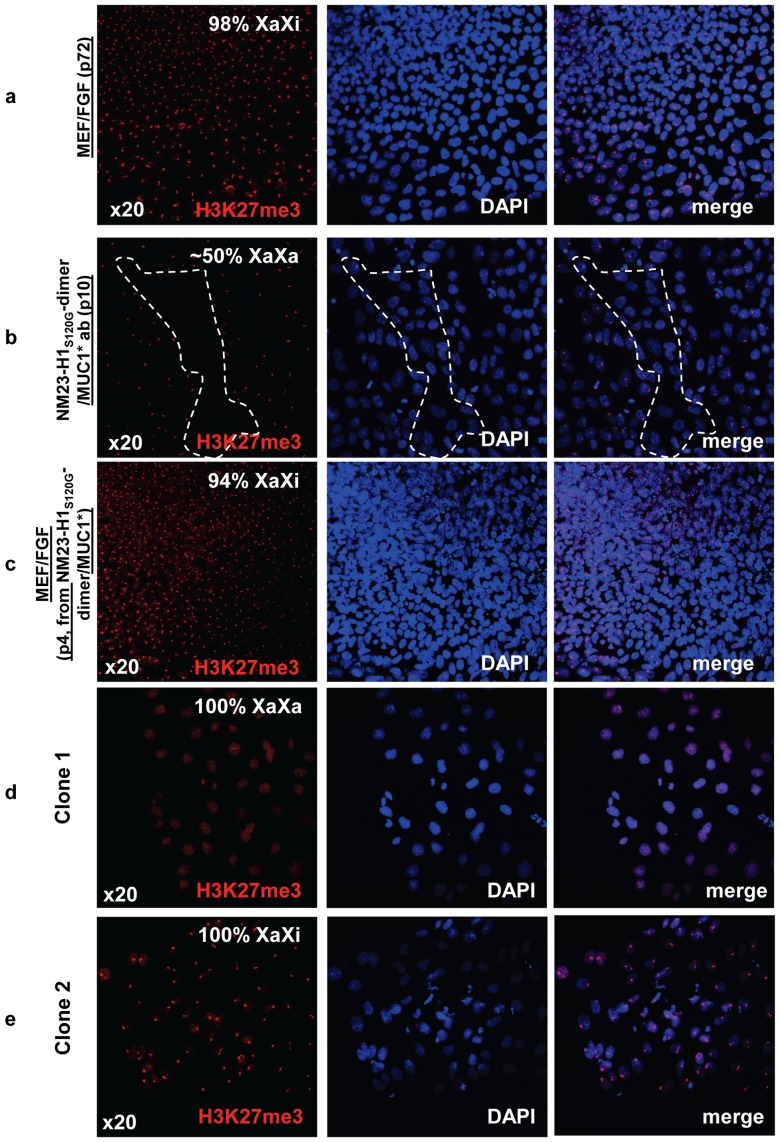
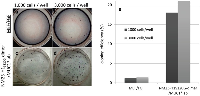
We report that a single growth factor, NM23-H1, enables serial passaging of both human ES and iPS cells in the absence of feeder cells, their conditioned media or bFGF in a fully defined xeno-free media on a novel defined, xeno-free surface. Stem cells cultured in this system show a gene expression pattern indicative of a more "naïve" state than stem cells grown in bFGF-based media. NM23-H1 and MUC1* growth factor receptor cooperate to control stem cell self-replication. By manipulating the multimerization state of NM23-H1, we override the stem cell's inherent programming that turns off pluripotency and trick the cells into continuously replicating as pluripotent stem cells. Dimeric NM23-H1 binds to and dimerizes the extra cellular domain of the MUC1* transmembrane receptor which stimulates growth and promotes pluripotency. Inhibition of the NM23-H1/MUC1* interaction accelerates differentiation and causes a spike in miR-145 expression which signals a cell's exit from pluripotency.


Conflict of interest statement

Competing Interests: CB is CEO and a share holder of Minerva Biotechnologies. KSK is a share holder and board member of Minerva Biotechnologies and a consultant to Amgen and Noscira. BJS, LMS, MGC, EJH, KJB, BAD, AKS, AKC, VMH and BAK are employees and hold stock options for shares in Minerva Biotechnologies. This does not alter the authors' adherence to all the PLOS ONE policies on sharing data and materials.

Figures

Figure 1
Figure 1. Conditioned media depleted of NM23-H1 no longer supports pluripotent stem cell growth.
a) Conditioned media from either murine (MEFs) or human (HS27) embryonic fibroblast cells was immuno-depleted of NM23-H1. Western blot confirmed that the depleted media no longer contains NM23-H1. b) NM23-H1 was captured by affinity chromatography, eluted and its concentration quantified by the optical density at 280nm. Human conditioned media contains 5-times more NM23-H1 than murine conditioned media. Human ES cells on Matrigel were cultured with: c, d) NM23-H1-depleted media, e, f) bFGF plus complete conditioned media. NM23-H1-depleted conditioned media induced differentiation (c, d).
Figure 2
Figure 2. Different NM23-H1 multimers are generated and assayed for function.
a) Recombinant NM23-H1 wt (wild type) and NM23-H1S120G mutant were expressed using different protocols that resulted in the formation of different multimerization states, which were characterized and then purified by size exclusion chromatography. Simple expression and collection of the soluble protein of NM23-H1 -wt and NM23-H1S120G mutant results in a population that is essentially all hexamer. Denaturation and refolding of the NM23-H1S120G produces a stable dimer population. A mixture of hexamers, tetramers and dimers was generated such that it contained ∼50% dimer, NM23-H1S120G-mixed. b) NM23-H1S120G or wild type multimers were tested by Surface Plasmon Resonance (SPR) to determine their ability to bind to a synthetic MUC1* extra cellular domain (ecd) peptide immobilized on the chip surface. The amount of NM23-H1 binding to the MUC1* peptide corresponds to the concentration of dimer present in each sample. c) Nanoparticles presenting the MUC1*ecd peptide were mixed with NM23-H1-wt, NM23-H1S120G-dimer or NM23-H1S120G-hexamer containing the Strep-tag II. A nanoparticle color change from pink to blue/gray indicates binding. NM23-H1 dimer binds to the MUC1*ecd peptide at 64nM while the hexamer, whether wild type or S120G mutant, does not. The interaction was competitively inhibited by an anti-MUC1* Fab, showing that the color change was due to the specific interaction between NM23-H1dimers and MUC1*ecd. d, g) H9 hES cells on Matrigel were cultured in: d) NM23-H1S120G-dimer, e) NM23-H1S120G-hexamer, f) NM23-H1-wt or g) NM23-H1S120G-dimer plus a synthetic MUC1*ecd peptide (1 µM). Only NM23-H1S120G-dimers supported pluripotent stem cell growth. Hexamers or inhibition of the NM23-H1S120G-dimer-MUC1* interaction resulted in immediate differentiation. All images 4X. h) H9 hES cells were cultured in either bFGF plus conditioned media or in NM23-H1S120G-dimer, and then allowed to differentiate by withholding the growth factor. Some cells cultured in NM23-H1S120G-dimer continued to receive the growth factor but also received the MUC1*ecd peptide (1 µM) to competitively inhibit the NM23-H1-MUC1* interaction. miR-145, a marker for exit from pluripotency, is measured by RT-PCR as a function of time. Competitive inhibition of the NM23-H1-MUC1* interaction caused an earlier spike in miR-145 than that by merely withholding the growth factor, demonstrating that interrupting the NM23-H1-MUC1* interaction induces differentiation.
Figure 3
Figure 3. Human ES cells serially passaged in NM23-H1-MM express pluripotency markers, differentiate down all three germlines and display coordinated differentiation.
H9 hES cells on Matrigel were cultured for at least six passages in either NM23-H1-MM (minimal stem cell media) or bFGF plus MEF conditioned media. a) NM23-H1-MM cultured cells stained positive for the presence of the typical pluripotency markers.. NM23-H1 cultured cells were allowed to differentiate by the embryoid body method (Methods S1) then stained with nuclear marker DAPI and antibodies against markers of the three germlines: b, e) endoderm - alpha feto protein, c, f) ectoderm - nestin, and d, g) mesoderm - smooth muscle actin. Cells cultured in either NM23-H1-MM or bFGF-CM both differentiated down all three germlines. Cells that had been cultured in NM23-H1-MM displayed apparently coordinated differentiation with most cells in a cluster differentiating down the same germline (b–d), whereas cells cultured in bFGF did not (e–g). h) The percentage of cells in each cluster that expressed the same germline marker was quantified (n = 5 to 7). Cells that had been cultured in NM23-H1-MM have a higher percentage of cells in the same cluster differentiating down the same germline than cells that had been cultured in bFGF. i) iPS cells on Matrigel, cultured for six passages in NM23-H1-MM stained positive for the presence of the typical pluripotency markers. All images 4X
Figure 4
Figure 4. Anti-MUC1* antibodies are a novel surface coating that enables stem cell attachment and growth.
a) An anti-MUC1* rabbit polyclonal antibody or a control IgG antibody were adsorbed at varying concentrations onto a tissue culture treated multi-well plate. BGO1V/hOG hES cells were plated onto the surfaces and allowed to grow for 48 hours. A Calcein AM assay to quantify cell number was performed. Anti-MUC1* antibody, but not the control antibody, enabled stem cell attachment and growth. BGO1V/hOG hES cells were cultured for 20 passages in NM23-H1-MM without a decrease in pluripotency or change in karyotype (data not shown). b–e) H9 hES cells (b, c) or iPS cells (d, e) were plated onto VitaTM multi-well plates, reported to have higher protein binding capability, that were coated with either 3.12 µg/mL (b, d) or 12.5 µg/mL (c, e) of a monoclonal anti-MUC1* antibody, MN-C3. Cells attached and proliferated as a function of antibody concentration, with maximal attachment observed at 12.5 µg/mL. f) Doubling times forhuman ES cells (H9 or HES-3), human iPS cells and mouse ES cells were measured as a function of various culture media and surfaces. The traces marked by red circles/dotted line and red circles/dashed line (human H9 ES and iPS cells, respectively) show the change in doubling time as cells transition from bFGF-based media to NM23-H1 media on anti-MUC1* surface.
Figure 5
Figure 5. ES and iPS cells cultured long-term in NM23-H1-MM on Anti-MUC1* surfaces express pluripotency markers and differentiate down all three germlines.
a, b) H9 ES cells and iPS cells serially passaged on a monoclonal anti-MUC1* antibody surface in NM23-H1-MM stained positive for typical pluripotency markers. c, h) After more than 20 passages (H9, P20; iPS, P28), cells were allowed to differentiate by embryoid body method (see Methods S1). Staining with nuclear marker DAPI and antibodies against markers of the three germlines, endoderm - alpha feto protein (c, f), ectoderm–beta-III-tubulin (d, g), and mesoderm - smooth muscle actin (e, h) shows that the cells differentiate normally down all three germlines. (All images 4X). i–q) ES cells serially passaged (p10) on a monoclonal anti-MUC1* antibody surface in NM23-H1-MM were injected in the kidney capsule and in the testis of mice for teratoma formation analysis (Applied Stem Cell, Menlo Park, CA). Tumors were fixed, embedded in paraffin, cut into sections and stained (Hematoxylin and eosin) to detect embryonic germ cell layers (endoderm, mesoderm and ectoderm). Typical structures from each germ layer were detected. All images x200.
Figure 6
Figure 6. Human stem cells cultured in NM23-H1-MM over anti-MUC1* antibody surfaces express higher levels of naïve markers and lower levels of primed markers.
RT-PCR was used to quantify expression of a subset of naïve markers that included Oct4, Nanog, Klf4 and Klf2, which should be high in the naïve state, and a subset of primed markers that included FoxA2, XIST, Otx2 and Lhx2, which are high in the primed state but low in the naïve state. Measurements were normalized to housekeeping gene GAPDH and expressed as fold change to H9 ES cells cultured in 4ng/ml bFGF over MEFs (control, n = 3). a) H9 ES cells cultured in NM23-H1-MM on anti-MUC1* antibody (MN-C3) surfaces, on average, showed increased expression of naïve markers and decreased expression of primed markers (n = 6). Conversely, H9 cells cultured in mTeSR over Matrigel showed decreased expression of naïve markers and increased expression of primed markers (n = 4). b) Individual measurements of the subset of naïve or primed markers are plotted as a function of passage number for NM23-H1-MM over anti-MUC1* antibody surfaces and c) for mTeSR over Matrigel. The trend toward the naïve state increased with successive passage in NM23-H1-MM but not with mTeSR. Large standard error for some experiments may be due to contamination of visually pluripotent stem cells with newly differentiating stem cells. For statistical analysis see Fig. S10 and S11.
Figure 7
Figure 7. hES cells cultured in NM23-H1-MM on Anti-MUC1* surfaces are pre X-inactivation, characteristic of naïve state.
a) Staining with nuclear marker DAPI and antibodies against tri-methylated (Lys27) Histone 3 shows that ES cells cultured in bFGF over MEFs are XaXi or post X-inactivation, characteristic of primed state. b) The cells shown in (a) were then transferred to anti-MUC1* antibody surfaces and cultured in NM23-H1 media for 10 passages then stained for X-activation status as in (a). The confocal images show a 50/50 mix of regions that are XaXa (dotted region) and others that are XaXi. c) The ES cells shown in (b) were then transferred back again to culture in bFGF over MEFs for 4 passages and images show 95% reversion to the XaXi, characteristic of primed state. d, e) ES cells cultured in NM23-H1 media over an anti-MUC1* antibody surface for 14 passages were serially diluted and allowed to grow until isolated colonies were observed. Cells were stained with nuclear marker DAPI and antibodies against tri-methylated (Lys27) Histone 3 to measure Chromosome-X status. X-activation status was clonal as we isolated clones with 100% X-inactivated (XaXi) and clones with 100% X-activated (XaXa).
Figure 8
Figure 8. hES cells cultured in NM23-H1-MM on Anti-MUC1* surfaces have a single cell cloning efficiency that is 15 times higher than the cloning efficiency of ES cells cultures in bFGF over MEFs.
a-d) Cloning efficiency of ES cells cultured in NM23-H1 media over anti-MUC1* antibody surfaces or cultured in bFGF over MEFs. Cells were plated at density of 1,000 (a and c) or 3,000 (b and d) cells/well and cultured for six days and formed colonies were stained with alkaline phosphatase. e) Stained colonies were counted and cloning efficiency was calculated. ES cells cultured in the NM23-H1 media over an anti-MUC1* antibody surface have a cloning efficiency of 21%.

Similar articles

Cited by

References

    1. Nelson TJ, Martinez-Fernandez A, Terzic A (2010) Induced pluripotent stem cells: developmental biology to regenerative medicine. Nat Rev Cardiol 7: 700–710. - PubMed
    1. Nishikawa S-i, Goldstein RA, Nierras CR (2008) The promise of human induced pluripotent stem cells for research and therapy. Nat Rev Mol Cell Biol 9: 725–729. - PubMed
    1. Ludwig T, Thomson JA (2007) Defined, feeder-independent medium for human embryonic stem cell culture. Curr Protoc Stem Cell Biol Chapter 1: Unit 1C 2 - PubMed
    1. Unger C, Skottman H, Blomberg P, Dilber MS, Hovatta O (2008) Good manufacturing practice and clinical-grade human embryonic stem cell lines. Hum Mol Genet 17: R48–53. - PubMed
    1. Akopian V, Andrews PW, Beil S, Benvenisty N, Brehm J, et al. (2010) Comparison of defined culture systems for feeder cell free propagation of human embryonic stem cells. In Vitro Cell Dev Biol Anim 46: 247–258. - PMC - PubMed

Publication types

MeSH terms