

A Novel Core-Shell Microcapsule for Encapsulation and 3D Culture of Embryonic Stem Cells
- PMID: 23505611
- PMCID: PMC3596163
- DOI: 10.1039/C2TB00058J
A Novel Core-Shell Microcapsule for Encapsulation and 3D Culture of Embryonic Stem Cells
Abstract
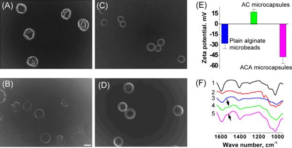
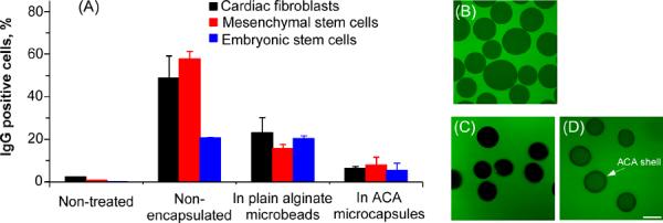
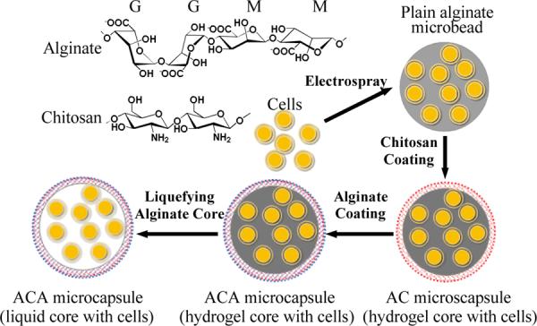
In this study, we report the preparation of a novel microcapsule of ~ 100 μm with a liquid (as compared to solid-like alginate hydrogel) core and an alginate-chitosan-alginate (ACA) shell for encapsulation and culture of embryonic stem (ES) cells in the miniaturized 3D space of the liquid core. Murine R1 ES cells cultured in the microcapsules were found to survive (> 90%) well and proliferate to form either a single aggregate of pluripotent cells or embryoid body (EB) of more differentiated cells in each microcapsule within 7 days, dependent on the culture medium used. This novel microcapsule technology allows massive production of the cell aggregates or EBs of uniform size and controllable pluripotency, which is important for the practical application of stem cell based therapy. Moreover, the semipermeable ACA shell was found to significantly reduce immunoglobulin G (IgG) binding to the encapsulated cells by up to 8.2 times, compared to non-encapsulated cardiac fibroblasts, mesenchymal stem cells, and ES cells. This reduction should minimize inflammatory and immune responses induced damage to the cells implanted in vivo becasue IgG binding is an important first step of the undesired host responses. Therefore, the ACA microcapsule with selective shell permeability should be of importance to advance the emerging cell-based medicine.
Figures

Similar articles
-
One-step microfluidic generation of pre-hatching embryo-like core-shell microcapsules for miniaturized 3D culture of pluripotent stem cells.Lab Chip. 2013 Dec 7;13(23):4525-33. doi: 10.1039/c3lc50678a. Lab Chip. 2013. PMID: 24113543 Free PMC article.
-
Coaxial electrospray of liquid core-hydrogel shell microcapsules for encapsulation and miniaturized 3D culture of pluripotent stem cells.Integr Biol (Camb). 2014 Sep;6(9):874-84. doi: 10.1039/c4ib00100a. Integr Biol (Camb). 2014. PMID: 25036382 Free PMC article.
-
Generation of core-shell microcapsules with three-dimensional focusing device for efficient formation of cell spheroid.Lab Chip. 2011 Jan 21;11(2):246-52. doi: 10.1039/c0lc00036a. Epub 2010 Oct 21. Lab Chip. 2011. PMID: 20967338
-
Cell Encapsulation Within Alginate Microcapsules: Immunological Challenges and Outlook.Front Bioeng Biotechnol. 2019 Dec 3;7:380. doi: 10.3389/fbioe.2019.00380. eCollection 2019. Front Bioeng Biotechnol. 2019. PMID: 31850335 Free PMC article. Review.
-
Progress technology in microencapsulation methods for cell therapy.Biotechnol Prog. 2009 Jul-Aug;25(4):946-63. doi: 10.1002/btpr.226. Biotechnol Prog. 2009. PMID: 19551901 Review.
Cited by
-
Electrohydrodynamic atomization: A two-decade effort to produce and process micro-/nanoparticulate materials.Chem Eng Sci. 2015 Mar 24;125:32-57. doi: 10.1016/j.ces.2014.08.061. Chem Eng Sci. 2015. PMID: 25684778 Free PMC article.
-
Effect of high cyclic hydrostatic pressure on osteogenesis of mesenchymal stem cells cultured in liquefied micro-compartments.Mater Today Bio. 2023 Nov 15;23:100861. doi: 10.1016/j.mtbio.2023.100861. eCollection 2023 Dec. Mater Today Bio. 2023. PMID: 38058695 Free PMC article.
-
The crucial role of mechanical heterogeneity in regulating follicle development and ovulation with engineered ovarian microtissue.Biomaterials. 2014 Jun;35(19):5122-8. doi: 10.1016/j.biomaterials.2014.03.028. Epub 2014 Apr 2. Biomaterials. 2014. PMID: 24702961 Free PMC article.
-
Conformal Nanoencapsulation of Allogeneic T Cells Mitigates Graft-versus-Host Disease and Retains Graft-versus-Leukemia Activity.ACS Nano. 2016 Jun 28;10(6):6189-200. doi: 10.1021/acsnano.6b02206. Epub 2016 May 31. ACS Nano. 2016. PMID: 27224853 Free PMC article.
-
All-in-one 3D printed microscopy chamber for multidimensional imaging, the UniverSlide.Sci Rep. 2017 Feb 10;7:42378. doi: 10.1038/srep42378. Sci Rep. 2017. PMID: 28186188 Free PMC article.
References
-
- Chang TM. Science. 1964;146:524–525. - PubMed
-
- Lim F, Sun AM. Science. 1980;210:908–910. - PubMed
-
- Ding HF, Liu R, Li BG, Lou JR, Dai KR, Tang TT. Biochem Biophys Res Commun. 2007;362:923–927. - PubMed
-
- Stensvaag V, Furmanek T, Lonning K, Terzis AJ, Bjerkvig R, Visted T. Cell Transplant. 2004;13:35–44. - PubMed
-
- Tobias CA, Han SS, Shumsky JS, Kim D, Tumolo M, Dhoot NO, Wheatley MA, Fischer I, Tessler A, Murray M. J Neurotrauma. 2005;22:138–156. - PubMed
Grants and funding
LinkOut - more resources
Full Text Sources
Other Literature Sources