

A detailed molecular analysis of complete bovine leukemia virus genomes isolated from B-cell lymphosarcomas
- PMID: 23506507
- PMCID: PMC3618307
- DOI: 10.1186/1297-9716-44-19
A detailed molecular analysis of complete bovine leukemia virus genomes isolated from B-cell lymphosarcomas
Abstract
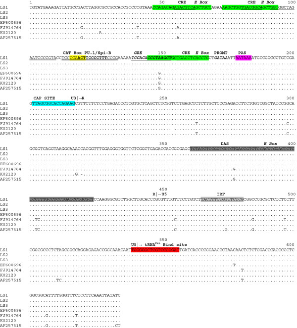
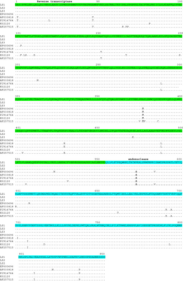
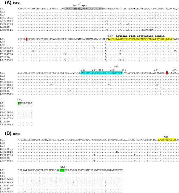
It is widely accepted that the majority of cancers result from multiple cellular events leading to malignancy after a prolonged period of clinical latency, and that the immune system plays a critical role in the control of cancer progression. Bovine leukemia virus (BLV) is an oncogenic member of the Retroviridae family. Complete genomic sequences of BLV strains isolated from peripheral blood mononuclear cells (PBMC) from cattle have been previously reported. However, a detailed characterization of the complete genome of BLV strains directly isolated from bovine tumors is much needed in order to contribute to the understanding of the mechanisms of leukemogenesis induced by BLV in cattle. In this study, we performed a molecular characterization of BLV complete genomes from bovine B-cell lymphosarcoma isolates. A nucleotide substitution was found in the glucocorticoid response element (GRE) site of the 5' long terminal repeat (5'LTR) of the BLV isolates. All amino acid substitutions in Tax previously found to be related to stimulate high transcriptional activity of 5'LTR were not found in these studies. Amino acid substitutions were found in the nucleocapsid, gp51 and G4 proteins. Premature stop-codons in R3 were observed. Few mutations or amino acid substitutions may be needed to allow BLV provirus to achieve silencing. Substitutions that favor suppression of viral expression in malignant B cells might be a strategy to circumvent effective immune attack.
Figures


Similar articles
-
Characterization of novel Bovine Leukemia Virus (BLV) antisense transcripts by deep sequencing reveals constitutive expression in tumors and transcriptional interaction with viral microRNAs.Retrovirology. 2016 May 3;13(1):33. doi: 10.1186/s12977-016-0267-8. Retrovirology. 2016. PMID: 27141823 Free PMC article.
-
A nucleotide deletion causing a translational stop in the protease reading frame of bovine leukaemia virus (BLV) results in modified protein expression and loss of infectivity.J Vet Med B Infect Dis Vet Public Health. 2000 Jun;47(5):361-71. doi: 10.1046/j.1439-0450.2000.00355.x. J Vet Med B Infect Dis Vet Public Health. 2000. PMID: 10900827
-
Detection and molecular characterization of bovine leukemia virus in Philippine cattle.Arch Virol. 2015 Jan;160(1):285-96. doi: 10.1007/s00705-014-2280-3. Epub 2014 Nov 16. Arch Virol. 2015. PMID: 25399399
-
Epidemiology and genetic diversity of bovine leukemia virus.Virol J. 2017 Nov 2;14(1):209. doi: 10.1186/s12985-017-0876-4. Virol J. 2017. PMID: 29096657 Free PMC article. Review.
-
A complex network of transcription factors and epigenetic regulators involved in bovine leukemia virus transcriptional regulation.Retrovirology. 2023 Jun 2;20(1):11. doi: 10.1186/s12977-023-00623-w. Retrovirology. 2023. PMID: 37268923 Free PMC article. Review.
Cited by
-
Interaction between Bovine leukemia virus (BLV) infection and age on telomerase misregulation.Vet Res Commun. 2015 Jun;39(2):97-103. doi: 10.1007/s11259-015-9629-2. Epub 2015 Feb 11. Vet Res Commun. 2015. PMID: 25665900
-
Bovine leukemia virus long terminal repeat variability: identification of single nucleotide polymorphisms in regulatory sequences.Virol J. 2018 Oct 25;15(1):165. doi: 10.1186/s12985-018-1062-z. Virol J. 2018. PMID: 30359262 Free PMC article.
-
Bovine Leukaemia Virus: Current Epidemiological Circumstance and Future Prospective.Viruses. 2021 Oct 27;13(11):2167. doi: 10.3390/v13112167. Viruses. 2021. PMID: 34834973 Free PMC article. Review.
-
Sequencing and phylogenetic analysis of the gp51 gene from Korean bovine leukemia virus isolates.Virol J. 2015 Apr 15;12:64. doi: 10.1186/s12985-015-0286-4. Virol J. 2015. PMID: 25879943 Free PMC article.
-
A target enrichment high throughput sequencing system for characterization of BLV whole genome sequence, integration sites, clonality and host SNP.Sci Rep. 2021 Feb 25;11(1):4521. doi: 10.1038/s41598-021-83909-3. Sci Rep. 2021. PMID: 33633166 Free PMC article.
References
-
- Burny A, Cleuter Y, Kettmann R, Mammerickx M, Marbaix G, Portetelle D, Van den Broeke A, Willems L, Thomas R. Bovine leukaemia: facts and hypothesis derived from the study of an infectious cancer. Cancer Surv. 1987;6:139–159. - PubMed
-
- Kettmann R, Deschamps J, Cleuter Y, Couez D, Burny A, Marbaix G. Leukemogenesis by bovine leukemia virus: proviral DNA integration and lack of RNA expression of viral long terminal repeat and 3′ proximate cellular sequences. Proc Natl Acad Sci U S A. 1982;79:2465–2469. doi: 10.1073/pnas.79.8.2465. - DOI - PMC - PubMed
-
- Kettmann R, Cleuter Y, Mammerickx M, Meunier-Rotival M, Bernardi G, Burny A, Chantrenne H. Genomic integration of bovine leukemia provirus: comparison of persistent lymphocytosis with lymph node tumor from of enzootic. Proc Natl Acad Sci U S A. 1980;77:2577–2581. doi: 10.1073/pnas.77.5.2577. - DOI - PMC - PubMed
-
- Pierard V, Guiguen A, Colin L, Wijmeersch G, Vanhulle C, Van Driessche B, Dekoninck A, Blazkova J, Cardona C, Merimi M, Vierendeel V, Calomme C, Nguyen T, Nuttinck M, Twizere JC, Kettmann R, Portetelle D, Burny A, Hirsch I, Rohr O, Van Lint C. DNA cytosine methylation in the Bovine Leukemia Virus promoter is associated with latency in a lymphoma-derived B-cell line. J Biol Chem. 2010;285:19434–19449. doi: 10.1074/jbc.M110.107607. - DOI - PMC - PubMed
-
- Merimi M, Klener P, Szynal M, Cleuter Y, Bagnis C, Kerkhofs P, Burny A, Martiat P, Van den Broeke A. Complete suppression of viral gene expression is associated with the onset and progression of lymphoid malignancy: observations in Bovine Leukemia Virus-infected sheep. Retrovirology. 2007;4:e51. doi: 10.1186/1742-4690-4-51. - DOI - PMC - PubMed
Publication types
MeSH terms
Associated data
- Actions
- Actions
- Actions
LinkOut - more resources
Full Text Sources
Other Literature Sources
