. 2013 Mar 18;44(1):20.
doi: 10.1186/1297-9716-44-20.

Pathogenic mechanisms implicated in the intravascular coagulation in the lungs of BVDV-infected calves challenged with BHV-1

Affiliations

Pathogenic mechanisms implicated in the intravascular coagulation in the lungs of BVDV-infected calves challenged with BHV-1

María A Risalde et al. Vet Res. .

Abstract

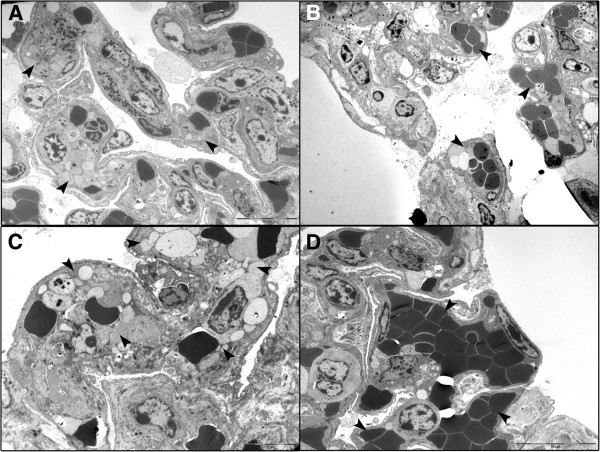
Resistance to respiratory disease in cattle requires host defense mechanisms that protect against pathogens which have evolved sophisticated strategies to evade them, including an altered function of pulmonary macrophages (MΦs) or the induction of inflammatory responses that cause lung injury and sepsis. The aim of this study was to clarify the mechanisms responsible for vascular changes occurring in the lungs of calves infected with bovine viral diarrhea virus (BVDV) and challenged later with bovine herpesvirus type 1 (BHV-1), evaluating the role of MΦs in the development of pathological lesions in this organ. For this purpose, pulmonary lesions were compared between co-infected calves and healthy animals inoculated only with BHV-1 through immunohistochemical (MAC387, TNFα, IL-1α, iNOS, COX-2 and Factor-VIII) and ultrastructural studies. Both groups of calves presented important vascular alterations produced by fibrin microthrombi and platelet aggregations within the blood vessels. These findings were earlier and more severe in the co-infected group, indicating that the concomitance of BVDV and BHV-1 in the lungs disrupts the pulmonary homeostasis by facilitating the establishment of an inflammatory and procoagulant environment modulated by inflammatory mediators released by pulmonary MΦs. In this regard, the co-infected calves, in spite of presenting a greater number of IMΦs than single-infected group, show a significant decrease in iNOS expression coinciding with the presence of more coagulation lesions. Moreover, animals pre-inoculated with BVDV displayed an alteration in the response of pro-inflammatory cytokines (TNFα and IL-1), which play a key role in activating the immune response, as well as in the local cell-mediated response.


Figures

Figure 1
Figure 1
Fibrin microthrombi in pulmonary venules of animals pre-infected with BVDV and challenged later with BHV-1.1. Co-infected animals presented vascular alterations that facilitate the occlusion of the pulmonary venules as fibrin microthrombi observed with Fraser Lendrum technique (A) and TEM (B) at 4 dpi, as well as PIM enlarged and rounded engulfing cell debris (arrowheads) observed with TEM at 4 dpi (C).
Figure 2
Figure 2
Immunohistochemical detection of pulmonary platelet clusters in calves with and without pre-existing BVDV challenged with BHV-1.1. Immunohistochemical study of the lung in these animals revealed that the calves of the BVDV/BHV-1 group showed a great quantity of Factor VIII-positive clusters of platelets in some pulmonary venules and capillaries at 4 dpi (A), compared with minor changes present in the BHV-1 group (B), where IMΦs and periarterial MΦs were observed engulfing positive granular material. Moreover, the calves of the BVDV/BHV-1 group showed lower quantities of platelet clusters at 14 dpi (C), with this lesion in the BHV-1 group being almost inexistent at this time (D).
Figure 3
Figure 3
Representative graphic of the valuation of factor VIII-positive platelet clusters detected by immunohistochemical method. These score values were obtained for the analysis of 100 fields (0.2 mm2 each) from the lung of calves (n = 2 per time point) inoculated with BHV-1.1 (a) versus calves inoculated with BVDV and BHV-1.1 (b). The results are given as follows: absent (−), mild (+), moderate (++) and abundant (+++), and scored from absent to severe (0 to 3). (UI, BHV-1.1 un-infected: negative controls for the BHV-1 group and BVDV infection controls for the BVDV/BHV-1 group).
Figure 4
Figure 4
Ultrastructural detection of pulmonary platelet clusters in calves with and without pre-existing BVDV challenged with BHV-1.1. The analysis of the samples with transmission electron microscopy confirmed the presence of multiple aggregations of platelets within the blood vessels with some vascular lumina completely occluded (arrowheads) in the lungs of the BVDV/BHV-1 group at 4 dpi (A), versus a mild lesion (arrowheads) observed in the BHV-1 group (B). Pulmonary parenchyma showed recovery signs of this lesion (arrowheads) in the co-infected group at 14 dpi (C), in comparison with a total recuperation (arrowheads) in the single infection (D).
Figure 5
Figure 5
Septal MΦs and PAM (means ± standard errors) positive for MAC387 by immunohistochemical method. The values were evaluated in the lung of calves co-infected experimentally with BVDV and BHV-1.1 compared with calves inoculated only with BHV-1.1 (n = 2 per time point). (UI, BHV-1.1 un-infected: negative controls for the BHV-1 group and BVDV infection controls for the BVDV/BHV-1 group. *p < 0.05 significant differences in the same group at various time points; **p < 0.05 significant differences between inoculated groups at the same time point).
Figure 6
Figure 6
Pro-inflammatory cytokines in the lungs of calves with and without pre-existing BVDV challenged with BHV-1.1. An immunohistochemical study revealed the presence of septal MΦs and PAM positive for TNFα (A) and IL-1α (C) associated with sites of inflammation in the pulmonary parenchyma of the BVDV/BHV-1 group at 2 dpi. However, the calves of the BHV-1 group presented a higher number of IMΦs reactive to TNFα (B) and IL-1α (D) in the peribronchial areas of the lung at 2 dpi.
Figure 7
Figure 7
Septal MΦs and PAM (mean ± standard error) positive for TNFα, IL-1α, iNOS and COX-2. Immunohistochemical study revealed changes in the lungs of calves co-infected experimentally with BVDV and BHV-1.1 compared with calves inoculated only with BHV-1.1 (n = 2 per time point). (UI, BHV-1.1 un-infected: negative controls for the BHV-1 group and BVDV infection controls for the BVDV/BHV-1 group. *p < 0.05 significant differences in the same group at various time points; **p < 0.05 significant differences between inoculated groups at the same time point).
Figure 8
Figure 8
Immunohistochemical detection of iNOS and COX-2 in the lungs of BVDV pre-infected calves challenged with BHV-1.1. Numerous immunolabeled septal MΦs iNOS-positive were observed in the lungs of calves inoculated only with BVDV (0 dpi of BVDV/BHV-1 group) (A), versus a minor number of these cells at 4 dpi BHV-1.1 (B). Pulmonary parenchyma of calves inoculated only with BVDV (0 dpi of BVDV/BHV-1 group) showed an absence of MΦs positive for COX-2 Ab (C), compared with numerous immunolabeled MΦs COX-2-positive observed in the sites of inflammation at 4 dpi (D).

Similar articles

Cited by

References

    1. Barrett DC. Bovine respiratory disease-A clinician’s perspective. Cattle Pract. 1998;6:251–255.
    1. Caldow G, Nettleton P. Pneumonia: identifying the causal agent. Cattle Pract. 2000;8:131–134.
    1. Griffin D. Economic impact associated with respiratory disease in beef cattle. Vet Clin North Am Food Anim Pract. 1997;13:367–377. - PubMed
    1. Fulton RW, Purdy CW, Confer AW, Saliki JT, Loan RW, Briggs RE, Burge LJ. Bovine viral diarrhea viral infections in feeder calves with respiratory disease: interactions with Pasteurella spp., parainfluenza-3 virus, and bovine respiratory syncytial virus. Can J Vet Res. 2000;64:151–159. - PMC - PubMed
    1. Fulton RW, Blood KS, Panciera RJ, Payton ME, Ridpath JF, Confer AW, Saliki JT, Burge LT, Welsh RD, Johnson BJ, Reck A. Lung pathology and infectious agents in fatal feedlot pneumonias and relationship with mortality, disease onset, and treatments. J Vet Diagn Invest. 2009;21:464–477. doi: 10.1177/104063870902100407. - DOI - PubMed

Publication types

MeSH terms

LinkOut - more resources