

Vaccinia virus GLV-1h153 is a novel agent for detection and effective local control of positive surgical margins for breast cancer
- PMID: 23506710
- PMCID: PMC3672815
- DOI: 10.1186/bcr3404
Vaccinia virus GLV-1h153 is a novel agent for detection and effective local control of positive surgical margins for breast cancer
Abstract
Introduction: Surgery is currently the definitive treatment for early-stage breast cancer. However, the rate of positive surgical margins remains unacceptably high. The human sodium iodide symporter (hNIS) is a naturally occurring protein in human thyroid tissue, which enables cells to concentrate radionuclides. The hNIS has been exploited to image and treat thyroid cancer. We therefore investigated the potential of a novel oncolytic vaccinia virus GLV1h-153 engineered to express the hNIS gene for identifying positive surgical margins after tumor resection via positron emission tomography (PET). Furthermore, we studied its role as an adjuvant therapeutic agent in achieving local control of remaining tumors in an orthotopic breast cancer model.
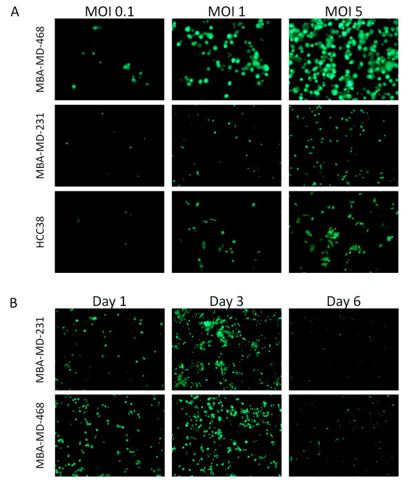
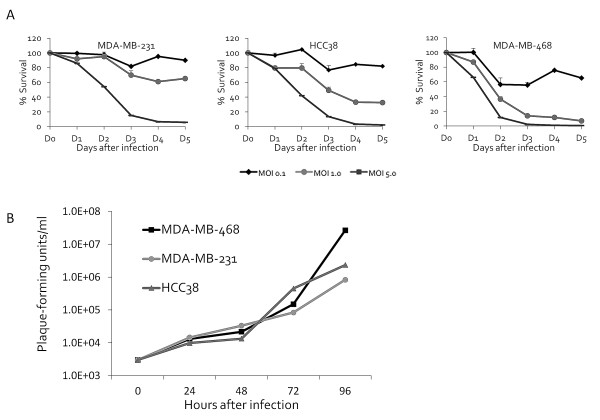
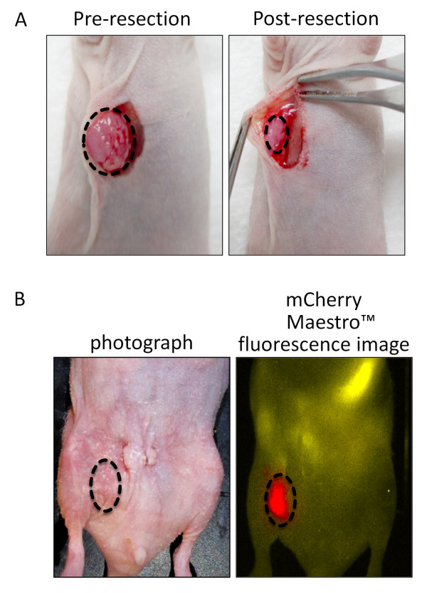
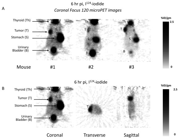
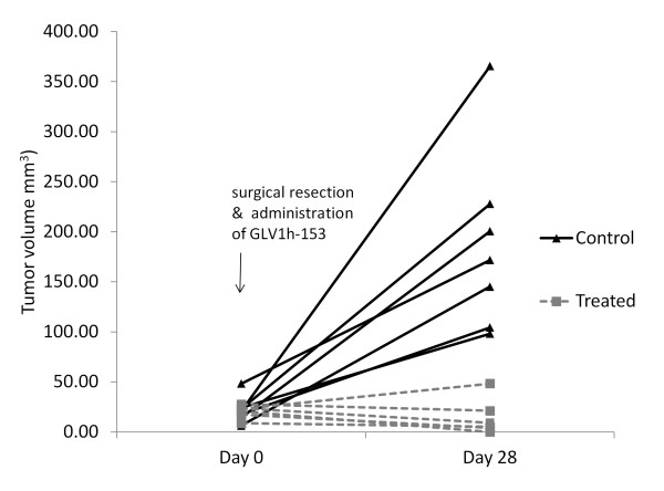
Methods: GLV-1h153, a replication-competent vaccinia virus, was tested against breast cancer cell lines at various multiplicities of infection (MOIs). Cytotoxicity and viral replication were determined. Mammary fat pad tumors were generated in athymic nude mice. To determine the utility of GLV-1h153 in identifying positive surgical margins, 90% of the mammary fat pad tumors were surgically resected and subsequently injected with GLV-1h153 or phosphate buffered saline (PBS) in the surgical wound. Serial Focus 120 microPET images were obtained six hours post-tail vein injection of approximately 600 μCi of 124I-iodide.
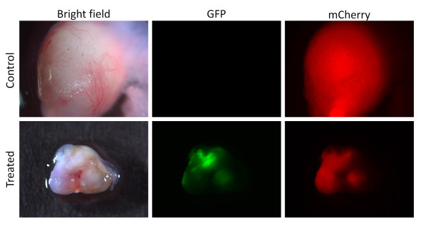
Results: Viral infectivity, measured by green fluorescent protein (GFP) expression, was time- and concentration-dependent. All cell lines showed less than 10% of cell survival five days after treatment at an MOI of 5. GLV-1h153 replicated efficiently in all cell lines with a peak titer of 27 million viral plaque forming units (PFU) ( <10,000-fold increase from the initial viral dose ) by Day 4. Administration of GLV-1h153 into the surgical wound allowed positive surgical margins to be identified via PET scanning. In vivo, mean volume of infected surgically resected residual tumors four weeks after treatment was 14 mm3 versus 168 mm3 in untreated controls (P < 0.05).
Conclusions: This is the first study to our knowledge to demonstrate a novel vaccinia virus carrying hNIS as an imaging tool in identifying positive surgical margins of breast cancers in an orthotopic murine model. Moreover, our results suggest that GLV-1h153 is a promising therapeutic agent in achieving local control for positive surgical margins in resected breast tumors.
Figures
Similar articles
-
Insertion of the human sodium iodide symporter to facilitate deep tissue imaging does not alter oncolytic or replication capability of a novel vaccinia virus.J Transl Med. 2011 Mar 31;9:36. doi: 10.1186/1479-5876-9-36. J Transl Med. 2011. PMID: 21453532 Free PMC article.
-
Imaging characteristics, tissue distribution, and spread of a novel oncolytic vaccinia virus carrying the human sodium iodide symporter.PLoS One. 2012;7(8):e41647. doi: 10.1371/journal.pone.0041647. Epub 2012 Aug 17. PLoS One. 2012. PMID: 22912675 Free PMC article.
-
Novel therapy for anaplastic thyroid carcinoma cells using an oncolytic vaccinia virus carrying the human sodium iodide symporter.Surgery. 2011 Dec;150(6):1040-7. doi: 10.1016/j.surg.2011.09.010. Surgery. 2011. PMID: 22136819
-
Gene therapy using therapeutic and diagnostic recombinant oncolytic vaccinia virus GLV-1h153 for management of colorectal peritoneal carcinomatosis.Surgery. 2015 Feb;157(2):331-7. doi: 10.1016/j.surg.2014.09.008. Surgery. 2015. PMID: 25616946 Free PMC article.
-
Close/positive margins after breast-conserving therapy: additional resection or no resection?Breast. 2013 Aug;22 Suppl 2:S115-7. doi: 10.1016/j.breast.2013.07.022. Breast. 2013. PMID: 24074771 Review.
Cited by
-
The Sodium/Iodide Symporter (NIS): Molecular Physiology and Preclinical and Clinical Applications.Annu Rev Physiol. 2017 Feb 10;79:261-289. doi: 10.1146/annurev-physiol-022516-034125. Annu Rev Physiol. 2017. PMID: 28192058 Free PMC article. Review.
-
Oncolytic Poxviruses.Annu Rev Virol. 2014 Sep 1;1(1):119-141. doi: 10.1146/annurev-virology-031413-085442. Annu Rev Virol. 2014. PMID: 25839047 Free PMC article.
-
Oncolytic vaccinia virus synergizes with irinotecan in colorectal cancer.Mol Oncol. 2015 Oct;9(8):1539-52. doi: 10.1016/j.molonc.2015.04.009. Epub 2015 May 6. Mol Oncol. 2015. PMID: 26004084 Free PMC article.
-
Oncolytic viruses: A novel treatment strategy for breast cancer.Genes Dis. 2021 Dec 16;10(2):430-446. doi: 10.1016/j.gendis.2021.11.011. eCollection 2023 Mar. Genes Dis. 2021. PMID: 37223527 Free PMC article. Review.
-
Oncolytic Virotherapy for Cancer: Clinical Experience.Biomedicines. 2021 Apr 13;9(4):419. doi: 10.3390/biomedicines9040419. Biomedicines. 2021. PMID: 33924556 Free PMC article. Review.
References
-
- Balch GC, Mithani SK, Simpson JF, Kelley MC. Accuracy of intraoperative gross examination of surgical margin status in women undergoing partial mastectomy for breast malignancy. Am Surg. 2005;71:22–27; discussion 27-28. - PubMed
-
- Cabioglu N, Hunt KK, Sahin AA, Kuerer HM, Babiera GV, Singletary SE, Whitman GJ, Ross MI, Ames FC, Feig BW, Buchholz TA, Meric-Bernstam F. Role for intraoperative margin assessment in patients undergoing breast-conserving surgery. Ann Surg Oncol. 2007;14:1458–1471. doi: 10.1245/s10434-006-9236-0. - DOI - PubMed
-
- Buzdar AU, Ibrahim NK, Francis D, Booser DJ, Thomas ES, Theriault RL, Pusztai L, Green MC, Arun BK, Giordano SH, Cristofanilli M, Frye DK, Smith TL, Hunt KK, Singletary SE, Sahin AA, Ewer MS, Buchholz TA, Berry D, Hortobagyi GN. Significantly higher pathologic complete remission rate after neoadjuvant therapy with trastuzumab, paclitaxel, and epirubicin chemotherapy: results of a randomized trial in human epidermal growth factor receptor 2-positive operable breast cancer. J Clin Oncol. 2005;23:3676–3685. doi: 10.1200/JCO.2005.07.032. - DOI - PubMed
Publication types
MeSH terms
Substances
LinkOut - more resources
Full Text Sources
Other Literature Sources
Medical
