Review
. 2013 Apr-Jun;34(2-3):121-38.
doi: 10.1016/j.mam.2012.07.001.

The SLC2 (GLUT) family of membrane transporters

Affiliations
Review

The SLC2 (GLUT) family of membrane transporters

Mike Mueckler et al. Mol Aspects Med. 2013 Apr-Jun.

Abstract

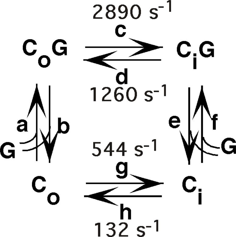
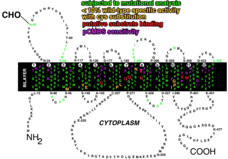
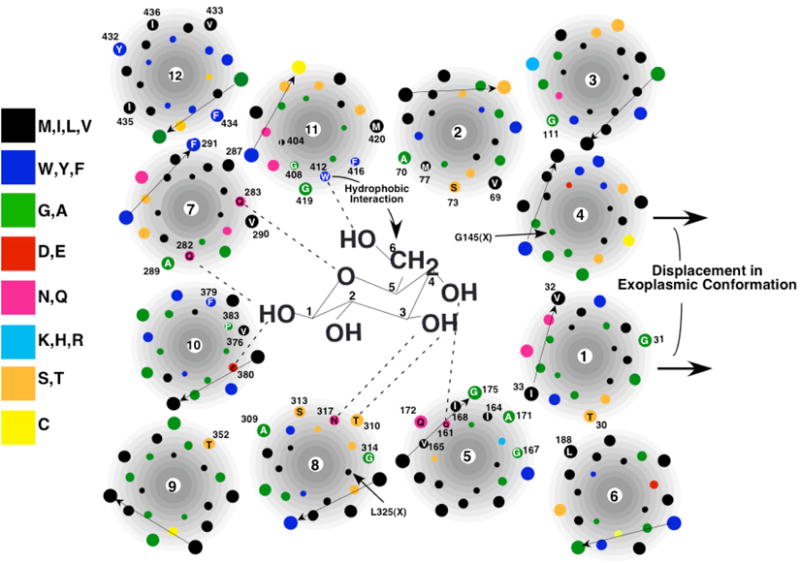
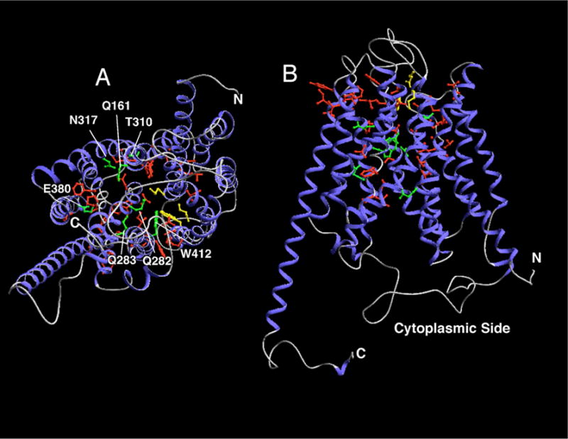
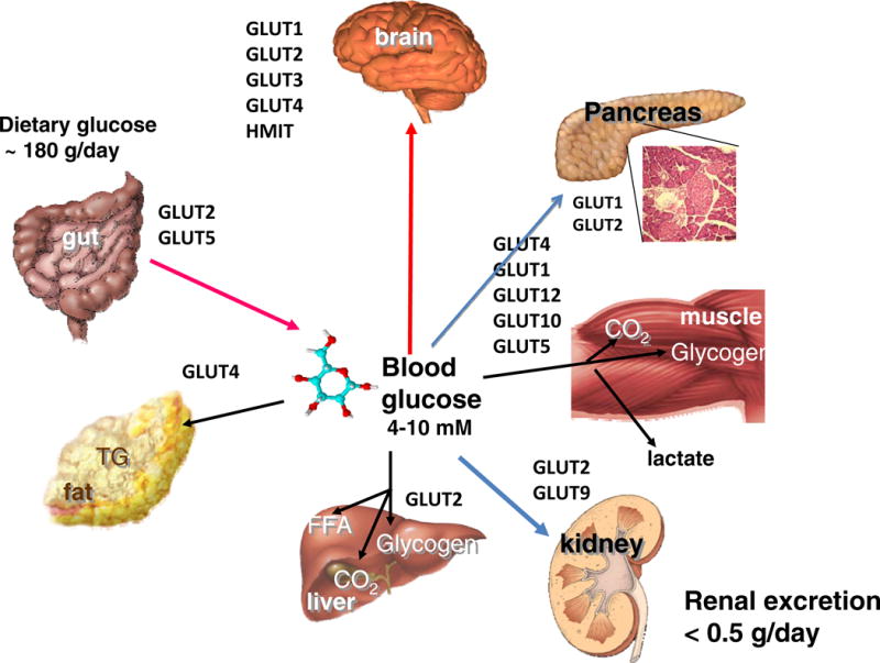
GLUT proteins are encoded by the SLC2 genes and are members of the major facilitator superfamily of membrane transporters. Fourteen GLUT proteins are expressed in the human and they are categorized into three classes based on sequence similarity. All GLUTs appear to transport hexoses or polyols when expressed ectopically, but the primary physiological substrates for several of the GLUTs remain uncertain. GLUTs 1-5 are the most thoroughly studied and all have well established roles as glucose and/or fructose transporters in various tissues and cell types. The GLUT proteins are comprised of ∼500 amino acid residues, possess a single N-linked oligosaccharide, and have 12 membrane-spanning domains. In this review we briefly describe the major characteristics of the 14 GLUT family members.


Figures

Figure 1
Figure 1. Simple carrier model for the mechanism of glucose transport
Co and Ci represent the transporter in the outward and inward-facing conformations, respectively, G is glucose, and a–h are the fundamental rate constants governing the conformational changes or the glucose binding and release steps. Estimates for rate constants c,d,g, and h at 20° C were calculated from the data of Lowe and Walmsley (Lowe and Walmsley, 1989).
Figure 2
Figure 2. Amino acid sequence and membrane topology of human GLUT1
Amino acid residues are designated by the single letter code. The transmembrane segments are numbered 1–12. The linkage of the N-linked oligosaccharide at N45 is shown. Positions that have been analyzed by site-directed mutagenesis are in green. Residues that are believed to play a direct role in substrate binding are shown in red. Residues that are accessible to pCMBS from the external solvent are shown in purple. Residues that appear to be critical for transport activity are shown in yellow. Adapted from (Mueckler and Makepeace, 2009).
Figure 3
Figure 3. Model of the exoplasmic substrate-binding site of GLUT1 (Mueckler and Makepeace, 2009)
Glucose is not drawn to scale. The arrangement of helices is shown in a simplistic fashion for clarity. Amino acid residues that are in contact with solvent in the aqueous cavity are numbered and identified by the single-letter code. Dotted lines represent putative hydrogen bonds.
Figure 4
Figure 4. Localization of pCMBS-reactive and putative substrate-binding residues in a homology based model of GLUT1
The molecular diagrams were created using DeepView (Swiss Institute of Bioinformatics) based on the coordinates published in (Salas-Burgos et al., 2004). The homology model is based on the crystal structure of the glycerol-3-P antiporter of E. coli in its cytoplasmic-facing conformation. The 12 transmembrane helices are drawn as blue ribbons and non-helical regions as gray lines. pCMBS-reactive side chains are shown in green. The 3 side chains near the exoplasmic face of the membrane that were identified as a putative glucose docking site are shown in yellow. Possible substrate-binding side chains based on mutagenesis experiments are presented in green and identified by the single letter amino acid code. A) View perpendicular to the membrane from inside the cell. B) Transverse view.
Figure 5
Figure 5. Colocalization of GFP-tagged wild type GLUT4 and HA-tagged IRM mutant GLUT4 by immunofluorescence laser confocal microscopy
3T3L1 adipocytes were co-infected with recombinant adenoviruses encoding the GFP-tagged wild-type GLUT4 and a HA-tagged IRM mutant (see text). 48 h later the cells were serum-starved for two hours, and then either exposed to insulin for 30 min or maintained in the basal state. Tagged wild-type GLUT4 is shown in the middle panels in green, the tagged mutants are shown in the left panels in red, and the merged images are shown in the right panels with colocalization between the two coexpressed proteins presented in yellow. The scale bars represent 10 μM. Note the redistribution of wild-type GLUT4 to the plasma membrane after insulin treatment and the lack of redistribution in the IRM mutant. Additionally, the mutant and wild type transporter are largely present in distinct intracellular membranes in the absence or presence of insulin. Adapted from (Song et al., 2008).
Figure 6
Figure 6. Role of GLUT Proteins in the Maintenance of glucose homeostasis
Glucose enters the hepatic portal system by transport across the gut via SGLT1 and GLUT2. In humans about one-third of blood glucose is carried within the red cell cytoplasm due to the very high level of expression of Glut1 and subsequent equilibration that occurs across the red cell membrane. Fructose crosses the gut via GLUT5. Most absorbed glucose escapes permanent catabolism by the hepatic system (which it enters via GLUT2) except when hepatic glycogen levels are low. Dietary fructose is mostly metabolized in the gut and liver and consequently circulating levels are very low. In the resting state most circulating glucose is oxidized by the central nervous after crossing the blood brain barrier via GLUT1 and enters parenchymal cells of the brain via GLUT3 (neurons) and GLUT1 (astrocytes). HMIT transports inositol against its concentration gradient in many brain cells. Other Glut proteins are expressed at lower levels and/or in smaller numbers of brain parenchymal cells and some, such as GLUT2 and GLUT4, may participate in fuel sensing by individual neurons. During exercise skeletal muscle consumes the bulk of circulating glucose via uptake by Glut1 in the endothelium and hence via Glut4 into muscle fibers. Glucose entering resting muscle is mostly converted to glycogen. Most glucose taken up into fat depots (via GLUT4) provides the glycerol moieties for the synthesis of triglycerides. Adipose tissue is also a critical endocrine organ with respect to glucose homeostasis and secretes numerous adipokines and cytokines that regulate this process. The most important organ of all with respect to the regulation of whole body glucose homeostasis is the endocrine pancreas, especially the insulin-secreting beta cells that sense blood glucose levels after initial uptake via GLUT1 (humans) or GLUT2 (rodents). Glucose is efficiently retained by the human body and very little is lost to urinary excretion, due to the combined actions of SGLT2 and GLUT2. (Figure is courtesy of Ernest Wright, UCLA Medical School).

References

    1. Abel ED, Peroni O, Kim JK, Kim YB, Boss O, Hadro E, Minnemann T, Shulman GI, Kahn BB. Adipose-selective targeting of the GLUT4 gene impairs insulin action in muscle and liver. [see comments] Nature. 2001;409(6821):729–733. - PubMed
    1. Abramson J, Smirnova I, Kasho V, Verner G, Kaback HR, Iwata S. Structure and mechanism of the lactose permease of Escherichia coli.[see comment] Science. 2003;301(5633):610–615. - PubMed
    1. Aerni-Flessner LB, Otu MC, Moley KH. The amino acids upstream of NH(2)-terminal dileucine motif play a role in regulating the intracellular sorting of the Class III transporters GLUT8 and GLUT12. Mol Membr Biol. 2011;28(1):30–41. - PMC - PubMed
    1. Angulo C, Rauch MC, Droppelmann A, Reyes AM, Slebe JC, Delgado-Lopez F, Guaiquil VH, Vera JC, Concha II. Hexose transporter expression and function in mammalian spermatozoa: cellular localization and transport of hexoses and vitamin C. Journal of Cellular Biochemistry. 1998;71(2):189–203. - PubMed
    1. Anzai N, Ichida K, Jutabha P, Kimura T, Babu E, Jin CJ, Srivastava S, Kitamura K, Hisatome I, Endou H, Sakurai H. Plasma urate level is directly regulated by a voltage-driven urate efflux transporter URATv1 (SLC2A9) in humans. J Biol Chem. 2008;283(40):26834–26838. - PubMed

Publication types

MeSH terms