Review
. 2013 Apr-Jun;34(2-3):197-219.
doi: 10.1016/j.mam.2012.07.002.

SLC6 transporters: structure, function, regulation, disease association and therapeutics

Affiliations
Review

SLC6 transporters: structure, function, regulation, disease association and therapeutics

Akula Bala Pramod et al. Mol Aspects Med. 2013 Apr-Jun.

Abstract

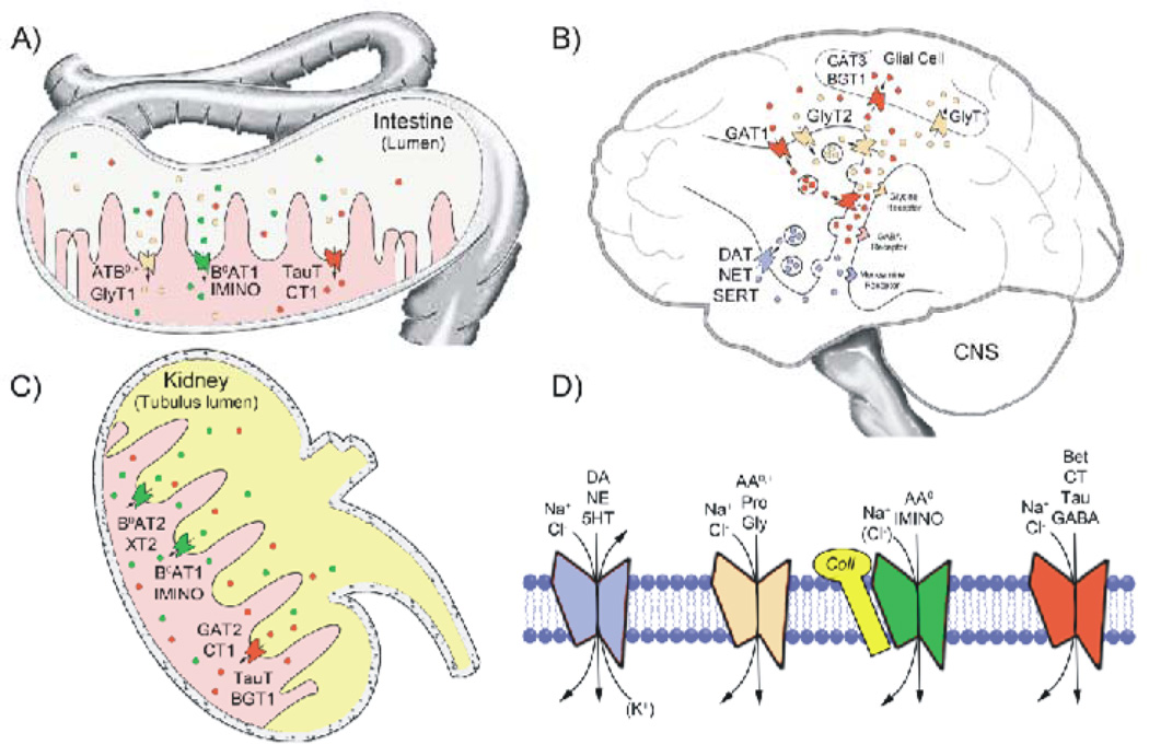
The SLC6 family of secondary active transporters are integral membrane solute carrier proteins characterized by the Na(+)-dependent translocation of small amino acid or amino acid-like substrates. SLC6 transporters, which include the serotonin, dopamine, norepinephrine, GABA, taurine, creatine, as well as amino acid transporters, are associated with a number of human diseases and disorders making this family a critical target for therapeutic development. In addition, several members of this family are directly involved in the action of drugs of abuse such as cocaine, amphetamines, and ecstasy. Recent advances providing structural insight into this family have vastly accelerated our ability to study these proteins and their involvement in complex biological processes.


Figures

Fig. 1
Fig. 1
Phylogenetic Tree Diagram of Human SLC6 Members. Phylogenetic tree was generated in Geneious Pro Software (Biomatters Ltd) using Blosum 62 scoring matrix, open gap penalty of 12 and gap extension penalty of 3, the Jukes-Cantor genetic distance model, and Neighbor-Joining tree building method. The genes are given as A1, A2, etc along with the common name. The sub-groupings within the tree are designated by different colored ovals and named as indicated. SLC6A21P which is a pseudogene is not shown.
Fig. 2
Fig. 2
Tissue distribution, physiologic function and substrate specificity of several SLC6 family transporters. A schematic representation of the prevalent tissue distribution and physiologic function of several SLC6 transporters is shown in A–C. The prominent roles of these transporters in intestinal nutrient absorption (A), renal reabsorption (C) of several amino acid and osmolyte substrates, and the role of transporters in synaptic transmission in the central nervous system (B) are depicted. Substrate specificity and ion and binding partner dependence for the four subclasses of SLC6 transporters is shown in panel D. Substrate abbreviations are: dopamine (DA), norepinephrine (NE), serotonin (5-HT), neutral and cationic amino acids (AA0,+), proline (Pro), glycine (Gly), neutral amino acids (AA0), proline and hydroxyproline (IMINO), betaine (Bet), creatine (CT), taurine (Tau), and -aminobutyric acid (GABA). All SLC6 transporters are Na+-dependent and most are Cl-dependent with the exception of B0AT2/SBAT1, NTT4/XT1 and B0AT1. Additionally, SERT utilizes the antiport of K+ in the translocation of 5-HT. Although SLC6 transporters have numerous binding partners that influence transport activity, B0AT1, B0AT3, and IMINO require expression of collectrin/TMEM27 or angiotensin-converting enzyme 2 (ACE2) for activity. For a detailed listing of transporter tissue distribution, substrate specificity and disease association, see Table 1.
Fig. 3
Fig. 3
Structural aspects of the SLC6 family. (A) Helical architecture of the LeuT fold transporters. The 5+5 (LeuT fold) motif is represented as 2D where TMs 1 and 3 and 6 and 8 form the substrate binding helices (red cylinders) and combine with TMs 2, 4 and 5 and 7, 9 and 10 (blue), respectively, to form the antiparallel heptahelical domains (demarcated by the dashed boxes). TMs 11 and 12 (dark gray cylinders, labeled +1 and +2, respectively) are located peripheral to the 5+5 motif. The black lines represent densities resolved in the LeuT crystal structures. Gray solid lines represent additional regions found in eukaryotic SLC6 proteins. Other LeuT fold proteins, such as vSGLT, possess the 5+5 core but also have additional N and C terminal helices (dashed gray lines and light gray cylinders). Substrate (S), Na+ (purple spheres) and Cl (green sphere) are also represented. (B) Illustration of alternating access and channel modes of transport. Substrate and ions access the binding site of the outward facing transporter (i) via the open outer gate (black sticks), outer gate closes yielding the occluded state (ii) which then transitions to the inward facing state (iii) releasing substrate and ions to cytosol (though controversial, this transition is proposed to be driven by a second substrate molecule (dashed line hexagon) binding to the S2 site). The transporter then rectifies to the outward facing structure (i). The transporter may undergo a lower probability transition (dashed lines) to a channel state (iv) where both gates are open simultaneously.
Fig. 4
Fig. 4
SLC6 transporter regulatory components. A representative topological diagram of SLC6 transporters is shown indicting major and common regulatory features and their location or site of action within the carrier protein. Identified Ser and Thr phosphorylation (S or T), ubiquitination (Ub), glycosylation (branched lines) and palmitoylation (Pal) sites as well as the location of the internalization motif (FREK) and selected known coding variants that alter transport function are shown. Common interacting proteins with their functional binding sites that are known to alter transport activity and trafficking are also indicted. The transmembrane domain of Syn 1A has been omitted for clarity.
Fig. 5
Fig. 5
Modes of SLC6 transporter down-regulation. Plasma membrane residing SLC6 transporters are depicted with the many factors that influence their activity and internalization. Two modes of PKC-dependent transporter down-regulation that result in functional loss of substrate transport are depicted. This includes well-known clathrin/dynamin-dependent transporter endocytosis and the more recently uncovered endocytosis-independent mode that is dependent on membrane cholesterol. It is postulated that loss of substantial transport activity occurs prior to endocytosis, which removes transporters from the cell surface. Several post-translational modifications and binding partner interactions as well as cholesterol-rich membrane raft localization and PKC-dependent raft to non-raft transition are depicted with hypothetical roles for these participants and events in the down-regulation process. The transmembrane domain of Syn 1A and extended dimerization domain of Flot1 have been omitted for clarity.

References

    1. Accardi A, Miller C. Secondary active transport mediated by a prokaryotic homologue of ClC Cl-channels. Nature. 2004;427:803–807. - PubMed
    1. Adams SV, DeFelice LJ. Flux coupling in the human serotonin transporter. Biophys J. 2002;83:3268–3282. - PMC - PubMed
    1. Adkins EM, Samuvel DJ, Fog JU, Eriksen J, Jayanthi LD, Vaegter CB, Ramamoorthy S, Gether U. Membrane mobility and microdomain association of the dopamine transporter studied with fluorescence correlation spectroscopy and fluorescence recovery after photobleaching. Biochemistry. 2007;46:10484–10497. - PubMed
    1. Agoston GE, Vaughan R, Lever JR, Izenwasser S, Terry PD, Newman AH. A novel photoaffinity label for the dopamine transporter based on N-substituted 3 -[bis(4'-fluorophenyl)methoxy]tropane. Bioorg Med Chem Lett. 1997;7:3027–3032.
    1. Amara SG, Sonders MS. Neurotransmitter transporters as molecular targets for addictive drugs. Drug Alcohol Depend. 1998;51:87–96. - PubMed

Publication types

MeSH terms

Substances

LinkOut - more resources