Review
. 2013 Jun;34(6):259-68.
doi: 10.1016/j.it.2013.02.004. Epub 2013 Mar 15.

Ras and extracellular signal-regulated kinase signaling in thymocytes and T cells

Affiliations
Review

Ras and extracellular signal-regulated kinase signaling in thymocytes and T cells

Robert L Kortum et al. Trends Immunol. 2013 Jun.

Abstract

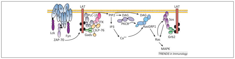
Extracellular signal-regulated kinase (ERK) activation is important for both thymocyte development and T cell function. Classically, signal transduction from the T cell antigen receptor (TCR) to ERK is thought to be regulated by signaling from Ras guanine nucleotide exchange factors (GEFs), through the small G protein Ras, to the three-tiered Raf-MAPK/ERK kinase (MEK)-ERK kinase cascade. Developing and mature T cells express four members of two RasGEF families, RasGRP1, RasGRP4, son of sevenless 1 (Sos1), and Sos2, and several models describing combined signaling from these RasGEFs have been proposed. However, recent studies suggest that existing models need revision to include both distinct and overlapping roles of multiple RasGEFs during thymocyte development and novel, Ras-independent signals to ERK that have been identified in peripheral T cells.


Figures

Figure 1
Figure 1
Canonical T cell receptor (TCR) signal transduction to Ras–mitogen-activated protein kinase (MAPK). After TCR binding and engagement of the upstream kinases lymphocyte-specific protein tyrosine kinase (Lck) and feline yes-related protein (Fyn), activated ζchain-associated protein kinase of 70 kDa (ZAP70) triggers phosphorylation of linker for activation of T cells (LAT). Phosphorylated LAT associates with two molecular complexes that regulate Ras activation: phospholipase C (PLC)-γ1–Grb2-related adaptor protein involved in T cell signaling (GADS)–SH2 domain-containing leukocyte phosphoprotein of 76 kDa (SLP-76) and growth factor receptor-bound protein 2 (Grb2)-son of sevenless (Sos). On LAT, activated PLC-γ1 cleaves phosphatidylinositol 4,5 bisphosphate (PIP2) generating inositol 1,4,5-trisphosphate (IP3), which stimulates release of calcium from intracellular stores and diacylglycerol (DAG). DAG then recruits protein kinase C (PKC)θ and RasGRP1 to the membrane, and the combined actions of Ca2+, DAG, and PKCθ activate Ras guanine nucleotide-releasing protein 1 (RasGRP1). The Ras guanine nucleotide exchange factors (GEFs) Sos1 and Sos2 are also recruited to LAT via the adapter Grb2. Sos proteins have basal RasGEF activity, which can be enhanced by the binding of activated Ras (Ras-GTP) to an allosteric binding pocket on Sos. Ras-GTP binding to Sos potentially allows for the engagement of a positive feedback loop, primed by either RasGRP1 or basal Sos activity, to produce high levels of Ras activation. Ras signals to multiple downstream effector pathways including the Raf–MAPK/ERK kinase (MEK)–extracellular signal-regulated kinase (ERK) kinase cascade.
Figure 2
Figure 2
Integrated model of Ras activation during thymocyte development. At the CD4CD8 (DN)3 stage, ligand-independent pre-T cell receptor (TCR) signals are transmitted to Ras and extracellular signal-regulated kinase (ERK) by the combined actions of son of sevenless 1 (Sos1), Ras guanine nucleotide-releasing protein 1 (RasGRP1), and RasGRP4 to stimulate proliferation and differentiation to the CD4+CD8+ (DP) stage. In the thymic cortex, positively selecting signals are transmitted from the TCR to Ras via RasGRP1. Downstream of Ras, ERK activation is required for efficient positive selection. In the medulla, higher-potency ligands trigger Ras activation via either RasGRP1 or Sos1 to promote negative selection. Here, activation of ERK seems to be dispensable, and activation of other mitogen-activated protein kinases [MAPKs; c-jun N-terminal kinase (JNK), p38, and ERK5] are likely more important. Ras guanine nucleotide exchange factors (GEFs) whose individual deletion blocks thymocyte development for a given signal are shown in red, whereas RasGEFs that require combined deletion with a second RasGEF to cause a developmental block are shown in blue. Proteins whose role at a given developmental checkpoint have not been demonstrated by developmental studies are shown in green.
Figure 3
Figure 3
Ras-dependent versus Ras-independent extracellular signal-regulated kinase (ERK) signaling. In peripheral T lymphocytes, two molecular complexes, both of which are capable of signaling to the Raf–MAPK/ERK kinase (MEK)–ERK kinase cascade, compete for phospholipase C (PLC)-γ1 binding. Association of PLC-γ1 with linker for activation of T cells (LAT) promotes canonical T cell receptor (TCR) signaling (Figure 1) and activation of the Raf–MEK–ERK kinase cascade via Ras. This pathway requires PLC-γ1 catalytic activity (red). By contrast, association of PLC-γ1 with the adapter B cell lymphocyte adaptor molecule of 32 kDa (Bam32) and the kinase p21-activated kinase 1 (PAK1) causes dissociation of PAK1 dimers promoting PAK1 activation. PAK1 can then signal to the Raf–MEK–ERK kinase cascade by phosphorylating both Raf and MEK. In this alternative pathway, PLC-γ1 is catalytically inactive (blue) and instead acts as a scaffold to dissociate pre-existing PAK1 dimers. Normally, both pathways act together to promote ERK activation, as MEK1 phosphorylation on S298 (by PAK1) potently enhances its phosphorylation on S217/S221 by Raf. Changes in the balance between expression of the adapters LAT and Bam32 can sequester PLC-γ1 and alter the relative activities of these two pathways. These two PLC-γ1-dependent pathways are likely to be responsible for the majority of TCR-dependent ERK activation, although LAT–son of sevenless (Sos) and lymphocyte-specific protein tyrosine kinase (Lck)–protein kinase C (PKC)θ–Ras guanine nucleotide-releasing protein 1 (RasGRP1) pathways also contribute to TCR-dependent ERK signaling.

References

    1. Downward J. Targeting RAS signalling pathways in cancer therapy. Nat Rev Cancer. 2003;3:11–22. - PubMed
    1. Gysin S, et al. Therapeutic strategies for targeting ras proteins. Genes Cancer. 2011;2:359–372. - PMC - PubMed
    1. Logue JS, Morrison DK. Complexity in the signaling network: insights from the use of targeted inhibitors in cancer therapy. Genes Dev. 2012;26:641–650. - PMC - PubMed
    1. Roberts PJ, Der CJ. Targeting the Raf-MEK-ERK mitogen-activated protein kinase cascade for the treatment of cancer. Oncogene. 2007;26:3291–3310. - PubMed
    1. Aguado E, et al. Induction of T helper type 2 immunity by a point mutation in the LAT adaptor. Science. 2002;296:2036–2040. - PubMed

Publication types

MeSH terms

Substances