. 2013 Jun;94(3):445-52.
doi: 10.1016/j.yexmp.2013.03.001. Epub 2013 Mar 16.

Deficit of p66ShcA restores redox-sensitive stress response program in cisplatin-induced acute kidney injury

Affiliations

Deficit of p66ShcA restores redox-sensitive stress response program in cisplatin-induced acute kidney injury

Rungwasee Rattanavich et al. Exp Mol Pathol. 2013 Jun.

Abstract

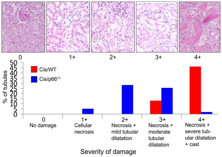
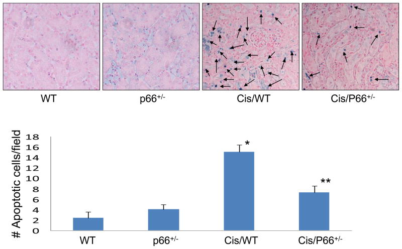
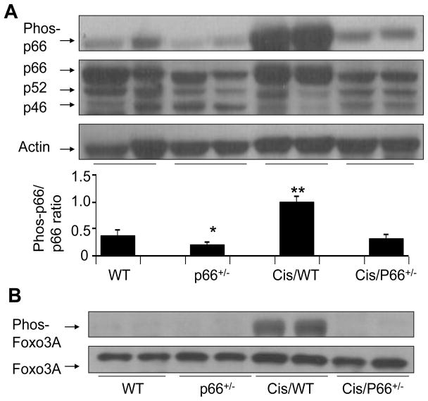
Overwhelming oxidative stress and compromised tubular cell antioxidant response have been incriminated for cisplatin (Cis)-induced acute kidney injury (AKI). We hypothesized that Cis-induced AKI was the outcome of the deactivated redox-sensitive stress response program (RSSRP). Wild type (WT) and heterozygous p66ShcA(p66(+/-)) mice in groups of six were administered either normal saline (WT) or Cis (12.5 mg/kg, intraperitoneal, Cis/WT). Renal biomarkers were collected and kidneys were harvested for renal histology. Cis/WT showed elevated blood urea nitrogen levels and enhanced tubular cell apoptosis, necrosis, and dilated tubules filled with casts when compared to Cis/p66(+/-). Cis/p66(+/-) developed only a clinically occult AKI (normal blood urea levels and only microscopic alterations). Immunoblots from the lysates of renal tissues of Cis/WT displayed enhanced expression of phospho-p66ShcA, and phospho-Foxo3A but attenuated expression of MnSOD and catalase; conversely, p66 deficit prevented these alterations in Cis milieu. In in vitro studies, Cis treated mouse proximal tubular cells (MPTCs) displayed enhanced phosphorylation of p66ShcA and no increase in tubular cell expression of MnSOD. In addition, renal tissues of Cis/WT and Cis-treated MPTCs displayed enhanced phosphorylation of p53 and Bax expression. However, MPTC partially silenced for p66ShcA displayed partial inhibition of Cis-induced tubular cell apoptosis as well as necrosis. These findings indicate that Cis-induced AKI is the outcome of the deactivated RSSRP (attenuated anti-oxidant response) and activation of pro-apoptotic (p53-induced Bax expression) pathway.


Figures

Fig. 1
Fig. 1. Cis/p66+/− mice display attenuated tubular cell injury
A. Representative microphotographs showing different grading of tubular cell injury. B. Cis/WT and Cis/WT mice (n=6) were graded for their tubular cell injury. Percentages of tubules displaying variable severity of injury are shown displayed massive tubular cell necrosis and dilatation of tubules. Approximately 50% tubules displayed 4+ injury in Cis/WT; whereas, only 1.5% of tubules in Cis/p66+/− displayed 4+ injury.
Fig. 2
Fig. 2. Cis/p66+/− mice display decreased injury score and only occult AKI
A. Representative microphotographs of WT, P66+/−, Cis/WT., and Cis/p66+/− mice are shown. B. Cumulative data (n=6) on injury score is shown *P<0.01 compared to Cis/WT. C. Mean BUN levels in WT, P66+/−, Cis/WT., and Cis/P66+/− mice are shown. *P<0.01 compared to other variables. D. mRNA expression for p66 as assayed by real time PCR studies from control mice and p66+/− mice (n=4). *P<0.01 compared to WT.
Fig. 3
Fig. 3. P66 deficiency partially rescues from Cis-induced apoptosis
Renal cortical sections of WT, P66+/−, Cis/WT, and Cis/P66+/− mice were labeled for TUNEL +ve cells. Representative Renal cortical sections of WT, P66+/−, Cis/WT., and Cis/P66+/− mice displaying TUNEL +ve cells (blue stained cells). *P<0.001 compared to WT and P66+/− mice. **P<0.01 compared to Cis/WT mice.
Fig. 4
Fig. 4. Cis enhances renal tissue p66ShcA and deactivates redox sensitive stress response program
A. Immunoblots of WT, Cis/WT, P66+/− and Cis/P66+/− were probed for phospho-p66ShcA (n=4). The same nitrocellulose blots were stripped and reprobed for total p66 as well as for actin. Representative gels of the lysates two different animals are shown. Cumulative data of four different mice are shown in the form of bar graphs. *P<0.05 compared to WT; **P<0.01 compared to Cis/p66+/− B. Renal tissue lysates of WT, Cis/WT, p66+/− and Cis/p66+/− were electrophoresed and then probed for phospho-Foxo3A (n=4). The same Western blots were stripped and reprobed for total Foxo3A.
Fig. 5
Fig. 5. p66 deficiency restores the renal tissue antioxidant generation
A. Protein blots of renal tissues of WT, Cis/WT, p66+/− and Cis/p66+/− were probed for MnSOD and catalase (n=4). The same blots were stripped and reprobed for actin. Representative gels from two different mice are shown. Cumulative data of four mice are shown in the form of a bar diagram. *P<0.05 compared with Cis/WT. B. Renal tissue lysates of WT, Cis/WT, p66+/− and Cis/p66+/− were electrophoresed and then probed for catalase and reprobed for actin (n=4). Representative gels of two different mice are shown. Renal tissues Cis/WT display attenuated expression of catalase; whereas, renal tissues of Cis/p66+/− display enhanced expression of catalase.
Fig. 6
Fig. 6. Cis enhances activation of p53 pathway
A. Renal tissue lysates of WT, Cis/WT, p66+/− and Cis/p66+/− were electrophoresed and probed for phospho-p53 (n=4). The same blots were stripped and reprobed for total p53. Representative gels of two different mice are shown. Cumulative data of four different mice are shown in bar graphs. *P<0.001 compared with other variables B. Immunoblots of renal tissues of WT, Cis/WT, p66+/− and Cis/p66+/− probed for the expression of Bax and actin. Renal tissues of Cis/WT displayed enhanced expression of Bax.
Fig. 7
Fig. 7. Cis deactivates redox-sensitive stress response pathway in mouse tubular cells
MPTCs were incubated in media containing either buffer or variable concentrations of Cis (25 or 50 μM) for 12 or 20 hours. Subsequently, protein blots were probed for phospho-p53, phospho-p66ShcA, or MnSOD. The same blots were reprobed for actin. A. Representative gels displaying MPTC expression of phospho-p53 and actin under control and Cis-treated conditions. B. Representative gels displaying MPTC expression of phospho-p66 and actin under control and Cis-treated conditions. C. Representative gels displaying MPTC expression of MnSOD and actin under control and Cis-treated conditions.
Fig. 8
Fig. 8. Cis enhances tubular cell expression of Bax but down regulates expression of Bcl2
MPTCs were incubated in media containing either buffer or Cis (25 μM or 50 μM) for 12 hours. Subsequently, cellular lysates were immunoelectrophoresed and probed for Bax and Bcl2. The same western blots were reprobed for actin. Representative gels are shown.
Fig. 9
Fig. 9. p66 is critical for Cis-induced tubular cell apoptosis
A. MPTCs were transfected with either scrambled (SCR) siRNA or p66-siRNA. The upper lane displayed representative immunoblots of control MPTCs, SCR/MPTCs and p66-siRNA/MPTCs. The lower lane shows stripped and reprobed blots for actin. B. Densitometric data is displayed in the form of bar graphs/ C. Control MPTCs. SCR/MPTCs and p66-siRNA/MPTCs were incubated in medium containing either buffer or Cis (25 μM) for 24 hours (n=3). Subsequently cells were assayed for apoptosis, primary and secondary necrosis. *P<0.01 compared to control and siRNA/p66/Cis; **P<0.05 compared to control; ***P<0.001 compared to control, siRNA/p66/Cis
Fig. 10
Fig. 10
Proposed schematic pathway for Cis-induced tubular cell apoptosis.

References

    1. Arany I, Safirstein RL. Cisplatin nephrotoxicity. Semin Nephrol. 2003;23:460–464. - PubMed
    1. Baliga R, Zhang Z, Baliga M, Ueda N, Shah SV. In vitro and in vivo evidence suggesting a role for iron in cisplatin-induced nephrotoxicity. Kidney Int. 1998;53:394–401. - PubMed
    1. Camici GG, Schiavoni M, Francia P, Bachschmid M, Martin-Padura I, Hersberger M, Tanner FC, Pelicci P, Volpe M, Anversa P, Luscher TF, Cosentino F. Genetic deletion of p66(Shc) adaptor protein prevents hyperglycemia-induced endothelial dysfunction and oxidative stress. Proc Natl Acad Sci USA. 2007;104:5217–5222. - PMC - PubMed
    1. Chintapalli J, Yang S, Opawumi D, Goyal SR, Shamsuddin N, Malhotra A, Reiss K, Meggs LG. Inhibition of wild-type p66ShcA in mesangial cells prevents glycooxidant-dependent FOXO3a regulation and promotes the survival phenotype. Am J Physiol Renal Physiol. 2007;292:F523–530. - PubMed
    1. Clark JS, Faisal A, Baliga R, Nagamine Y, Arany I. Cisplatin induces apoptosis through the ERK-p66shc pathway in renal proximal tubule cells. Cancer Lett. 2010;297:165–70. - PubMed

Publication types

MeSH terms

LinkOut - more resources