. 2014 Dec;1842(12 Pt A):2517-27.
doi: 10.1016/j.bbadis.2013.03.004. Epub 2013 Mar 16.

Cyclophilin D deficiency rescues Aβ-impaired PKA/CREB signaling and alleviates synaptic degeneration

Affiliations

Cyclophilin D deficiency rescues Aβ-impaired PKA/CREB signaling and alleviates synaptic degeneration

Heng Du et al. Biochim Biophys Acta. 2014 Dec.

Abstract

The coexistence of neuronal mitochondrial pathology and synaptic dysfunction is an early pathological feature of Alzheimer's disease (AD). Cyclophilin D (CypD), an integral part of mitochondrial permeability transition pore (mPTP), is involved in amyloid beta (Aβ)-instigated mitochondrial dysfunction. Blockade of CypD prevents Aβ-induced mitochondrial malfunction and the consequent cognitive impairments. Here, we showed the elimination of reactive oxygen species (ROS) by antioxidants probucol or superoxide dismutase (SOD)/catalase blocks Aβ-mediated inactivation of protein kinase A (PKA)/cAMP regulatory-element-binding (CREB) signal transduction pathway and loss of synapse, suggesting the detrimental effects of oxidative stress on neuronal PKA/CREB activity. Notably, neurons lacking CypD significantly attenuate Aβ-induced ROS. Consequently, CypD-deficient neurons are resistant to Aβ-disrupted PKA/CREB signaling by increased PKA activity, phosphorylation of PKA catalytic subunit (PKA C), and CREB. In parallel, lack of CypD protects neurons from Aβ-induced loss of synapses and synaptic dysfunction. Furthermore, compared to the mAPP mice, CypD-deficient mAPP mice reveal less inactivation of PKA-CREB activity and increased synaptic density, attenuate abnormalities in dendritic spine maturation, and improve spontaneous synaptic activity. These findings provide new insights into a mechanism in the crosstalk between the CypD-dependent mitochondrial oxidative stress and signaling cascade, leading to synaptic injury, functioning through the PKA/CREB signal transduction pathway.

Keywords: Alzheimer's disease; Amyloid beta; Mitochondrial permeability transition; Oxidative stress; PKA/CREB signaling; Synaptic alteration.


Conflict of interest statement

Conflict of interest: The authors have no conflict of interest to disclose.

Figures

Fig. 1
Fig. 1
Antioxidants attenuated Aβ-induced inhibitory effects on pPKA C and pCREB levels. (A) The treatment of SOD (200 U/ml)/catalase (250 U/ml) or probucol (10 μM) significantly attenuated Aβ (5 μM, 2 h)-induced decrease in pPKA C level. Densitometry of the pPKA C immunoreactive bands relative to the total PKA C was shown in the indicated groups of cells. The lower panel showed representative immunoblots for pPKA C and total PKA C in the indicated groups. (B) The treatment of antioxidants attenuated Aβ-induced reduction in PKA activity. (C1–C2) Densitometry of the immunoreactive bands for phospho-CREB (pCREB) relative to the total CREB in the indicated groups of cells. Levels of pCREB were increased in cells treated with SOD/catalase (C1) and probucol (C2) in the presence of Aβ. The lower panels in C1 and C2 showed the representative immunoblots for pCREB and total of CREB. Results were derived from 3 to 5 independent experiments.
Fig. 2
Fig. 2
Effects of CypD-deficiency on mitochondrial ROS production in neurons exposed to Aβ. (A) Aβ-treated neurons were subjected to MitoSox Red staining and the percentage of MitoSox Red-positive cells was quantified by flow cytometry. (B) CsA treatment significantly attenuated MitoSox Red intensity in Aβ-exposed neurons. Scale bar = 10 μm.
Fig. 3
Fig. 3
Effects of CypD-deficiency on PKA activity, phosphorylation of PKA C (p-PKA C), and pCREB in neurons exposed to Aβ. (A) PKA activity in cultured nonTg and Ppif−/− neurons exposed to oligomeric Aβ for 2, 6, 12 and 24 h, respectively. KT5720 (1 μM), a PKA inhibitor, was added to neurons. PKA activity in the indicated cell lysates was determined by PKA Kinase Activity Assay Kit. The data were expressed as fold increased relative to the PKA activity in vehicle-treated nonTg neurons. (B) NonTg and Ppif−/− neurons were treated with 5 μM Aβ for 2 h, then subjected to immunoblotting for phosphorylation of PKA C Thr197, and total PKA C. Quantification of intensity of phospho-PKA C immunoreactive bands normalized to total PKA C is presented in the upper panel. The lower panels represent the sample immunoblots for phospho-PKA C Thr197 and PKA C subunit. (C) Effect of CypD deficiency on glutamate-induced phosphorylation of CREB. Cortical neurons were exposed to 5 μM Aβ for 2 h in the presence or absence of glutamate stimulation, and then subjected for immunoblotting for pCREB Ser133 and total CREB. Densitometry of pCREB immunoreactive bands normalized to total CREB is presented in the upper panel and the lower panel shows representative immunoblots for pCREB and CREB. Rolipram (3 μM) and Forskolin (5 μM) were added to cultured neurons.
Fig. 4
Fig. 4
Effects of CypD deficiency and antioxidant on Aβ-induced synaptic loss. (A) Expression levels of synaptophysin in Aβ-treated nonTg and Ppif−/− neurons. Cultured neurons were treated with 5 μM oligomer Aβ1–42 for 2 h. Cell lysates were subjected to immunoblotting with synaptophysin antibody. Densitometry of the synaptophysin immunoreactive bands was normalized by α-tubulin using NIH Image J software. The lower panels are representative immunoblots for synaptophysin and α-tubulin. Data were collected from 3 to 4 independent experiments. (B) After exposure of 5 μM oligomer Aβ for 2 h, Ppif−/− neurons exhibited preserved synaptic positive clusters compared with nonTg neurons. PKA activator, forskolin (5 μM), protected nonTg neurons against Aβ-induced synaptic loss. Data were collected from 17 to 20 neurons per group. The lower panel shows representative images of synaptophysin clusters (synaptophysin, red) and dendrite (MAP2, green) staining. Scale bar = 10 μm. (C) Antioxidants attenuated Aβ-induced synaptic loss. Data are derived from 16 to 23 neurons per group in 3 independent experiments. The right panel shows representative images of synaptic staining. Synapses were visualized by synaptophysin staining (red) and dendrites were shown by MAP2 staining (green). Scale bar = 10 μm.
Fig. 5
Fig. 5
Effect of CypD deletion on brain oxidative stress, PKA activity, phosphorylation of PKA C and CREB in mAPP mice. (A) Cerebral cortexes from the indicated Tg mice were subjected to 4-HNE assay. CypD-deficient mAPP mice demonstrated significantly lowered 4-HNE level than mAPP mice. (B) PKA activity in the cerebral cortex of the indicated Tg mice. Data are shown in fold-increase relative to the PKA activity in nonTg mice. (C) Densitometry of immunoreactive bands for p-PKA C, total PKA C, and α-tubulin in the cerebral cortex homogenates of the indicated Tg mice. Data were expressed as fold-increase relative to pPKA C level (normalized to total PKA C) in nonTg mice. The representative immunoblots were demonstrated in lower panel. (D) CREB phosphorylation in Tg mice. Data were shown as fold-increase relative to total CREB.
Fig. 6
Fig. 6
Effect of CypD on spontaneous synaptic activity in mAPP mice. mAPP mice showed decreased mEPSC amplitude (A) and frequency (B), which were restored by CypD deficiency. (C) The representative traces of mEPSC recorded for CA1 neurons from the indicated Tg mice. Scale = 20 pA, 500 ms.
Fig. 7
Fig. 7
Effect of CypD deletion on dendritic spine architecture in mAPP mice. (A–D) Quantification of spine density (A) and types of synaptic morphology (B–D). (A) The comparison of spine density. N = 4–6 mice per group, 10, 11, 15 and 12 neurons from nonTg, Ppif−/−, mAPP mice, and mAPP/Ppif−/− mice, respectively. (B–D) The comparison of the percentage of mushroom (B1–2), stubby (C1–2), and thin (D1–2) type spines occupied by the total dendritic spines. (E) Representative images of basal dendritic spines of CA1 neurons in Tg mice. Dendritic spines (green) and senile plaques (red) were visualized by Lucifer yellow injection and 3D6 immunostaining, respectively. The panels i, ii, iii and iv show representative images of dendritic spines from nonTg, Ppif−/−, mAPP and mAPP/Ppif−/− mice, respectively. The lower panels present outlines of the dendritic spines corresponding to the images above. Scale bar = 5 μm.
Fig. 8
Fig. 8
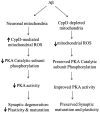
Schematic figure of CypD deficiency protects synaptic plasticity and maturation against Aβ toxicity. In the presence of CypD, Aβ augments mitochondrial ROS production/accumulation, subsequently decreases phosphorylation of PKA catalytic subunit and the resultant depression in PKA/CREB signaling transduction, eventually leading to synaptic degeneration. In contrast, the deficiency of CypD attenuates Aβ-instigated mitochondrial ROS production and thereby ameliorates PKA/CREB perturbation and improves synaptic plasticity and maturation in Aβ rich scenario.

Similar articles

Cited by

References

    1. von Gunten A, Kovari E, Bussiere T, Rivara CB, Gold G, Bouras C, Hof PR, Giannakopoulos P. Cognitive impact of neuronal pathology in the entorhinal cortex and CA1 field in Alzheimer's disease. Neurobiol Aging. 2006;27:270–277. - PubMed
    1. Cummings BJ, Pike CJ, Shankle R, Cotman CW. Beta-amyloid deposition and other measures of neuropathology predict cognitive status in Alzheimer's disease. Neurobiol Aging. 1996;17:921–933. - PubMed
    1. Scheff SW, Price DA, Schmitt FA, Mufson EJ. Hippocampal synaptic loss in early Alzheimer's disease and mild cognitive impairment. Neurobiol Aging. 2006;27:1372–1384. - PubMed
    1. Selkoe DJ. Alzheimer's disease is a synaptic failure. Science. 2002;298:789–791. - PubMed
    1. Yao J, Irwin RW, Zhao L, Nilsen J, Hamilton RT, Brinton RD. Mitochondrial bio-energetic deficit precedes Alzheimer's pathology in female mouse model of Alzheimer's disease. Proc Natl Acad Sci U S A. 2009;106:14670–14675. - PMC - PubMed

Publication types

MeSH terms

LinkOut - more resources