Review
. 2013 May;24(5):490-8.
doi: 10.1016/j.semcdb.2013.03.001. Epub 2013 Mar 15.

A balance of form and function: planar polarity and development of the vestibular maculae

Affiliations
Review

A balance of form and function: planar polarity and development of the vestibular maculae

Michael R Deans. Semin Cell Dev Biol. 2013 May.

Abstract

The mechanosensory hair cells of the inner ear have emerged as one of the primary models for studying the development of planar polarity in vertebrates. Planar polarity is the polarized organization of cells or cellular structures in the plane of an epithelium. For hair cells, planar polarity is manifest at the subcellular level in the polarized organization of the stereociliary bundle and at the cellular level in the coordinated orientation of stereociliary bundles between adjacent cells. This latter organization is commonly called Planar Cell Polarity and has been described in the greatest detail for auditory hair cells of the cochlea. A third level of planar polarity, referred to as tissue polarity, occurs in the utricular and saccular maculae; two inner ear sensory organs that use hair cells to detect linear acceleration and gravity. In the utricle and saccule hair cells are divided between two groups that have opposite stereociliary bundle polarities and, as a result, are able to detect movements in opposite directions. Thus vestibular hair cells are a unique model system for studying planar polarity because polarization develops at three different anatomical scales in the same sensory organ. Moreover the system has the potential to be used to dissect functional interactions between molecules regulating planar polarity at each of the three levels. Here the significance of planar polarity on vestibular system function will be discussed, and the molecular mechanisms associated with development of planar polarity at each anatomical level will be reviewed. Additional aspects of planar polarity that are unique to the vestibular maculae will also be introduced.


Figures

Figure 1
Figure 1. Planar Polarity and the organization of vestibular HCs in the utricle and saccule
(A) The vertebrate inner ear is composed of six sensory epithelia (blue shading) that contain the HCs mediating hearing and balance. Vestibular function is divided between the utricular and saccular maculae that detect linear acceleration, and three semi-circular canal cristae that detect head rotation. Hearing is mediated by HCs located in the organ of Corti of the cochlea. (B) Vestibular HCs are surrounded by supporting cells, and extend stereociliary bundles into the otoconial membrane; an extracellular matrix embedded with otoconia. Inertial movements of the membrane lead to bundle deflections. (C–E) The planar polarity of vestibular HCs is seen at three anatomical scales. (C) Subcellular Planar Polarity is evident in the polarized organization of the stereociliary bundles. Individual stereocilia are grouped in a staircase array of increasing heights with the tallest adjacent to a kinocilium. The kinocilium is always displaced to one side of the bundle. (D) Planar Cell Polarity (PCP) is the coordinated orientation of stereociliary bundles between adjacent HCs. (E) Tissue Polarity is the organization of PCP with respect to the anatomy of the maculae. This is best illustrated by HC patterning about the Line of Polarity Reversal (LPR, red line) in the utricle and saccule. In the utricle, groups of HCs (illustrated by black arrows) are oriented with their stereociliary bundles pointed towards the LPR while in the saccule stereociliary bundles point away from the LPR. In mammals HCs in the utricle and saccule are divided by a single LPR. However in some species of fish, as illustrated for the deep sea Elopomorph Synaphobranchus bathybius [20] the saccular maculae contains multiple lines of reversal and more elaborate patterns of Tissue Polarity. Lateral (L), Medial (M), Superior (S), Inferior (Inf.).
Figure 2
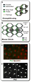
Figure 2. The subcellular distributions of core PCP proteins are likely conserved between mouse and Drosophila and are not altered at the LPR
(A) Core PCP proteins are asymmetrically localized in epidermal cells of the developing Drosophila wing with Frizzled and Dishevelled enriched at the distal cell boundary near the site of hair formation, and Van Gogh and Prickle enriched at the proximal side. Flamingo is present at both cell boundaries. (B) In the developing mouse utricle the core PCP proteins Fz6 and Pk2 are located on opposite sides of HCs and supporting cells, similar to the relative distribution of Frizzled and Prickle in Drosophila, suggesting that other PCP proteins are similarly distributed. The remaining core PCP proteins in mouse are asymmetrically localized at cell boundaries however proximal versus distal distributions have not been established. (C) Pk2 (green) is enriched at the same side of vestibular HCs regardless of stereociliary bundle orientation or cellular position relative to the LPR. Bundle orientation is illustrated using antibodies against Spectrin (red) which labels the cuticular plate beneath the stereocilia but not the basal body of the kinocilium. (C′) Annotated stereociliary bundle orientation (arrowheads) for HCs illustrated in (C) relative to Pk2 protein distribution. The dashed line indicates the position of the LPR.
Figure 3
Figure 3. Theoretical Model for LPR formation in a common utricular/saccular precursor
(A) In this model, the subcellular distribution of PCP proteins like Pk2 (green) establishes a ground polarity throughout the utricular/saccular precursor prior to HC development. (B) The precursor is then patterned into two domains containing HCs that produce opposite stereociliary bundle orientations (arrows and arrowheads) in response to the ground polarity. In this simplified model the utricular/saccular precursor is drawn as an annulus though other configurations are possible. The two domains are indicated by blue and white shading. (C) Separation of the utricular/saccular precursor during inner ear morphogenesis produces the utricle and a saccule intermediate. Both sensory organs contain an LPR however HC patterning at the LPR is complimentary. (D) When this model is based upon an annular patterning event, additional morphogenic movements (illustrated by red arrows) are required in order produce the mature saccular conformation.
Figure 4
Figure 4. Afferent Innervation is Coordinated with Bundle Polarity and the LPR
(A) The maculae contain two types of HCs that are distinguished from each other by the synaptic structure formed with afferent neurons. Type I HCs receive large calyx endings that surround the cell body while Type II HCs make Bouton-like contacts. The two HC classes are innervated by three types of afferent neurons. The Calyx-only class of neurons (orange) contacts clusters of Type I HCs in the striola region. Dipmorphic neurons (blue) contact HCs throughout the sensory epithelia, forming calyxes with Type I HCs and boutons with Type II HCs. Bouton-only afferents (green) only contact Type II HCs located outside of the striola. (B) Each class of afferent neuron may contact multiple HCs, and the Dimorphic and Bouton-only neurons contact groups of HCs located on either side of the LPR. Despite the range of bundle orientations present in the utricle, afferent neurons only contact HCs with similar stereociliary bundle orientations. Developmental mechanisms coordinating neuronal innervation and stereociliary bundle orientation are not known. Bold arrows indicate the stereociliary bundle orientation.

References

    1. Goodrich LV, Strutt D. Principles of planar polarity in animal development. Development. 2011;138:1877–92. - PMC - PubMed
    1. Wang Y, Nathans J. Tissue/planar cell polarity in vertebrates: new insights and new questions. Development. 2007;134:647–58. - PubMed
    1. Wang J, Mark S, Zhang X, Qian D, Yoo SJ, Radde-Gallwitz K, et al. Regulation of polarized extension and planar cell polarity in the cochlea by the vertebrate PCP pathway. Nat Genet. 2005;37:980–5. - PMC - PubMed
    1. Jones C, Chen P. Planar cell polarity signaling in vertebrates. Bioessays. 2007;29:120–32. - PMC - PubMed
    1. Wallingford JB, Fraser SE, Harland RM. Convergent extension: the molecular control of polarized cell movement during embryonic development. Dev Cell. 2002;2:695–706. - PubMed

Publication types

MeSH terms

LinkOut - more resources