Review
. 2013 Nov-Dec;1827(11-12):1295-308.
doi: 10.1016/j.bbabio.2013.03.002. Epub 2013 Mar 16.

Transmembrane signaling and assembly of the cytochrome b6f-lipidic charge transfer complex

Affiliations
Review

Transmembrane signaling and assembly of the cytochrome b6f-lipidic charge transfer complex

S Saif Hasan et al. Biochim Biophys Acta. 2013 Nov-Dec.

Abstract

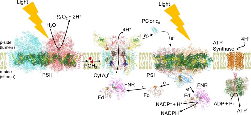
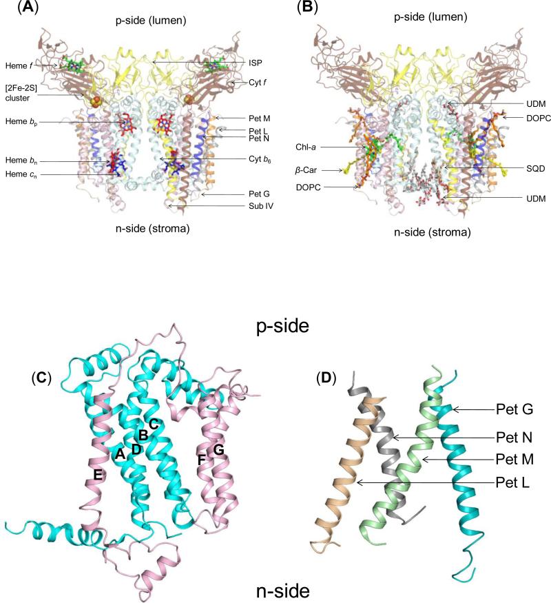
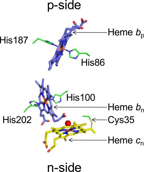
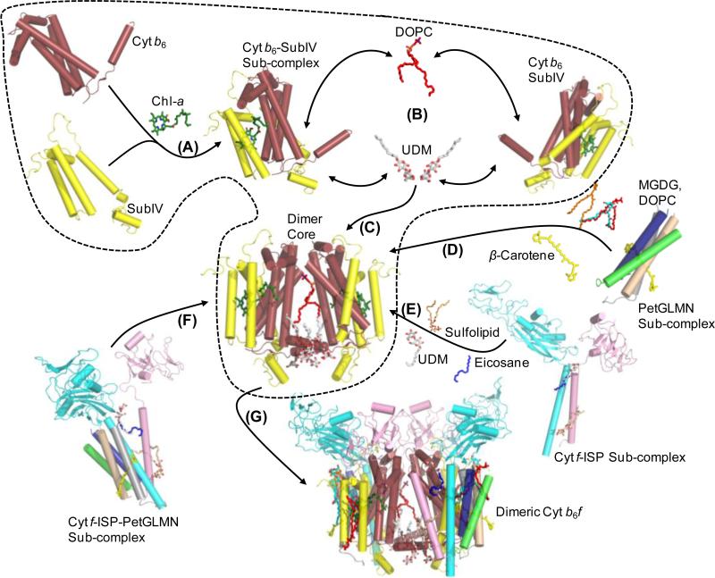
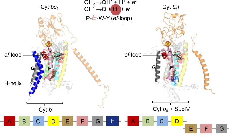
Structure-function properties of the cytochrome b6f complex are sufficiently unique compared to those of the cytochrome bc1 complex that b6f should not be considered a trivially modified bc1 complex. A unique property of the dimeric b6f complex is its involvement in transmembrane signaling associated with the p-side oxidation of plastoquinol. Structure analysis of lipid binding sites in the cyanobacterial b6f complex prepared by hydrophobic chromatography shows that the space occupied by the H transmembrane helix in the cytochrome b subunit of the bc1 complex is mostly filled by a lipid in the b6f crystal structure. It is suggested that this space can be filled by the domain of a transmembrane signaling protein. The identification of lipid sites and likely function defines the intra-membrane conserved central core of the b6f complex, consisting of the seven trans-membrane helices of the cytochrome b and subunit IV polypeptides. The other six TM helices, contributed by cytochrome f, the iron-sulfur protein, and the four peripheral single span subunits, define a peripheral less conserved domain of the complex. The distribution of conserved and non-conserved domains of each monomer of the complex, and the position and inferred function of a number of the lipids, suggests a model for the sequential assembly in the membrane of the eight subunits of the b6f complex, in which the assembly is initiated by formation of the cytochrome b6-subunit IV core sub-complex in a monomer unit. Two conformations of the unique lipidic chlorophyll a, defined in crystal structures, are described, and functions of the outlying β-carotene, a possible 'latch' in supercomplex formation, are discussed. This article is part of a Special Issue entitled: Respiratory complex III and related bc complexes.

Keywords: Assembly; Chl-a; Cyt; Cytochrome; Em7; LHC kinase; PC; PDB; PQ; PS; TMH; Transmembrane signaling; b(6)f; bc(1); chlorophyll-a; cytochrome; midpoint oxidation-reduction potential at pH7; n; p-sides, electrochemically negative and positive side of the membrane; photosystem; plastocyanin; plastoquinone; protein data bank; subIV; subunit IV of the b(6)f complex; transmembrane helix; transmembrane proton electrochemical potential gradient; Δμ̃H+; β-Car; β-carotene.


Figures

Figure 1
Figure 1. The electron transport chain of oxygenic photosynthesis
Formation of the transmembrane proton electrochemical gradient coupled to the electron transport extending from H2O oxidation to NADP+ reduction, in which H+ is translocated in the protein complexes of the PSII reaction center and cytochrome b6f; this H+ gradient is utilized for ATP synthesis by the ATP synthase. PDB accession for structure data: Cyt b6f (PDB ID 2E74), Fd (PDB ID 1EWY), ferredoxin; FNR (PDB ID 1EWY), ferredoxin- NADP+-reductase; PC (PDB ID 2Q5B), plastocyanin; PSII (PDB ID 3ARC) and PSI (PDB ID 1JB0), reaction center complexes.
Figure 2
Figure 2. Structure of dimeric b6f complex from M.laminosus (PDB ID 2E74)
(240,000 MW; per monomer, 8 subunits/7 prosthetic groups; 8 lipids) Subunit organization and lipid binding sites. (A) View along membrane plane showing the positions of the 8 subunits. Color code: cytochrome f (pet A), yellow; cytochrome b6 (pet B), cyan); Rieske [2Fe-2S] protein (Pet C), orange; subunit IV/Pet D (pink), Pet G (teal), Pet L (light brown), Pet M (green) and Pet N (gray). (B) Side view of M. laminosus b6f complex showing bound lipids, detergents and pigments. (C) Polytopic core of the b6f complex. Cyt b6 (4 TMH, cyan) and subunit IV (3 TMH, pink) form the core of the b6f monomer. The cytb6TMH form a four helix bundle. SubIV is organized around the bundle as a bipartite structure, with the E-helix separated from the F and G TMH. (D) Peripheral four helix bundle formed by the small Pet subunits, Pet G, L, M and N. In addition, including ferredoxin-NADP+-reductase (FNR), which may mediate electron transfer from PSI to cyt b6f. (Fig. 1) In addition, there are four soluble subunits that associate with purified b6f complex from plant or algal sources, but have not been seen in the crystal structures and have presumably dissociated and been lost during crystallization: the Pet P polypeptide seen in cyanobacteria, the light-harvesting LHCII chlorophyll protein kinase Stt7-STN7, the correlated phosphatase, and the PetO nuclear-encoded phosphorylatable subunit,.
Figure 3
Figure 3. Transmembrane heme organization within cyt b6f
Three hemes, bp, bn and cn, are located within the b6f hydrophobic core. Hemes bp and bn are axially ligated by histidines from cyt b6. Heme cn, a unique heme without an amino acid axial ligand, is linked to a conserved Cys35 residue of cyt b6, and is axially ligated by a water or hydroxide molecule (red sphere).
Figure 4
Figure 4. Family of cyt bc complexes
(A) Cyt b6f and the bc1 complex of mitochondria and anoxygenic photosynthetic bacteria constitute the cyt bc family. The transmembrane core (teal) of the bc complexes is highly conserved. The complexes catalyze quinone reduction-protonation and quinol deprotonation-oxidation, respectively, on the electrochemically negative and positive sides of the membrane to generate the proton electrochemical potential gradient. (B) Structural differences in the core: C-terminal domain of the bc1 cyt b subunit (4 TMH, yellow) has a TMH organization that differs from subIV (3 TMH, pink) of the b6f complex, although amino acid sequences are highly conserved.
Figure 5
Figure 5. Proposed model for assembly of the cyt b6f complex
(Step 1) The first step involves the assembly of the monomeric polytopic core of cyt b6 and subIV. (Step 2) The monomeric polytopic core then undergoes dimerization, mediated by the cross-linking interactions of lipids (DOPC, shown as red sticks; UDM shown as red/white sticks), which leads to the formation of the dimeric cyt b6-subIV polytopic core (Step 3). Formation of the ISP-cyt f sub-complex takes place via the stabilizing interactions of lipids (Step 4). Assembly of the PetGLMN sub-complex takes place around the β-carotene molecule (Step 5). Interaction of the PetGLMN sub-complex with the core polytopic core takes place via the β-carotene and lipids (sulfolipid, DOPC, MGDG). Alternatively, formation of an ISP-cyt f-PetGLMN sub-complex may take place prior to interaction with the dimeric core (Step 6). (Step 7) Interaction of the peripheral sub-complex with the dimeric core would lead to the formation of the fully assembled, functional dimeric cyt b6f complex.
Figure 6
Figure 6. Organization of the Qp-site in cyochrome bc complexes
The ef-loop bears the conserved PEWY sequence, whose Glu residue is involved in the second deprotonation reaction (highlighted in reaction sequence) of the substrate within the Qp-site. Left panel: In the cyt bc1 complex (PDB ID 3CX5), the ef-loop is inserted between the F and G transmembrane helices of the 8 helix cyt b polypeptide (shown in block diagram at the bottom). The space between the F and G TMH is stabilized by the H TMH of cyt b. Right panel: In the cyt b6f complex (PDB ID 2E74), the ‘H’ TMH is absent from the subIV polypeptide (shown as block diagram). This niche is occupied by a lipid and a chlorophyll-a molecule.
Figure 7
Figure 7. Photosynthetic state transitions
(A) The photosystem II (PSII) reaction center complex utilizes light energy harvested by accessory light harvesting complex II (LHC) molecules to reduce plastoquinone (PQ) to plastoquinol (PQH2). PQH2 undergoes diffusion to the Qp-site (blue circle) of the cyt b6f complex (brown cartoon), where it undergoes oxidation. (B) Slow PQH2 oxidation by cyt b6f causes PQH2 accumulation in the thylakoid membrane, which activates the LHCII kinase (shown in red). (C) In the phosphorylated state, LHC molecules migrate to photosystem I (PSI), thereby reducing the efficiency of PQ reduction by PSII. As PQH2 consumption by cyt b6f is not affected, a redox balance is restored in the quinone pool, which inactivates the LHCII kinase (shown in gray).
Figure 8
Figure 8. Alternate conformations of the chl-a phytyl-tail in cyt b6f
The chl-a chlorin-ring is inserted between the F and G TMH of subIV (light pink) while the tail is inserted between the F TMH and the C TMH of cyt b6 (pale cyan). In the crystal structure of cyt b6f (PDB ID 2E74) from the cyanobacterium M. laminosus, the chl-a phytyl-tail (green) is wrapped around the F TMH while in the structure obtained from C. reinhardtii, the chl-a phytyl-tail (red) is located proximal to the C TMH.
Figure 9
Figure 9. Proposed mechanism of chl-a mediated signal transduction in the cyt b6f complex (PDB ID 2E74) for activation of photosynthetic state transitions
(Step 1) Binding of the natural substrate plastoquinol (black/red sticks) within the Qp-site displaces the chl-a phytyl-tail (displacement shown as broken line with arrows) due to interaction with the long isoprenoid tail of the palstoquinol molecule. The displacement event generates a signal that is transduced to the n-side of the complex either (Step 2) via the F TMH or (Step 3) through a combination of the F TMH and the n-side lipid (yellow) present between the F and G TMH. The LHCII kinase is shown (gray) in cartoon format.
Figure 10
Figure 10. Potential lipid binding site in cyt b6f complex of C. reinhardtii
The cyt b6f structure (PDB ID 1Q90) was obtained in the presence of native lipids. Residual electron density is shown as green mesh (highlighted by broken black line) between the Fo-Fc map (3.0 σ). The electron density corresponds to the n-side lipid in the cyanbacterial cyt b6f complexes from M. laminosus (PDB ID 2E74) and Nostoc PCC 7120 (PDB ID 2ZT9). The electron density map was obtained from the Electron Density Server [96].
Figure 11
Figure 11. The lipidic mechanism of cyt b6f-LHCII kinase super-complex formation
It is proposed that the lipid DOPC (yellow/red/blue sticks) is bound weakly in the niche formed between the F and G TMH, which represents the LHCII kinase (shown as cartoon. gray) binding site. Recruitment of the LHCII kinase to the cyt b6f complex and binding results in the replacement of the lipid from the inter-helix niche.
Figure 12
Figure 12. Arrangement of β–carotene in the major photosynthetic integral membrane protein complexes (A) photosystem II (PDB ID 3ARC), (B) photosystem I (1JB0) and light harvesting complex II (PDB ID 2BHW)
The β–car molecules are shown as yellow sticks while the polypeptides are represented as thin green ribbons. The β-car molecules are located on the periphery of the complexes, providing potential interaction sites for the chl-a of cyt b6f. All three complexes are viewed along the normal to the membrane plane. For simplicity, other prosthetic groups have been omitted.
Figure 13
Figure 13. Binding of ligands to the Qn-site of cyt bc complexes and the role of the protein environment in selectivity
(A) In the cyt b6f structure (PDB ID 2E76) obtained from the cyanobacterium M. laminosus, the majority of the interactions to the quinone analog inhibitor TDS (green/red ball and stick model) are contributed through non-specific Van der Waals interaction. (B) On the other hand, binding of the natural ligand ubiquinone-6 (green/red ball and stick model) to the cyt bc1 complex (PDB ID 1KB9) Qn-site is mediated by polar interactions. For simplicity, the hydrophobic tails of TDS and ubiquinone-6 have been omitted.

References

    1. Nelson N, Ben-Shem A. The complex architecture of oxygenic photosynthesis, Nature reviews. Molecular cell biology. 2004;5:971–982. - PubMed
    1. Kurisu G, Zhang H, Smith JL, Cramer WA. Structure of the cytochrome b6f complex of oxygenic photosynthesis: tuning the cavity. Science. 2003;302:1009–1014. - PubMed
    1. Stroebel D, Choquet Y, Popot JL, Picot D. An atypical haem in the cytochrome b(6)f complex. Nature. 2003;426:413–418. - PubMed
    1. Allen JF. Cytochrome b6f: structure for signalling and vectorial metabolism. Trends in plant science. 2004;9:130–137. - PubMed
    1. Baniulis D, Yamashita E, Whitelegge JP, Zatsman AI, Hendrich MP, Hasan SS, Ryan CM, Cramer WA. Structure-Function, Stability, and Chemical Modification of the Cyanobacterial Cytochrome b6f Complex from Nostoc sp. PCC 7120. The Journal of biological chemistry. 2009;284:9861–9869. - PMC - PubMed

Publication types

MeSH terms

LinkOut - more resources