Review
. 2011 Oct-Dec;1(2):121-64.
doi: 10.4161/biom.18790.

Calcium orthophosphates: occurrence, properties, biomineralization, pathological calcification and biomimetic applications

Affiliations
Review

Calcium orthophosphates: occurrence, properties, biomineralization, pathological calcification and biomimetic applications

Sergey V Dorozhkin. Biomatter. 2011 Oct-Dec.

Abstract

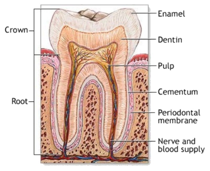
The present overview is intended to point the readers' attention to the important subject of calcium orthophosphates. This type of materials is of special significance for human beings, because they represent the inorganic part of major normal (bones, teeth and antlers) and pathological (i.e., those appearing due to various diseases) calcified tissues of mammals. For example, atherosclerosis results in blood vessel blockage caused by a solid composite of cholesterol with calcium orthophosphates, while dental caries and osteoporosis mean a partial decalcification of teeth and bones, respectively, that results in replacement of a less soluble and harder biological apatite by more soluble and softer calcium hydrogenphosphates. Therefore, the processes of both normal and pathological calcifications are just an in vivo crystallization of calcium orthophosphates. Similarly, dental caries and osteoporosis might be considered an in vivo dissolution of calcium orthophosphates. Thus, calcium orthophosphates hold a great significance for humankind, and in this paper, an overview on the current knowledge on this subject is provided.

Keywords: antlers; biomimetics; bones; calcification; calcium orthophosphates; crystallization; fluorapatite; hydroxyapatite; teeth.


Figures

None
Figure 1. Simplified schematic of the phosphorus cycle from apatitic igneous rock to phosphorite sedimentary rock through chemical or physical weathering. Life forms accumulate soluble phosphorus species and can produce apatite through biomineralization. Reprinted from reference with permission.
None
Figure 2. Polycrystalline (A) and single-crystalline (B) FA of a geological origin. The single crystal has a gray-green color due to incorporated ions of transition metals.
None
Figure 3. Phase diagram of the system CaO-P2O5 (C = CaO, p = P2O5) at elevated temperatures. Here: C7P5 means 7CaO·5P2O5; other abbreviations should be written out in the same manner. Reprinted from references and with permission.
None
Figure 4. pH variation of ionic concentrations in triprotic equilibrium for phosphoric acid solutions. Reprinted from reference with permission.
None
Figure 5. Various calcium orthophosphates obtained by neutralizing of orthophosphoric acid. Ca/P are reported in the figure. The solubility of calcium orthophosphates in water decreases drastically from left to right, HA being the most insoluble and stable phase. Reprinted from reference with permission.
None
Figure 6. Top: a 3D version of the classical solubility phase diagrams for the ternary system Ca(OH)2-H3PO4-H2O. Reprinted from reference with permission. Middle and bottom: solubility phase diagrams in two-dimensional graphs, showing two logarithms of the concentrations of (a) calcium and (b) orthophosphate ions as a function of the pH in solutions saturated with various salts. Reprinted from reference with permission.
None
Figure 7. A model of ACP structure. Reprinted from reference with permission.
None
Figure 8. A biomimetically grown aggregate of FA that was crystallized in a gelatin matrix. Its shape can be explained and simulated by a fractal growth mechanism. Scale bar: 10 µm. Reprinted from reference with permission.
None
Figure 10. General structure of a mammalian bone. Other very good graphical sketches of the mammalian bone structure are available in references and .
None
Figure 14. Scanning electron micrograph of the forming enamel of a continuously growing rat incisor showing ordered rods of calcium orthophosphates. Scale bar: 10 µm. Reprinted from reference with permission.
None
Figure 9. Left: crystal structure of a biological apatite. Powder X-ray diffraction patterns (center) and infrared spectra (right) of human enamel, dentine and bone. Reprinted from reference with permission.
None
Figure 11. The seven hierarchical levels of organization of the zebrafish skeleton bone. Level 1: Isolated crystals and part of a collagen fibril with the triple helix structure. Level 2: Mineralized collagen fibrils. Level 3: The array of mineralized collagen fibrils with a cross-striation periodicity of nearly 60–70 nm. Level 4: Two fibril array patterns of organization as found in the zebrafish skeleton bone. Level 5: The lamellar structure in one vertebra. Level 6: A vertebra. Level 7: Skeleton bone. Reprinted from reference with permission. Other good graphical sketches of the hierarchical structure of bones are available in references , and .
None
Figure 12. A schematic illustration of in vivo mineralization of a collagen fibril: top layer-calcium orthophosphate clusters (green) form complexes with biopolymers (orange line), forming stable mineral droplets; second top layer-mineral droplets bind to a distinct region on the collagen fibers and enter the fibril; second bottom layer-once inside the collagen, the mineral in a liquid state diffuses through the interior of the fibril and solidifies into a disordered phase of ACP (black); bottom layer-finally, directed by the collagen, ACP is transformed into oriented crystals of biological apatite (yellow). Reprinted from reference with permission.
None
Figure 13. A schematic drawing of a tooth. Other very good graphical sketches of the mammalian tooth structure, including the hierarchical levels, are available in references and .
None
Figure 15. Red deer stag at velvet shedding. The bare bone of the hard antlers is exposed. Reprinted from reference with permission. A good cross-sectional image of a deer antler is available in reference .
None
Figure 16. A schematic representation of the different stages of a surface-directed mineralization of calcium orthophosphates. In stage 1, aggregates of pre-nucleation clusters are in equilibrium with ions in solution. The clusters approach a surface with chemical functionality. In stage 2, pre-nucleation clusters aggregate near the surface, with loose aggregates still in solution. In stage 3, further aggregation causes densification near the surface. In stage 4, nucleation of spherical particles of ACP occurs at the surface only. In stage 5, crystallization occurs in the region of the ACP particles directed by the surface. Reprinted from references and with permission.

References

    1. Shepperd J. The early biological history of calcium phosphates. In: Epinette JA, Manley MT, (Eds.) Fifteen years of clinical experience with hydroxyapatite coatings in joint arthroplasty. Springer, France 2004; 3-8.
    1. Berzelius J. Ueber basische phosphorsaure kalkerde. Ann Chem Pharmac. 1845;53:286–8. doi: 10.1002/jlac.18450530212. - DOI
    1. Warington R., Jr. Researches on the phosphates of calcium, and upon the solubility of tricalcic phosphate. J Chem Soc. 1866;19:296–318. doi: 10.1039/js8661900296. - DOI
    1. Fresenius R. Ueber die Bestimmung der Phosphorsäure im Phosphorit nebst Mittheilung der Analysen des Phosphorits und Staffelits aus dem Lahnthal. Z Anal Chem. 1867;6:403–9. doi: 10.1007/BF01347651. - DOI
    1. Church AH. New analyses of certain mineral arseniates and phosphates. 1. Apatite; 2. Arseniosiderite; 3. Childrenite; 4. Ehlite; 5. Tyrolite; 6. Wavellite. J Chem Soc. 1873;26:101–11. doi: 10.1039/js8732600101. - DOI

LinkOut - more resources