Review
. 2013 Jun;10(6):362-70.
doi: 10.1038/nrgastro.2013.36. Epub 2013 Mar 19.

The role of protein synthesis and digestive enzymes in acinar cell injury

Affiliations
Review

The role of protein synthesis and digestive enzymes in acinar cell injury

Craig D Logsdon et al. Nat Rev Gastroenterol Hepatol. 2013 Jun.

Abstract

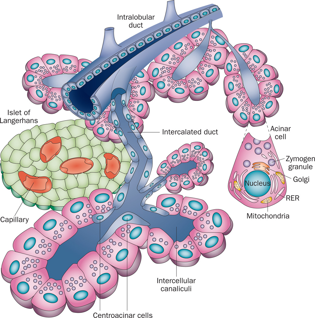
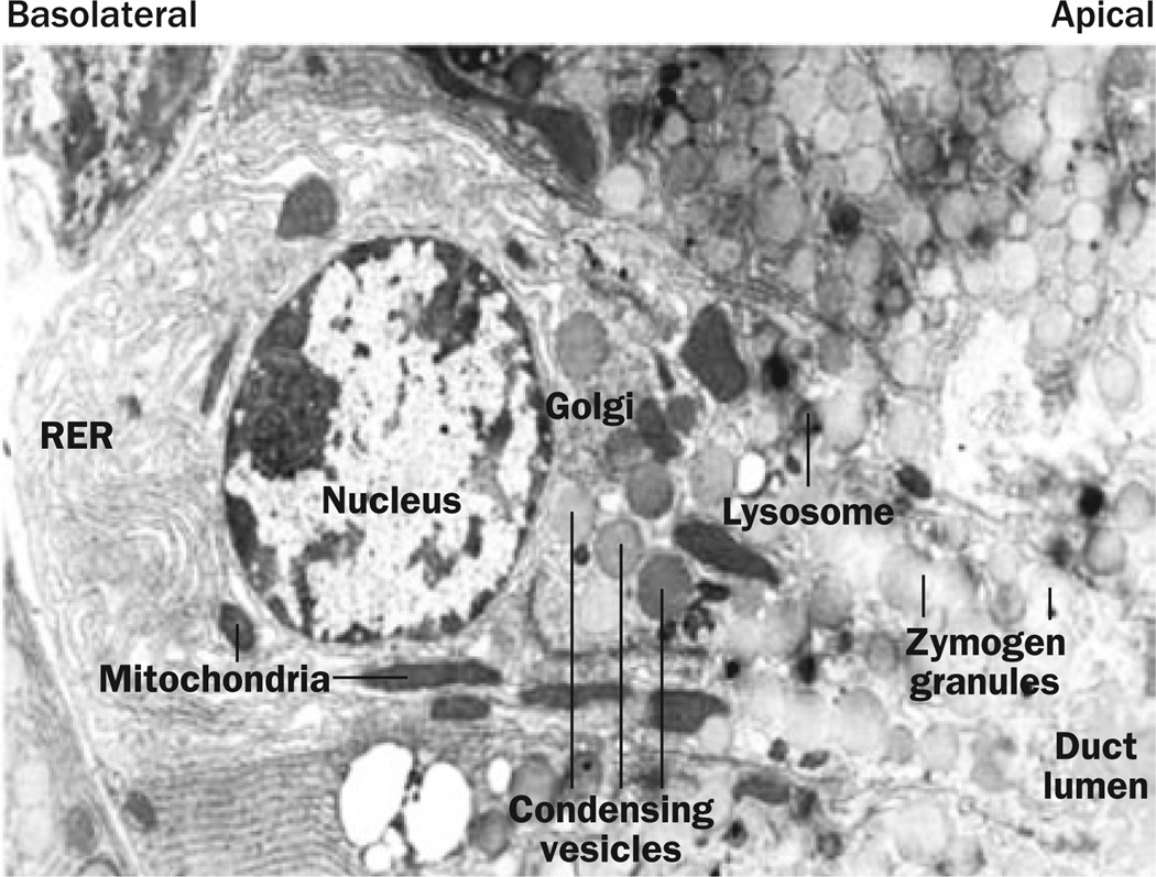
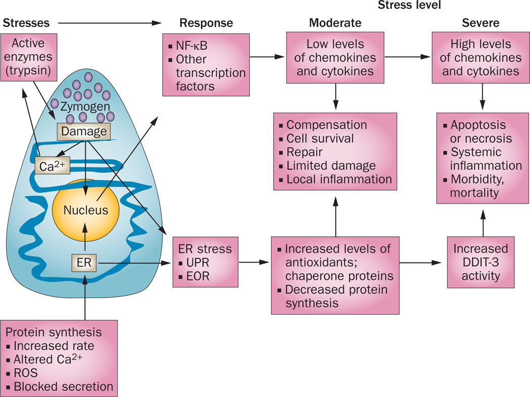
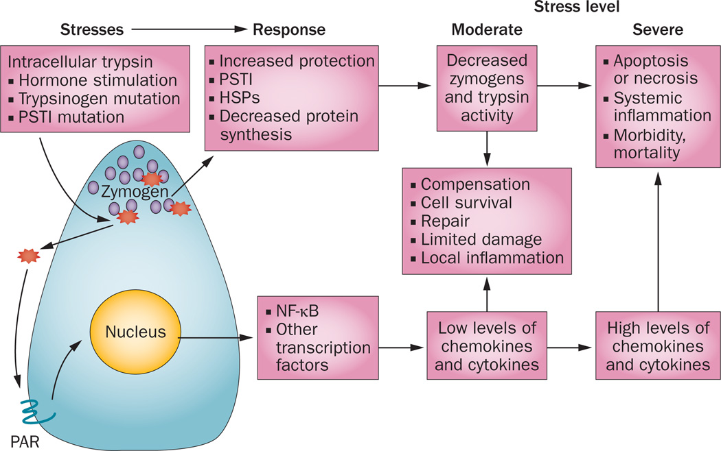
The exocrine pancreas is the organ with the highest level of protein synthesis in the adult--each day the pancreas produces litres of fluid filled with enzymes that are capable of breaking down nearly all organic substances. For optimal health, the pancreas must produce sufficient enzymes of the right character to match the dietary intake. Disruption of normal pancreatic function occurs primarily as a result of dysfunction of the acinar cells that produce these digestive enzymes, and can lead to acute or chronic diseases. For many years, the prevailing dogma has been that inappropriate intracellular activation of the digestive enzymes produced by acinar cells was the key to pancreatic inflammatory diseases, as digestive enzymes themselves are potentially harmful to the cells that secrete them. However, we now know that many stressors can affect pancreatic acinar cells, and that these stressors can independently trigger pancreatic pathology through various mechanisms. This Review focuses on protein synthesis and active digestive enzymes--two key stressors faced by the acinar cell that are likely to be the major drivers of pathology encountered in the pancreas.


Figures

Figure 1
Figure 1
The components of the pancreas. The pancreas consists of endocrine cells localized within structures named the Islets of Langerhans, which contain multiple endocrine cell types including the β cells that secrete isulin, and the exocrine pancreas, which is composed of acinar calls and ductal structures. Pancreatic acinar cells form a basic structure called an acinus that surrounds a central lumen open to the duct system. Pancreatic acinar cells produce, store and secrete enzymes necessary for the digestion and absorption of food in the small intestine. Digestive enzymes are secreted through the apical membrane of the acinar cell into small intercalated ducts that are directly connected to increasingly larger intralobular ducts that join the main pancreatic duct. The main pancreatic duct joins the common bile duct just prior to the ampulla of Vater, where both pancreatic and liver products enter the small intestine. Blockage of the passage of materials through the ampulla of Vater, for example by the lodging of a bile stone or by the growth of a tumor, leads to increased pressure in the duct system and gives rise to acute pancreatitis.
Figure 2
Figure 2
The structure of the pancreatic acinar cell. Pancreatic digestive enzymes are processed within the RER located primarily in the basal domain of these cells. The RER is where folding, glycosylation and disulphide bond formation occurs during protein synthesis. From here, the digestive enzymes are transported to the Golgi where they are sorted and packaged into condensing vesicles. The condensing vesicles are compacted to form mature zymogen granules and are transported to the apical end of the cell in position for regulated secretion into the duct lumen. Abbreviation: RER, rough endoplasmic reticulum. Permission obtained from Nature Publishing Group © Adapted from Whitcomb, D. C.
Figure 3
Figure 3
ER stress responses that occur in pancreatic acinar cells. The very high rate of protein synthesis in acinar cells generates a constant low level of ER stress. Under normal conditions, this ER stress is readily compensated for by the UPR and EOR, which reduce the rate of protein synthesis while increasing production of molecules that facilitate cell recovery and repair. However, under conditions of increased ER stress, EOR and UPR lead to apoptosis. Excessive acinar cell apoptosis can lead to necrosis, a severe systemic inflammatory response, acute respiratory distress, and life-threatening multiorgan failure. Altered Ca2+ release from the ER, and ROS, are thought to activate NF-κB, a key regulator of inflammatory gene expression. Abbreviations: DDIT-3, DNA damage-inducible transcript 3 protein; EOR, ER overload response; ER, endoplasmic reticulum; ROS, reactive oxygen species; UPR, unfolded protein response.
Figure 4
Figure 4
Digestive enzyme stress responses in pancreatic acinar cells. Digestive enzymes produced by acinar cells are normally inactive until exported, but their inappropriate intracellular activation can result in cellular damage. Trypsinogen is the key digestive enzyme as, once activated, it can activate other enzymes—initiating a cycle of intracellular activation of trypsin, intracellular and extracellular digestive enzyme activity, and further cell damage. Whereas intracellular trypsin activity does not seem to activate NF-κB, extracellular trypsin does, probably by activation of PAR proteins, which subsequently activate transcription factors, including NF-κB. Low levels of these transcription factors have a local inflammatory effect and induce expression of various protective and repair genes. However, high levels of inflammatory transcription factors generate a systemic inflammatory response. Abbreviations: HSP, heat shock protein; PAR, proteinase-activated receptor; PSTI, pancreatic secretory trypsin inhibitor.

References

    1. Case RM. Synthesis, intracellular transport and discharge of exportable proteins in the pancreatic acinar cell and other cells. Biol. Rev. Camb. Philos. Soc. 1978;53:211–354. - PubMed
    1. Siegel R, Naishadham D, Jemal A. Cancer statistics, 2012. CA Cancer J. Clin. 2012;62:10–29. - PubMed
    1. Jamieson JD, Palade GE. Synthesis, intracellular transport, and discharge of secretory proteins in stimulated pancreatic exocrine cells. J. Cell Biol. 1971;50:135–158. - PMC - PubMed
    1. Weng N, Thomas DD, Groblewski GE. Pancreatic acinar cells express vesicle-associated membrane protein 2- and 8-specific populations of zymogen granules with distinct and overlapping roles in secretion. J. Biol. Chem. 2007;282:9635–9645. - PubMed
    1. Schroder M, Kaufman RJ. The mammalian unfolded protein response. Annu. Rev. Biochem. 2005;74:739–789. - PubMed

Publication types

MeSH terms