

DOT corrected fluorescence molecular tomography using targeted contrast agents for small animal tumor imaging
- PMID: 23507851
- PMCID: PMC3785613
- DOI: 10.3233/XST-130365
DOT corrected fluorescence molecular tomography using targeted contrast agents for small animal tumor imaging
Abstract
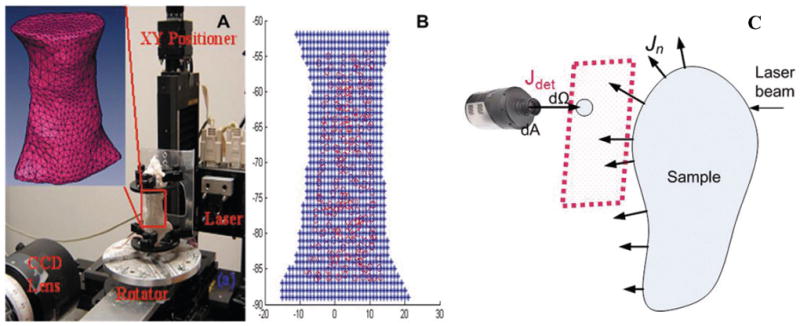
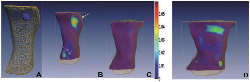
Purpose: To demonstrate diffuse optical tomography (DOT) corrected fluorescence molecular tomography (FMT) for quantitatively imaging tumor-targeted contrast agents in a 4T1 mouse mammary tumor model.
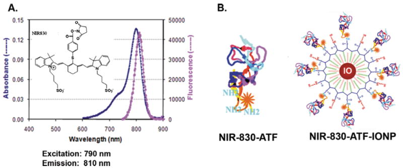
Procedures: In the first set of experiments, we validated our DOT corrected FMT method using subcutaneously injected 4T1 cells pre-labeled with a near-infrared (NIR) Cy 5.5 dye labeled recombinant amino-terminal fragment (ATF) of the receptor binding domain of urokinase plasminogen activator (uPA), which binds to uPA receptor (uPAR) that is highly expressed in breast cancer tissues. Next, we apply the DOT corrected FMT method to quantitatively evaluate the ability of sensitive tumor imaging after systemic delivery of new uPAR-targeted optical imaging probes in the mice bearing 4T1 mammary tumors. These uPAR-targeted optical imaging probes are ATF peptides labeled with a newly developed NIR-830 dye being conjugated to magnetic iron oxide nanoparticles (IONPs).
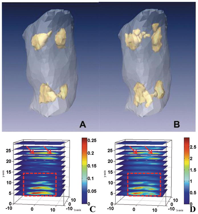
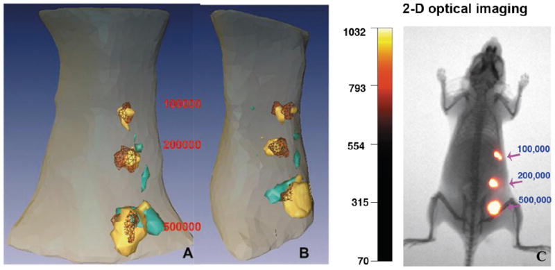
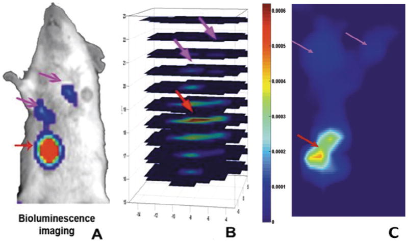
Results: Our results have shown that DOT corrected FMT can accurately quantify and localize the injected imaging probe labeled 4T1 cells. Following systemic delivery of the targeted imaging nanoprobes into the mice bearing orthotopic mammary tumors, specific accumulation of the imaging probes in the orthotopic mammary tumors was detected in the mice that received uPAR targeted NIR-830-ATF-IONP probes but not in the mice injected with non-targeted NIR-830-mouse serum albumin (MSA)-IONPs. Additionally, DOT corrected FMT also enables the detection of both locally recurrent tumor and lung metastasis in the mammary tumor model 72 hrs after systemic administration of the uPAR-targeted NIR-830-labeled ATF peptide imaging probes.
Conclusions: DOT corrected FMT and uPAR-targeted optical imaging probes have great potential for detection of breast cancer, recurrent tumor and metastasis in small animals.
Figures
Similar articles
-
uPAR-targeted optical imaging contrasts as theranostic agents for tumor margin detection.Theranostics. 2013 Dec 17;4(1):106-18. doi: 10.7150/thno.7409. eCollection 2013. Theranostics. 2013. PMID: 24396518 Free PMC article.
-
Molecular photoacoustic tomography of breast cancer using receptor targeted magnetic iron oxide nanoparticles as contrast agents.J Biophotonics. 2014 Jun;7(6):401-9. doi: 10.1002/jbio.201200155. Epub 2012 Nov 2. J Biophotonics. 2014. PMID: 23125139 Free PMC article.
-
Receptor-targeted nanoparticles for in vivo imaging of breast cancer.Clin Cancer Res. 2009 Jul 15;15(14):4722-32. doi: 10.1158/1078-0432.CCR-08-3289. Epub 2009 Jul 7. Clin Cancer Res. 2009. PMID: 19584158 Free PMC article.
-
[Mechanism of tumor cell-induced extracellular matrix degradation--inhibition of cell-surface proteolytic activity might have a therapeutic effect on tumor cell invasion and metastasis].Nihon Sanka Fujinka Gakkai Zasshi. 1996 Aug;48(8):623-32. Nihon Sanka Fujinka Gakkai Zasshi. 1996. PMID: 8808830 Review. Japanese.
-
Urokinase plasminogen activator receptor (uPAR) targeted nuclear imaging and radionuclide therapy.Theranostics. 2013 Jun 29;3(7):507-15. doi: 10.7150/thno.5557. Print 2013. Theranostics. 2013. PMID: 23843898 Free PMC article. Review.
Cited by
-
In Vitro/In Vivo Toxicity Evaluation and Quantification of Iron Oxide Nanoparticles.Int J Mol Sci. 2015 Oct 15;16(10):24417-50. doi: 10.3390/ijms161024417. Int J Mol Sci. 2015. PMID: 26501258 Free PMC article. Review.
References
-
- Ntziachristos V. Going deeper than microscopy: The optical imaging frontier in biology. Nat Methods. 2010;7:603–614. - PubMed
-
- Montet X, Figueiredo JL, Alencar H, Ntziachristos V, Mahmood U, Weissleder R. Tomographic fluorescence imaging of tumor vascular volume in mice. Radiology. 2007;242:751–758. - PubMed
-
- Tan Y, Novo M, Yao L, Zhou L, Jiang H. In Vivo monitoring of stem cells in drosophila pupae using the radiative transfer equation-based fluorescence molecular tomography. Mol Imaging and Bio. 2011;13:868–873. - PubMed
-
- Massoud T, Gambhir S. Molecular imaging in living subjects: Seeing fundamental biological processes in a new light. Gene Dev. 2003;17:545–580. - PubMed
Publication types
MeSH terms
Substances
Grants and funding
LinkOut - more resources
Full Text Sources
Other Literature Sources
Miscellaneous
