

Role of calpain in apoptosis
- PMID: 23507938
- PMCID: PMC3584455
Role of calpain in apoptosis
Abstract
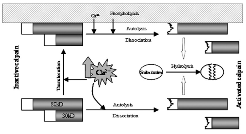
Apoptosis, a form of programmed cell death that occurs under physiological as well as pathological conditions, is characterized by morphological and biochemical features. While the importance of caspases in apoptosis is established, several noncaspase proteases (Ca(2+)-dependent proteases) such as calpain may play a role in the execution of apoptosis. The calpain family consists of two major isoforms, calpain I and calpain II which require µM and mM Ca(2+) concentrations to initiate their activity. An increase in intracellular Ca(2+) level is thought to trigger a cascade of biochemical processes including calpain activation. Once activated, calpains degrade membrane, cytoplasmic and nuclear substrates, leading to the breakdown of cellular architecture and finally apoptosis. The activation of calpain has been implicated in neuronal apoptosis following spinal cord injuries and neurodegenerative diseases. This review focuses on calpain with an emphasis on its key role in the proteolysis of cellular protein substrates following apoptosis.
Keywords: Apoptosis; Calpain; Calpain Substrates.
Figures


Similar articles
-
Caspase and calpain substrates: roles in synaptic plasticity and cell death.J Neurosci Res. 1999 Oct 1;58(1):167-90. J Neurosci Res. 1999. PMID: 10491581 Review.
-
Novel roles for ceramides, calpains and caspases in kidney proximal tubule cell apoptosis: lessons from in vitro cadmium toxicity studies.Biochem Pharmacol. 2008 Dec 1;76(11):1323-32. doi: 10.1016/j.bcp.2008.07.004. Epub 2008 Jul 11. Biochem Pharmacol. 2008. PMID: 18675256 Review.
-
The role of calpain in oncotic cell death.Annu Rev Pharmacol Toxicol. 2004;44:349-70. doi: 10.1146/annurev.pharmtox.44.101802.121804. Annu Rev Pharmacol Toxicol. 2004. PMID: 14744250 Review.
-
Conventional calpains and programmed cell death.Acta Biochim Pol. 2011;58(3):287-96. Epub 2011 Aug 29. Acta Biochim Pol. 2011. PMID: 21887410 Review.
-
Ischemic neuronal death in the rat hippocampus: the calpain-calpastatin-caspase hypothesis.Neurobiol Dis. 2003 Jul;13(2):75-88. doi: 10.1016/s0969-9961(03)00018-4. Neurobiol Dis. 2003. PMID: 12828932
Cited by
-
Role of the Alteration in Calcium Homeostasis in Cell Death Induced by Clostridioides difficile Toxin A and Toxin B.Biology (Basel). 2023 Aug 10;12(8):1117. doi: 10.3390/biology12081117. Biology (Basel). 2023. PMID: 37627001 Free PMC article. Review.
-
Protease signaling in animal and plant-regulated cell death.FEBS J. 2016 Jul;283(14):2577-98. doi: 10.1111/febs.13616. Epub 2015 Dec 31. FEBS J. 2016. PMID: 26648190 Free PMC article. Review.
-
Apoptotic gene loss in Cnidaria is associated with transition to parasitism.Sci Rep. 2023 May 17;13(1):8015. doi: 10.1038/s41598-023-34248-y. Sci Rep. 2023. PMID: 37198195 Free PMC article.
-
Calpastatin-Mediated Inhibition of Calpain Ameliorates Skin Scar Formation after Burn Injury.Int J Mol Sci. 2021 May 28;22(11):5771. doi: 10.3390/ijms22115771. Int J Mol Sci. 2021. PMID: 34071277 Free PMC article.
-
In vivo two-photon imaging of axonal dieback, blood flow, and calcium influx with methylprednisolone therapy after spinal cord injury.Sci Rep. 2015 May 19;5:9691. doi: 10.1038/srep09691. Sci Rep. 2015. PMID: 25989524 Free PMC article.
References
-
- Compton MM. A biochemical hallmark of apoptosis: internucleosomal degradation of the genome. Cancer Metastasis Rev. 1992;11(2):105–119. - PubMed
-
- Arends MJ, Wyllie AH. Apoptosis: mechanisms and roles in pathology. Int Rev Exp Pathol. 1991;32:223–254. - PubMed
-
- Sendtner M, Pei G, Beck M, Schweizer U, Wiese S. Developmental motoneuron cell death and neurotrophic factors. Cell Tissue Res. 2000;301(1):71–84. - PubMed
LinkOut - more resources
Full Text Sources
Other Literature Sources
Miscellaneous