

doi: 10.1038/nchembio.1211.
Determining target engagement in living systems
Affiliations
- PMID: 23508173
- PMCID: PMC4004587
- DOI: 10.1038/nchembio.1211
Item in Clipboard
Determining target engagement in living systems
Nat Chem Biol.
2013 Apr.
Abstract
Chemical probes are critical tools for elucidating the biological functions of proteins and can lead to new medicines for treating disease. The pharmacological validation of protein function requires verification that chemical probes engage their intended targets in vivo. Here we discuss technologies, both established and emergent, for measuring target engagement in living systems and propose that determining this parameter should become standard practice for chemical probe and drug discovery programs.
Figures
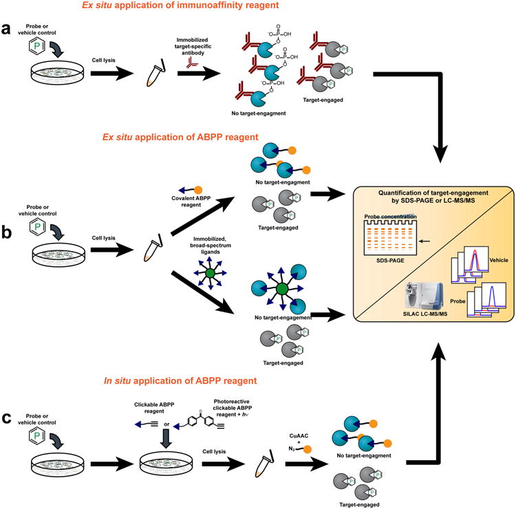
(a) Blockade of autophosphorylation as a proximal biomarker for kinase inhibition. Target engagement can be measured by immunoprecipitation of the kinase of interest from cells treated with a chemical probe (or vehicle control), followed by SILAC-LC-MS/MS to quantify changes in the phosphorylation status of autophosphorylated residues. (b) Ex situ evaluation of target engagement can be accomplished using two different approaches. In the upper pathway, an ABPP reagent is added to cell lysates following in situ treatment with a chemical probe (or vehicle control). The ABPP reagent is coupled to a reporter group (orange ball): either a fluorophore for target detection by SDS-PAGE coupled with in-gel fluorescence scanning or biotin for affinity-enrichment and target identification and quantification by LC-MS/MS. In the lower pathway, proteomes are incubated with bead-immobilized, broad-spectrum chemical ligands that affinity-enrich active (but not probe-engaged) enzymes. Target engagement is then monitored by observing loss-of-signal by LC-MS/MS. (c) Cell-permeant ABPP reagents can be generated by replacing reporter groups with latent affinity handles, such as an alkyne. If the ABPP reagent interacts with targets in a non-covalent manner, groups conferring photoreactivity (for example benzophenones or diazirines) can also be affixed. Following sequential treatment of cells with a chemical probe (or vehicle-control) and a cell-permeable ABPP reagent, cell lysis, and proteome preparation, a reporter group (orange ball) is covalently attached via a bioorthogonal reaction like CuAAC (click) chemistry and target engagement monitored by SDS-PAGE or LC-MS/MS.

Approaches for measuring target engagement in model organisms. (a) Target engagement can be monitored in vivo with radiotracer-based imaging methods such as microPET. For these methods, animals are usually pre-treated with a chemical probe or vehicle followed by administration of the radiotracer. Engaged targets do not bind the radiotracer; thus, loss of PET-signal can be used to assess target engagement. (b) Target engagement can be monitored ex situ by adding an ABPP reagent to tissue-lysates harvested from animals dosed with a chemical probe (or vehicle control). Depending on the choice of reporter group (orange ball), target engagement can then be measured via SDS-PAGE or LC-MS/MS (Fig. 1). (c) For in vivo observation of target engagement, cell-permeant ABPP reagents can be generated that bear a latent affinity handle, such as an alkyne. After dosing animals with chemical probe (or vehicle control), the ABPP reagent is administered in vivo. Tissues of interest are then harvested and homogenized and targets visualized by conjugation to a reporter group (orange ball) by a bioorthogonal reaction like CuAAC (click) chemistry.
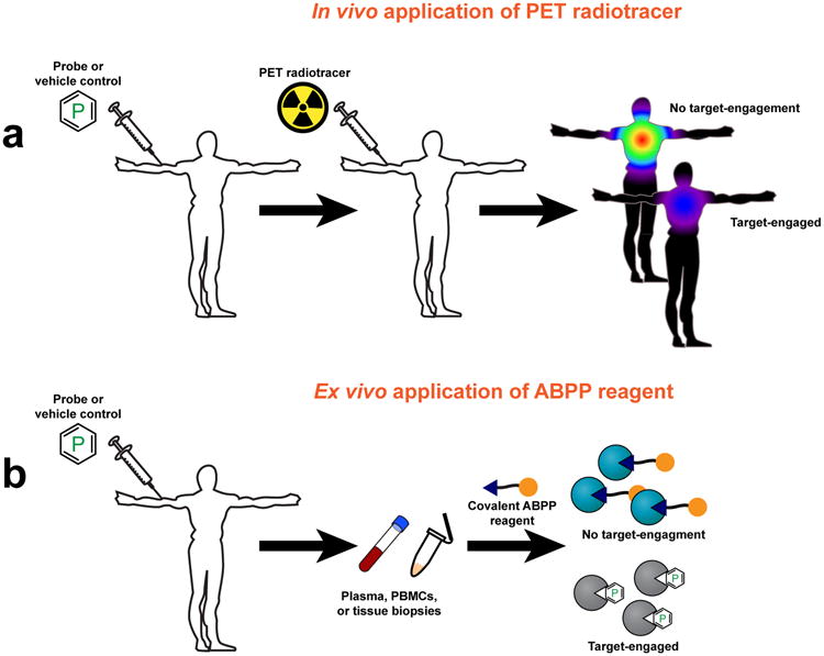
Approaches for measuring target engagement in humans. (a) Target engagement can be monitored in humans by PET, wherein human subjects are usually pre-treated with probe or vehicle, followed by administration of the radiotracer. Engaged targets do not bind the radiotracer; thus, loss of PET-signal can be used to assess target engagement. (b) Target engagement in humans can be monitored ex situ by harvesting plasma, PBMCs, or tissue biopsies from subjects that have been treated with a chemical probe (or vehicle control). An ABPP reagent is then added and, depending on the choice of reporter group (orange ball), target engagement can be measured via fluorescent SDS-PAGE or LC-MS/MS (Fig. 1).
References
-
- Grimwood S, Hartig PR. Target site occupancy: emerging generalizations from clinical and preclinical studies. Pharmacol Ther. 2009;122:281–301. - PubMed
-
- Wong DF, Tauscher J, Grunder G. The role of imaging in proof of concept for CNS drug discovery and development. Neuropsychopharmacology. 2009;34:187–203. - PubMed
Publication types
MeSH terms
Substances
Grants and funding
LinkOut - more resources
Full Text Sources
Other Literature Sources
