. 2013 Oct;22(10):2184-201.
doi: 10.1007/s00586-013-2738-0. Epub 2013 Mar 19.

AO spine injury classification system: a revision proposal for the thoracic and lumbar spine

Affiliations

AO spine injury classification system: a revision proposal for the thoracic and lumbar spine

Maximilian Reinhold et al. Eur Spine J. 2013 Oct.

Abstract

Purpose: The AO Spine Classification Group was established to propose a revised AO spine injury classification system. This paper provides details on the rationale, methodology, and results of the initial stage of the revision process for injuries of the thoracic and lumbar (TL) spine.

Methods: In a structured, iterative process involving five experienced spine trauma surgeons from various parts of the world, consecutive cases with TL injuries were classified independently by members of the classification group, and analyzed for classification reliability using the Kappa coefficient (κ) and for accuracy using latent class analysis. The reasons for disagreements were examined systematically during review meetings. In four successive sessions, the system was revised until consensus and sufficient reproducibility were achieved.

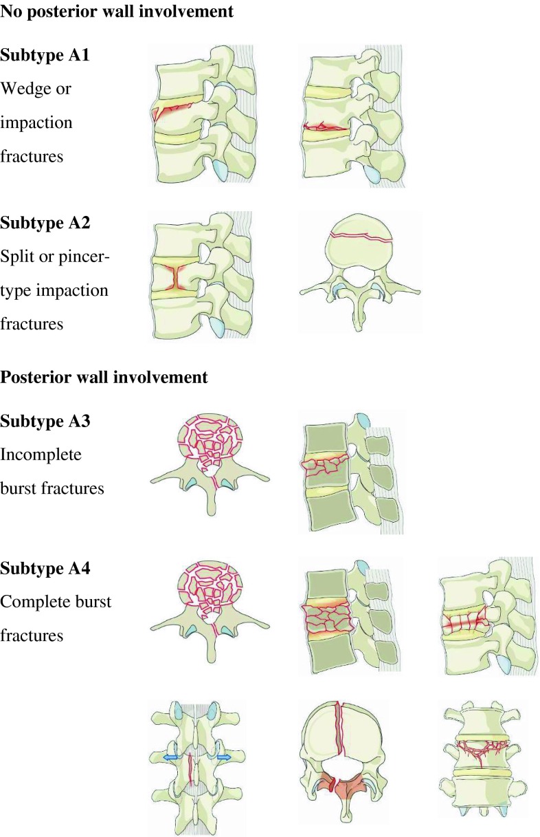
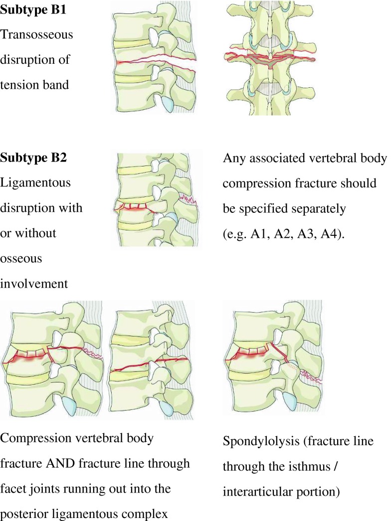
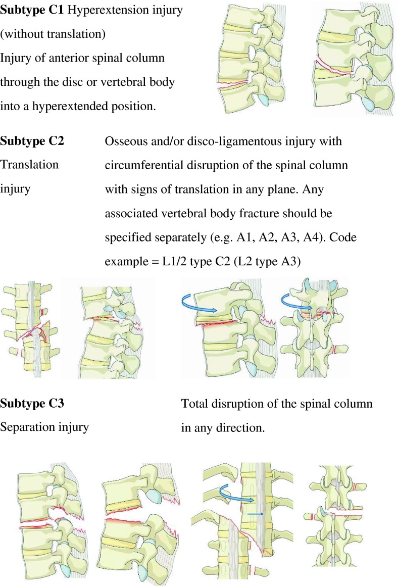
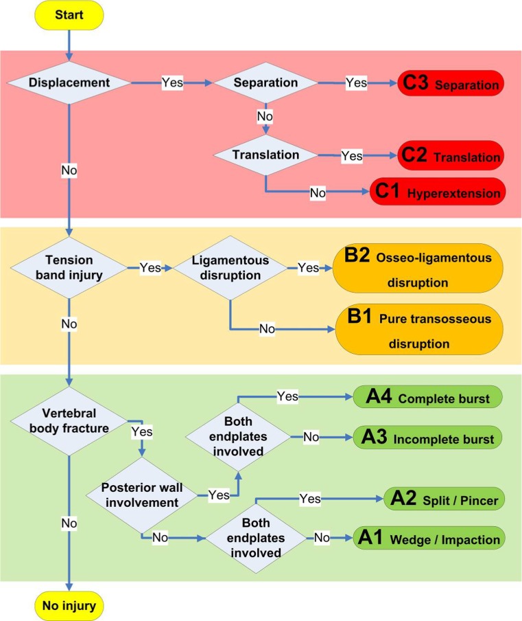
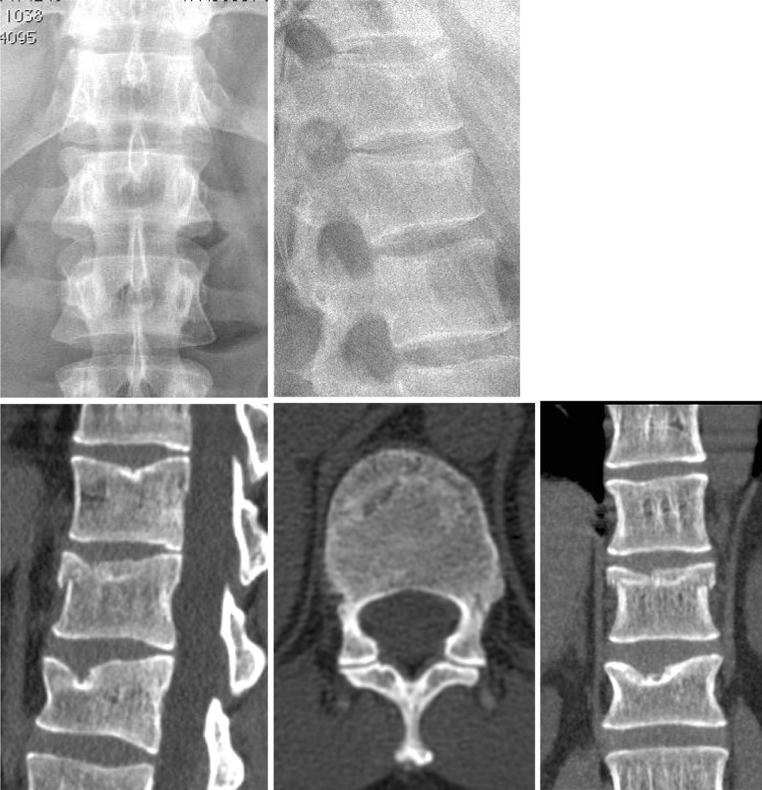
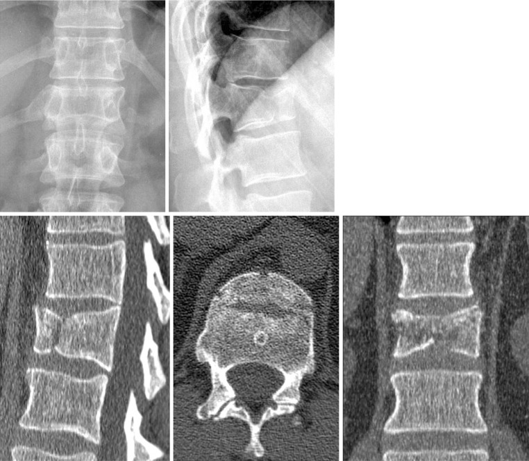
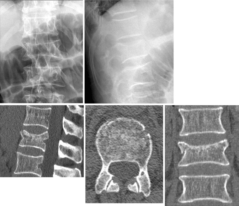
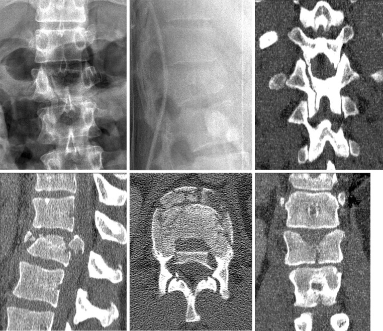
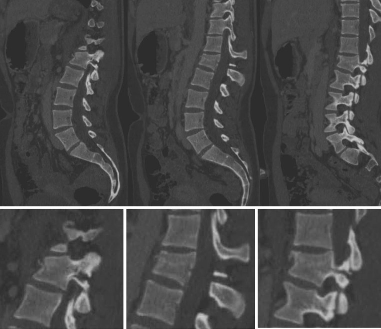
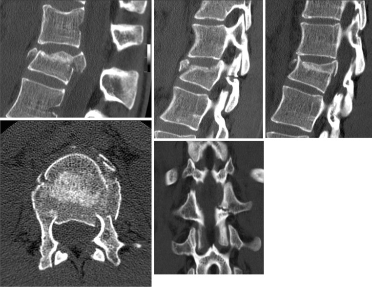
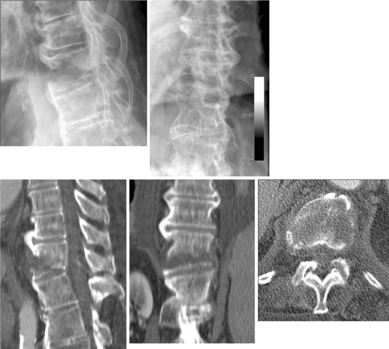
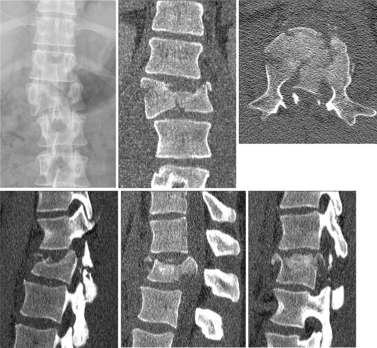
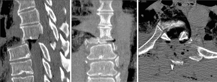
Results: The TL spine injury system is based on three main injury categories adapted from the original Magerl AO concept: A (compression), B (tension band), and C (displacement) type injuries. Type-A injuries include four subtypes (wedge-impaction/split-pincer/incomplete burst/complete burst); B-type injuries are divided between purely osseous and osseo-ligamentous disruptions; and C-type injuries are further categorized into three subtypes (hyperextension/translation/separation). There is no subgroup division. The reliability of injury types (A, B, C) was good (κ = 0.77). The surgeons' pairwise Kappa ranged from 0.69 to 0.90. Kappa coefficients κ for reliability of injury subtypes ranged from 0.26 to 0.78.

Conclusions: The proposed TL spine injury system is based on clinically relevant parameters. Final evaluation data showed reasonable reliability and accuracy. Further validation of the proposed revised AO Classification requires follow-up evaluation sessions and documentation by more surgeons from different countries and backgrounds and is subject to modification based on clinical parameters during subsequent phases.


Figures

Fig. 1
Fig. 1
Type-A compression injuries of the vertebral body
Fig. 2
Fig. 2
Type-B tension band injuries
Fig. 3
Fig. 3
Type-C displacement injuries
Fig. 4
Fig. 4
Diagnostic algorithm for the classification of TL injuries
Fig. 5
Fig. 5
Case example of a wedge/impaction fracture (A1). Code: L1–A1 [upper row conventional radiographs in AP (left) and lateral (right) views of the TL spine centered on the L1-type-A1 injury, lower row corresponding CT scans with sagittal (left), axial (middle), and coronal (right) reconstructions of L1]
Fig. 6
Fig. 6
Example of a split/pincer fracture (A2). Code: T12–A2 [upper row conventional radiographs in AP (left) and lateral (right) views of the TL spine centered on T12 with a type A2 (split/pincer fracture), lower row corresponding CT scans with sagittal (left), axial (middle), and coronal (right) reconstructions of the T12 injury]
Fig. 7
Fig. 7
Example of an incomplete burst fracture (A3). Code: L1–A3 [upper row conventional radiographs in AP (left) and lateral (right) views of the TL spine centered on L1, lower row corresponding CT scans with sagittal (left), axial (middle), and coronal (right) reconstructions of the L1 incomplete burst fracture]
Fig. 8
Fig. 8
Example of a complete burst fracture (A4). Code: T12–A4 [upper row conventional radiographs in AP (left) and lateral (middle) views of the TL spine centered on T12 with a type A4 (complete burst fracture), with a coronal CT reconstruction (right) of the posterior arch showing a vertical split, lower row corresponding CT scans with sagittal (left), axial (middle), and coronal (right) reconstructions of the T12-type-A4 injury]
Fig. 9
Fig. 9
Example of a trans-osseous disruption of a tension band injury (B1). Code: L1–B1 [upper row sagittal CT reconstructions of a pure trans-osseous disruption of tension band injury (B1) showing the extension of the fracture line through both pedicles (left and right images) and the L1 vertebral body (middle), lower scale-up of images above]
Fig. 10
Fig. 10
Example of an osseo-ligamentous disruption of a tension band injury (B2). Code: T12/L1–B2 (L1–A3) [upper row CT reconstructions of ligamentous disruption at the T12/L1 motion segment (left). Additional osseous component with left-sided horizontal facet fracture (middle) as well as right-sided facet joint incongruence with subluxation (right), lower row axial CT cut through the L1-type-A3 vertebral body fracture (left). Coronal reconstructions show the left-sided facet fracture extending through the pars interarticularis (middle)]
Fig. 11
Fig. 11
Example of a hyperextension injury (C1). Code: T12–C1 [upper row conventional radiographs of lateral (left) and AP (right) views of the TL spine centered on T12 with a type-C1 hyperextension injury. Note the bridging anterior ossification as commonly seen in DISH Syndrome, lower row corresponding CT scans with sagittal (left), coronal (middle), and axial reconstructions through the vertebral body of T12 showing the proximal and distal spinal column in a hyperextended position]
Fig. 12
Fig. 12
Example of a translation injury (C2). Code: T12/L1–C2 (L1–A4) [upper row translational injury as seen on an AP radiograph of a T12/L1 subtype C2 with L1 subtype A4 injury (left). Coronal (middle) and axial (right) CT cuts show marked set-off of the lateral vertebral body walls and a burst fracture with a large retropulsed bony posterior wall fragment into the spinal canal causing significant spinal canal encroachment at the L1 level, lower row corresponding sagittal CT reconstructions at the left facet joint (left), mid-sagittal (middle), and right facet joint (right). Failure of the anterior as well as posterior column with bilateral facet fractures]
Fig. 13
Fig. 13
Example of separation injury (C3). Code: T6/T7–C3 [sagittal (left), coronal (middle), and axial (right) CT cuts of a T6/T7 C3 injury of the thoracic spine. The spinal column is completely separated as seen in all three images]

References

    1. Holdsworth F. Fractures, dislocations, and fracture-dislocations of the spine. J Bone Jt Surg (Am) 1970;52:1534–1551. - PubMed
    1. Whitesides TE., Jr Traumatic kyphosis of the thoracolumbar spine. Clin Orthop Relat Res. 1977;128:78–92. - PubMed
    1. Denis F. The three column spine and its significance in the classification of acute thoracolumbar spinal injuries. Spine. 1983;8:817–831. doi: 10.1097/00007632-198311000-00003. - DOI - PubMed
    1. Wood KB, Khanna G, Vaccaro AR, Arnold PM, Harris MB, Mehbod AA. Assessment of two thoracolumbar fracture classification systems as used by multiple surgeons. J Bone Jt Surg (Am) 2005;87:1423–1429. doi: 10.2106/JBJS.C.01530. - DOI - PubMed
    1. Oner FC, Ramos LM, Simmermacher RK, Kingma PT, Diekerhof CH, Dhert WJ, Verbout AJ. Classification of thoracic and lumbar spine fractures: problems of reproducibility. A study of 53 patients using CT and MRI. Eur Spine J. 2002;11:235–245. doi: 10.1007/s00586-001-0364-8. - DOI - PMC - PubMed

Publication types

MeSH terms