. 2013 Oct;26(5):948-57.
doi: 10.1007/s10278-013-9587-6.

Automatic detection of the nipple in screen-film and full-field digital mammograms using a novel Hessian-based method

Affiliations

Automatic detection of the nipple in screen-film and full-field digital mammograms using a novel Hessian-based method

Paola Casti et al. J Digit Imaging. 2013 Oct.

Abstract

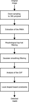
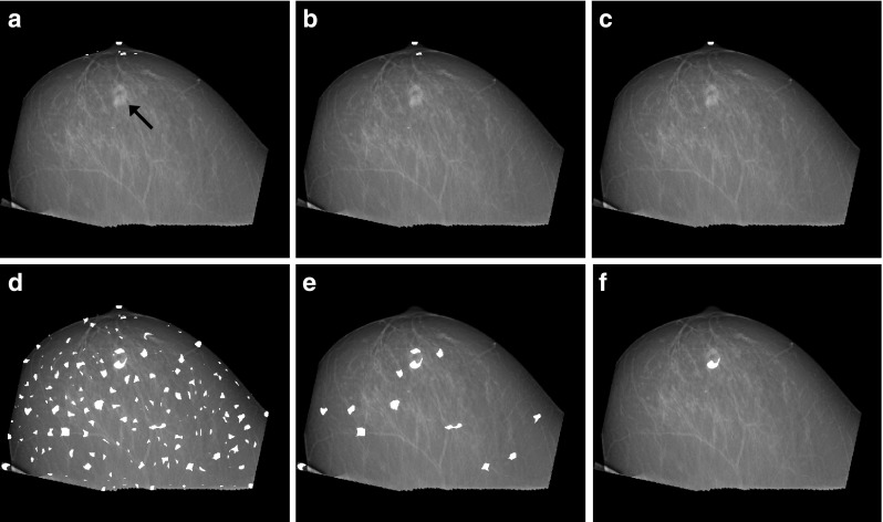
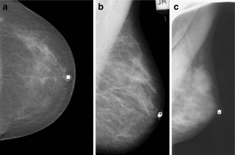
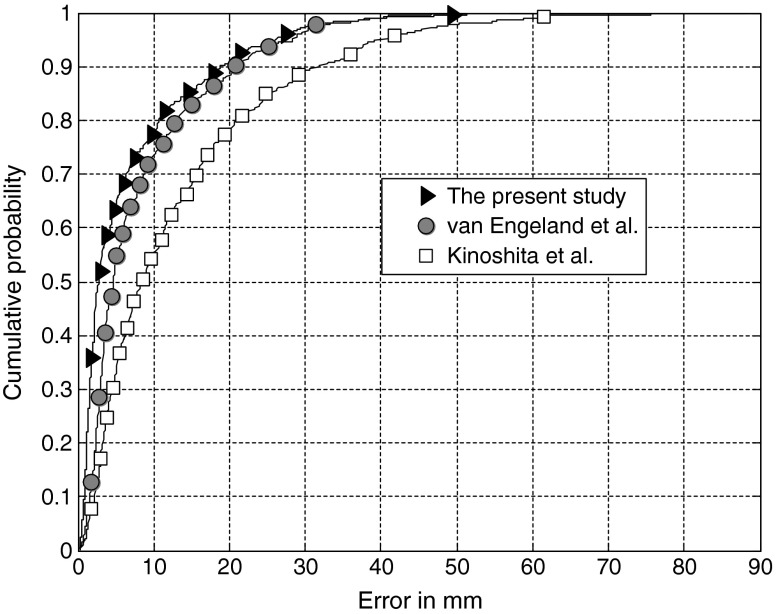
Automatic detection of the nipple in mammograms is an important step in computerized systems that combine multiview information for accurate detection and diagnosis of breast cancer. Locating the nipple is a difficult task owing to variations in image quality, presence of noise, and distortion and displacement of the breast tissue due to compression. In this work, we propose a novel Hessian-based method to locate automatically the nipple in screen-film and full-field digital mammograms (FFDMs). The method includes detection of a plausible nipple/retroareolar area in a mammogram using geometrical constraints, analysis of the gradient vector field by mean and Gaussian curvature measurements, and local shape-based conditions. The proposed procedure was tested on 566 mammographic images consisting of 372 randomly selected scanned films from two public databases (mini-MIAS and DDSM), and 194 digital mammograms acquired with a GE Senographe 2000D FFDM system. A radiologist independently marked the centers of the nipples for evaluation of the results. The average error obtained was 6.7 mm (22 pixels) with reference to the center of the nipple as identified by the radiologist. Only two out of the 566 detected nipples (0.35 %) had an error larger than 50 mm. The method was also directly compared with two other techniques for the detection of the nipple. The results indicate that the proposed method outperforms other algorithms presented in the literature and can be used to identify accurately the nipple on various types of mammographic images.


Figures

Fig. 1
Fig. 1
Examples of mammograms from a the mini-MIAS, b DDSM, and c FFDM databases
Fig. 2
Fig. 2
Flowchart of the procedures used to detect the nipple in mammograms. GVF: gradient vector field, PNRA: plausible nipple/retroareolar area
Fig. 3
Fig. 3
a Rotation of the image and the relative breast contour with reference to the pectoral muscle orientation. The straight-line fit to the pectoral muscle is shown by the black dashed line. b Selection of the inner and the outer contours. c Extraction of the PNRA. d Result of top-hat filtering. e Result of Gaussian smoothing filtering (σ = 10 pixels). f Magnitude of the gradient
Fig. 4
Fig. 4
a,b Zoomed view of the GVF superimposed on the structure of the nipple (a) and on a benign elongated structure in the mammogram (b). c,d Pixels satisfying conditions K > 0 and H < 0 (white and gray) and CN < 3 (white)
Fig. 5
Fig. 5
Maps of disjoint regions whose pixels satisfied the local shape-based conditions superimposed on the rotated version of mammogram with a malignant mass (indicated by the arrow). Results obtained with (a,b,c) and without (d,e,f) defining the PNRA. a,d Areas satisfying conditions K > 0 and H < 0 and with CN < 3. b,e Candidates after rejection of small regions. c,f Final candidate. The result in c is the desired response
Fig. 6
Fig. 6
Examples of detected nipples with the proposed method, one from each of the three databases of mammograms used in the present study: a FFDM image, error = 0 mm, b mini-MIAS image, error = 1.92 mm, and c DDSM image, error = 1.34 mm. Two points are shown on each mammogram corresponding to the nipple position detected automatically (square) and manually marked by the radiologist (star)
Fig. 7
Fig. 7
Histogram of the Euclidean distance between the detected location of the nipple and the center of the nipple as identified by the radiologist. Results obtained using the methods of van Engeland et al. [1] and Kinoshita et al. [11] are included for comparison
Fig. 8
Fig. 8
Cumulative distribution function for the Euclidean distance between the detected location of the nipple and the center of the nipple as identified by the radiologist. Results obtained with the methods of van Engeland et al. [1] and Kinoshita et al. [16] are included for comparison
Fig. 9
Fig. 9
Two cases of failure of the proposed method: a error = 51.40 mm, b error = 56.97 mm. Two points are shown on each mammogram corresponding to the position of the nipple detected automatically (square) and manually marked by the radiologist (star)

References

    1. Van Engeland S, Timp S, Karssemeijer N. Finding corresponding regions of interest in mediolateral oblique and craniocaudal mammographic views. Med Phys. 2006;33(9):3203–3212. doi: 10.1118/1.2230359. - DOI - PubMed
    1. Yuan Y, Giger ML, Li H, Sennett C. Correlative feature analysis on FFDM. Med Phys. 2008;35(12):5490–5500. doi: 10.1118/1.3005641. - DOI - PMC - PubMed
    1. Zheng B, Tan J, Ganott MA, Chough DM, Gur D. Matching breast masses depicted on different views: a comparison of three methods. Acad Radiol. 2009;16(11):1338–1347. doi: 10.1016/j.acra.2009.05.005. - DOI - PMC - PubMed
    1. Paquerault S, Petrick N, Chan HP, Sahiner B, Helvie MA. Improvement of computerized mass detection on mammograms: fusion of two-view information. Med Phys. 2002;29(2):238–247. doi: 10.1118/1.1446098. - DOI - PubMed
    1. Rangayyan RM, Banik S, Desautels JEL. Computer-aided detection of architectural distortion in prior mammograms of interval cancer. J Digit Imag. 2010;23(5):611–631. doi: 10.1007/s10278-009-9257-x. - DOI - PMC - PubMed

MeSH terms

LinkOut - more resources