. 2013 Aug;58(2):788-98.
doi: 10.1002/hep.26401. Epub 2013 Jul 1.

Hepatocyte divalent metal-ion transporter-1 is dispensable for hepatic iron accumulation and non-transferrin-bound iron uptake in mice

Affiliations

Hepatocyte divalent metal-ion transporter-1 is dispensable for hepatic iron accumulation and non-transferrin-bound iron uptake in mice

Chia-Yu Wang et al. Hepatology. 2013 Aug.

Abstract

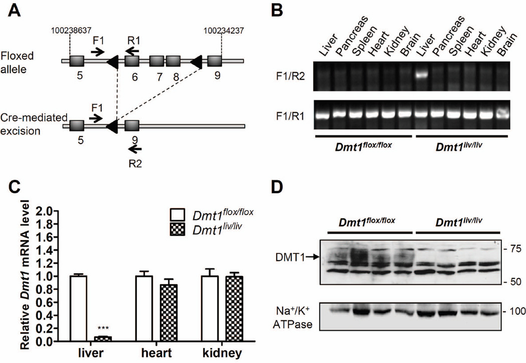
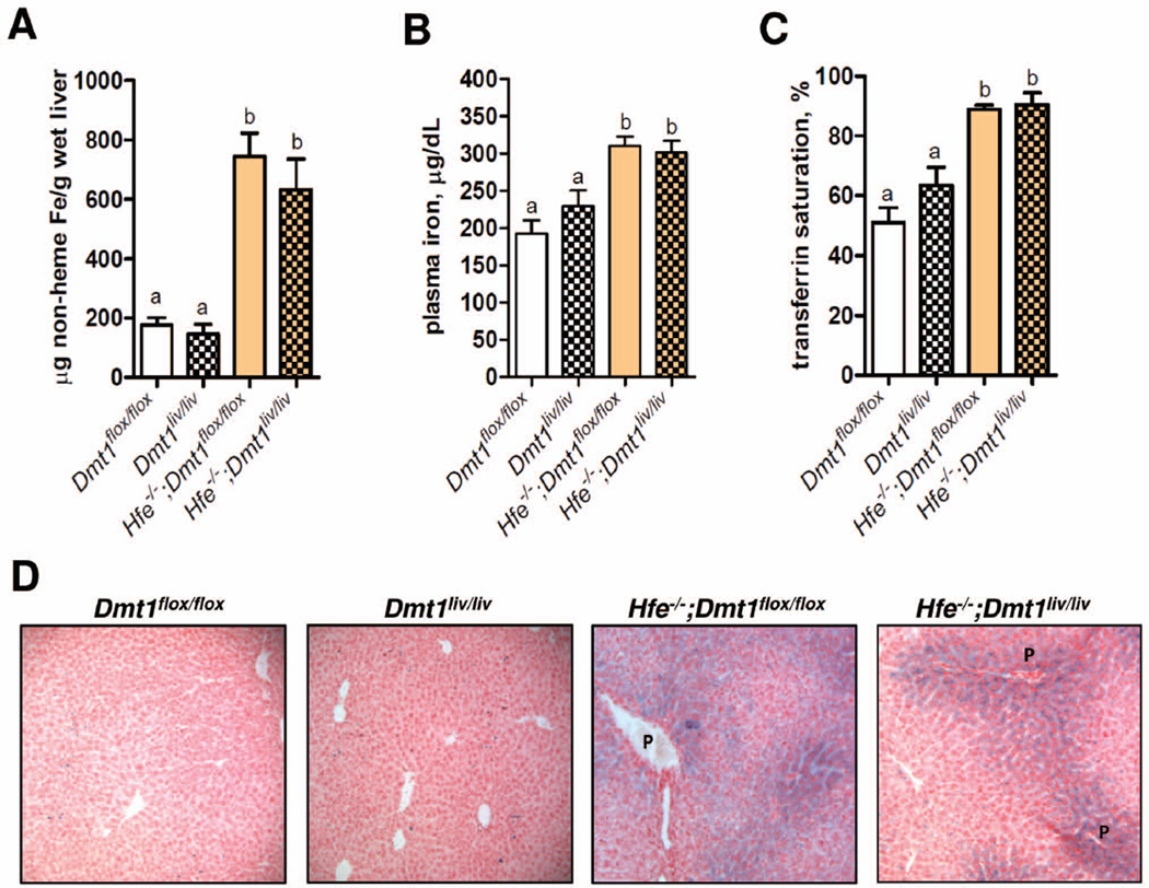
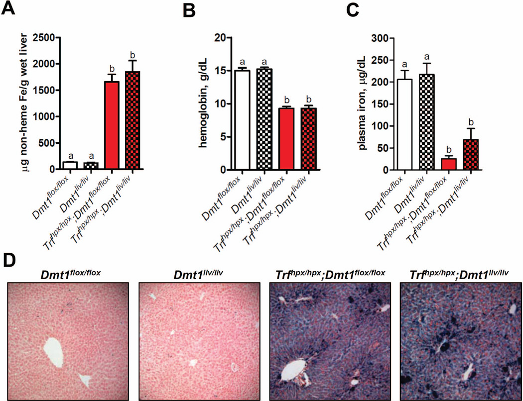
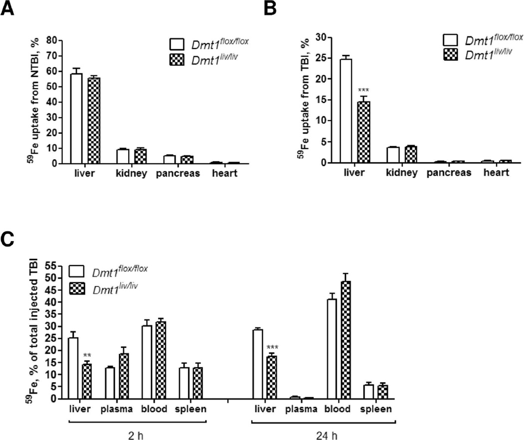
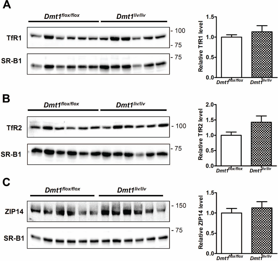
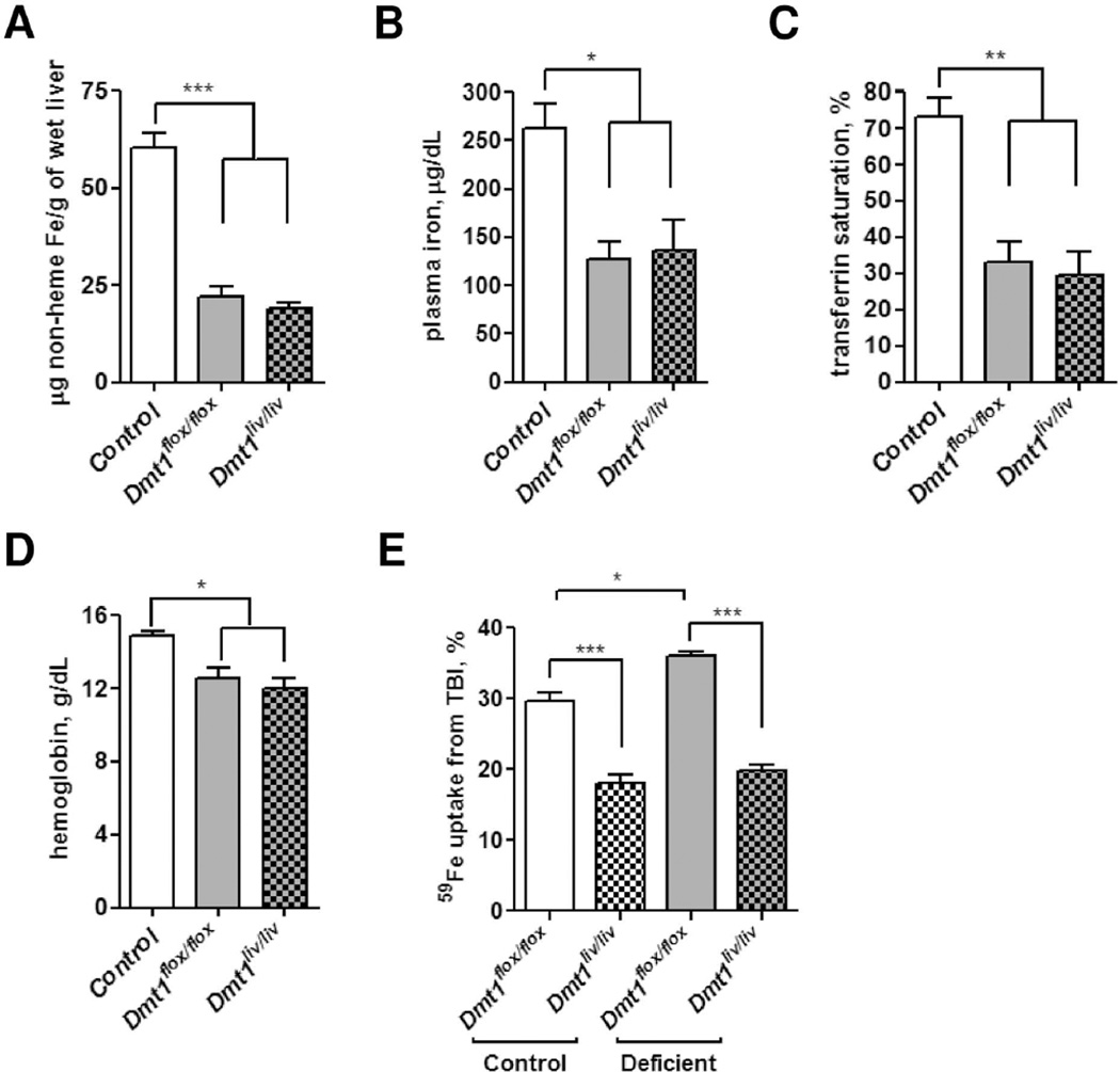
Divalent metal-ion transporter-1 (DMT1) is required for iron uptake by the intestine and developing erythroid cells. DMT1 is also present in the liver, where it has been implicated in the uptake of transferrin-bound iron (TBI) and non-transferrin-bound iron (NTBI), which appears in the plasma during iron overload. To test the hypothesis that DMT1 is required for hepatic iron uptake, we examined mice with the Dmt1 gene selectively inactivated in hepatocytes (Dmt1(liv/liv) ). We found that Dmt1(liv/liv) mice and controls (Dmt1(flox/flox) ) did not differ in terms of hepatic iron concentrations or other parameters of iron status. To determine whether hepatocyte DMT1 is required for hepatic iron accumulation, we crossed Dmt1(liv/liv) mice with Hfe(-) (/) (-) and hypotransferrinemic (Trf(hpx/hpx) ) mice that develop hepatic iron overload. Double-mutant Hfe(-) (/) (-) Dmt1(liv/liv) and Trf(hpx/hpx) ;Dmt1(liv/liv) mice were found to accumulate similar amounts of hepatic iron as did their respective controls. To directly assess the role of DMT1 in NTBI and TBI uptake, we injected (59) Fe-labeled ferric citrate (for NTBI) or (59) Fe-transferrin into plasma of Dmt1(liv/liv) and Dmt1(flox/flox) mice and measured uptake of (59) Fe by the liver. Dmt1(liv/liv) mice displayed no impairment of hepatic NTBI uptake, but TBI uptake was 40% lower. Hepatic levels of transferrin receptors 1 and 2 and ZRT/IRT-like protein 14, which may also participate in iron uptake, were unaffected in Dmt1(liv/liv) mice. Additionally, liver iron levels were unaffected in Dmt1(liv/liv) mice fed an iron-deficient diet.

Conclusion: Hepatocyte DMT1 is dispensable for hepatic iron accumulation and NTBI uptake. Although hepatocyte DMT1 is partially required for hepatic TBI uptake, hepatic iron levels were unaffected in Dmt1(liv/liv) mice, suggesting that this pathway is a minor contributor to the iron economy of the liver.


Conflict of interest statement

The authors have no conflicts of interest to declare.

Figures

Fig. 1
Fig. 1
Disruption of Dmt1 in mouse liver. (A) Schematic depictions of the loxP-flanked (floxed) Dmt1 allele and the allele after Cre recombinase-mediated excision. F1, R1, and R2 indicate forward (F) and reverse (R) primers used for PCR genotyping. Arrowheads denote loxP sites and shaded boxes indicate exons located between positions 10023867 and 10023437 on chromosome 15. (B) PCR analysis of genomic DNA extracted from tissues of mice at 8 weeks of age. (C) Relative Dmt1 mRNA levels in liver, heart, and kidney as determined by using quantitative RT-PCR with Rpl13a as an internal control gene. Values represent mean ± SE, n=3–4, ***P < 0.001. (D) Western blot analysis of DMT1 in crude membrane fractions isolated from livers of Dmt1flox/flox and Dmt1liv/liv mice. All analyses were performed on samples from 8-week-old mice.
Fig. 2
Fig. 2
Hepatic iron accumulation, plasma iron levels, and transferrin saturation are not affected by liver-specific inactivation of Dmt1 in Hfe knockout (Hfe−/−) mice. (A) Hepatic non-heme iron concentrations were determined colorimetrically after acid digestion of tissues. (B) Plasma iron and (C) transferrin saturation were determined by using standard methods. Values represent mean ± SE, n=6. Means without a common superscript differ significantly (P < 0.05). (D) Histological examination of iron loading in the liver by using Perls’ Prussian blue to stain for iron. Branches of the portal vein (P) are indicated. All analyses were performed on samples from 16-week-old mice.
Fig. 3
Fig. 3
Hepatic iron accumulation, hemoglobin levels, and plasma iron levels are not affected by liver-specific inactivation of Dmt1 in hypotransferrinemic (Trf hpx/hpx) mice. (A) Hepatic non-heme iron concentrations were determined colorimetrically after acid digestion of tissues. (B) Hemoglobin and (C) plasma iron levels were determined by using standard methods. Values represent mean ± SE, n=6, except for hemoglobin levels in (Trfhpx/hpx) mice (n=3–4). Means without a common superscript differ significantly (P < 0.05). (D) Histological examination of iron loading in the liver by using Perls’ Prussian blue to stain for iron. All analyses were performed on samples from 16-week-old mice.
Fig. 4
Fig. 4
Tissue uptake of 59Fe from NTBI or TBI injected into the plasma of Dmt1flox/flox and Dmt1liv/liv mice. (A) NTBI uptake by the liver (n=10), kidney (n=5), pancreas (n=5), and heart (n=5). Mice were injected with ferric citrate to transiently saturate plasma transferrin and then 59Fe-labeled ferric citrate was injected 10 minutes later. After 2 hours, mice were sacrificed and whole-body and tissue 59Fe cpm were measured by gamma counting. Tissue uptake of 59Fe from NTBI was calculated as a percentage of whole-body cpm. (B) TBI uptake by the liver (n=14), kidney (n=6), pancreas (n=6), and heart (n=6). Mice were injected with 59Fe-transferrin and sacrificed after 2 hours. Whole-body and tissue 59Fe cpm were determined by gamma counting. Tissue uptake of 59Fe from TBI was calculated as a percentage of whole-body cpm. (C) Distribution of 59Fe among liver, plasma, blood, and spleen 2 and 24 hours after injecting mice with 59Fe-transferrin (n=5–6 at each time point). Results are expressed as mean ± SE. All measurements were performed on mice at 7–8 weeks of age.
Fig. 5
Fig. 5
Effect of liver-specific inactivation of Dmt1 on hepatic levels of TfR1, TfR2, and ZIP14 (A–C) Western blot analyses of TfR1, TfR2, and ZIP14 in Dmt1liv/liv mice and controls (Dmt1flox/flox). Blots were stripped and reprobed for SR-B1 as a lane loading control. Relative band intensities determined by densitometry and normalized to SR-B1. Values represent mean ± SE, n=6. All analyses were performed on samples from 8-week-old mice.
Fig. 6
Fig. 6
Effect of liver-specific inactivation of Dmt1 on iron status and TBI uptake during iron deficiency. Dmt1flox/flox and Dmt1liv/liv mice were fed iron-deficient diet for 3 weeks to induce iron deficiency. Control values were obtained from age-matched Dmt1flox/flox mice fed with standard rodent diet for 3 weeks. Iron status was assessed by measuring (A) hepatic non-heme iron concentrations (B) plasma iron concentrations (C) transferrin saturation, and (D) hemoglobin. (E) TBI uptake by the liver in normal and iron-deficient conditions. Mice were injected with 59Fe-transferrin and sacrificed after 2 hours. Whole-body and tissue 59Fe cpm were determined by gamma counting. Tissue uptake of 59Fe from TBI was calculated as a percentage of whole-body cpm. Values represent mean ± SE, n=3. All analyses were performed on mice at 6 weeks of age.

References

    1. Bothwell TH, Charlton RW, Cook JD, Finch CA. Iron Metabolism in Man. Oxford: Blackwell Scientific Publications; 1979. p. 576.
    1. Pietrangelo A. Hereditary hemochromatosis: pathogenesis, diagnosis, and treatment. Gastroenterology. 2010;139:393–408. - PubMed
    1. Zhou XY, Tomatsu S, Fleming RE, Parkkila S, Waheed A, Jiang J, Fei Y, et al. HFE gene knockout produces mouse model of hereditary hemochromatosis. Proc Natl Acad Sci U S A. 1998;95:2492–2497. - PMC - PubMed
    1. Cheney BA, Lothe K, Morgan EH, Sood SK, Finch CA. Internal iron exchange in the rat. Am J Physiol. 1967;212:376–380. - PubMed
    1. Graham RM, Chua AC, Herbison CE, Olynyk JK, Trinder D. Liver iron transport. World J Gastroenterol. 2007;13:4725–4736. - PMC - PubMed

Publication types

MeSH terms

Substances

Supplementary concepts