

Ruptured conus medullaris dermoid cyst with fat droplets in the central canal [corrected]
- PMID: 23508636
- PMCID: PMC3596585
- DOI: 10.4184/asj.2013.7.1.50
Ruptured conus medullaris dermoid cyst with fat droplets in the central canal [corrected]
Erratum in
- Asian Spine J. 2013 Jun;7(2):158
-
Erratum: correction of title. Ruptured conus medullaris dermoid cyst with fat droplets in the central canal.Asian Spine J. 2013 Jun;7(2):158. doi: 10.4184/asj.2013.7.2.158. Asian Spine J. 2013. PMID: 23741558 Free PMC article. No abstract available.
Abstract
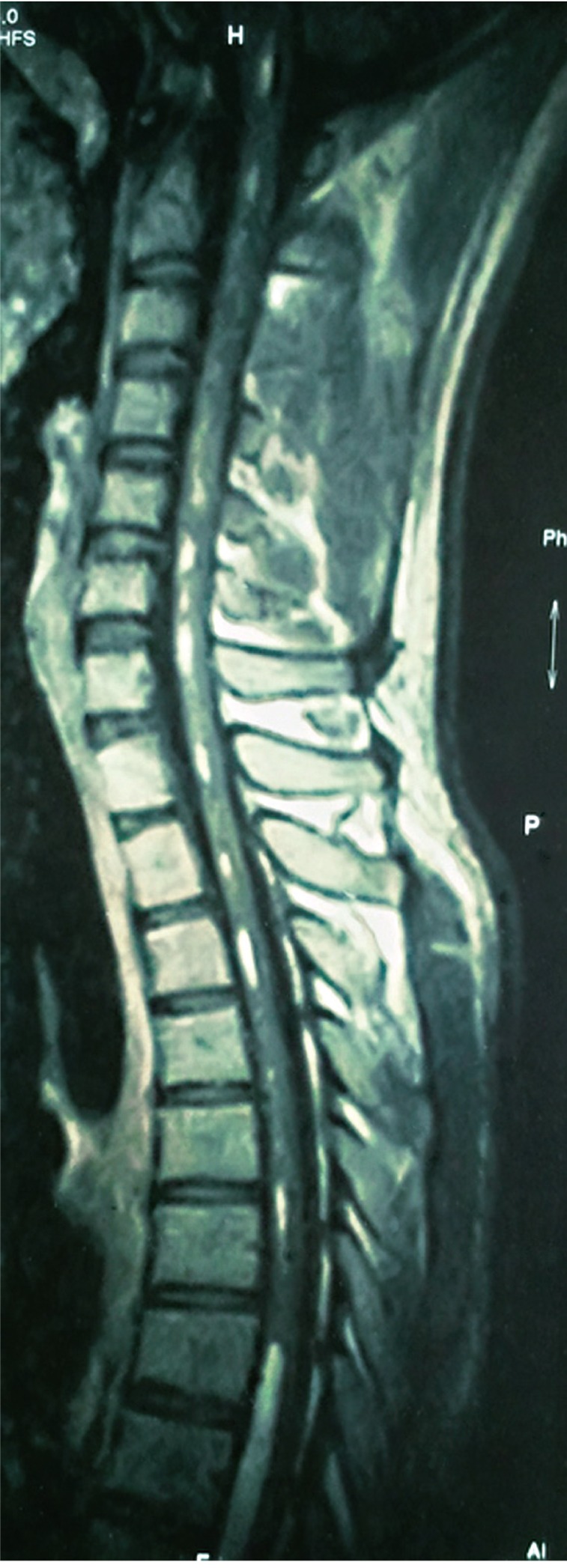
Spinal dermoid tumors are rare, benign, slow growing tumors. These tumors may become acutely symptomatic after rupture or infection. Excision of the lesion with long term close follow-up studies is required for the management of these lesions. We present a very rare case of ruptured conus medullaris dermoid cyst in a 22-year-old male presented with urinary retention and low back pain. Magnetic resonance imaging scan with contrast reveals a lesion in the cauda equina inseparable from conus medullaris with fat droplets within the central spinal canal extending up to the medulla. Patient was operated with laminectomy and near complete excision of the lesion was done. Patient's low back pain was relieved following surgery. However patient had persistent urinary incontinence and on clean intermittent self-catheterization. Histopathology was suggestive of dermoid cyst.
Keywords: Dermoid cyst; Ruptured; Spinal cord.
Conflict of interest statement
No potential conflict of interest relevant to this article was reported.
Figures


Similar articles
-
Dermoid cysts of the conus medullaris: Clinical review, case series and management strategies.J Clin Neurosci. 2018 Apr;50:247-251. doi: 10.1016/j.jocn.2018.01.049. Epub 2018 Feb 10. J Clin Neurosci. 2018. PMID: 29439906 Review.
-
Ruptured dermoid cyst of the conus medullaris in the myelomeningocele sac revealed at the initial repair surgery.Childs Nerv Syst. 2020 May;36(5):1061-1065. doi: 10.1007/s00381-019-04428-1. Epub 2019 Nov 28. Childs Nerv Syst. 2020. PMID: 31776717
-
Ruptured concomitant dermoid cysts of conus medullaris and cervico medullary junction.Asian J Neurosurg. 2015 Oct-Dec;10(4):313-5. doi: 10.4103/1793-5482.162716. Asian J Neurosurg. 2015. PMID: 26425163 Free PMC article.
-
Magnetic resonance imaging of ruptured spinal dermoid tumors with spread of fatty droplets in the central spinal canal and/or spinal subarachnoidal space.J Neuroimaging. 2013 Jan;23(1):71-4. doi: 10.1111/j.1552-6569.2012.00750.x. Epub 2012 Dec 13. J Neuroimaging. 2013. PMID: 23240791 Review.
-
Occult dural arteriovenous fistula causing rapidly progressive conus medullaris syndrome and paraplegia after lumbar microdiscectomy.Spine J. 2009 Sep;9(9):e8-12. doi: 10.1016/j.spinee.2009.03.015. Epub 2009 May 13. Spine J. 2009. PMID: 19442586
References
-
- Lunardi P, Missori P, Gagliardi FM, Fortuna A. Long-term results of the surgical treatment of spinal dermoid and epidermoid tumors. Neurosurgery. 1989;25:860–864. - PubMed
-
- Netsky MG. Epidermoid tumors. Review of the literature. Surg Neurol. 1988;29:477–483. - PubMed
-
- Graham DV, Tampieri D, Villemure JG. Intramedullary dermoid tumor diagnosed with the assistance of magnetic resonance imaging. Neurosurgery. 1988;23:765–767. - PubMed
-
- Kumar S, Gulati DR, Mann KS. Intraspinal dermoids. Neurochirurgia (Stuttg) 1977;20:105–108. - PubMed
-
- Egelhoff JC. Pediatric head and neck imaging. In: Haaga JR, editor. CT and MR imaging of the whole body. 4th ed. London: Mosby; 2003. p. 696.
LinkOut - more resources
Full Text Sources
Other Literature Sources
