. 2013 May;6(3):572-83.
doi: 10.1161/CIRCHEARTFAILURE.112.000200. Epub 2013 Mar 18.

Potential role of BNIP3 in cardiac remodeling, myocardial stiffness, and endoplasmic reticulum: mitochondrial calcium homeostasis in diastolic and systolic heart failure

Affiliations

Potential role of BNIP3 in cardiac remodeling, myocardial stiffness, and endoplasmic reticulum: mitochondrial calcium homeostasis in diastolic and systolic heart failure

Antoine H Chaanine et al. Circ Heart Fail. 2013 May.

Abstract

Background: We have shown that BNIP3 expression is significantly increased in heart failure (HF). In this study, we tested the effects of BNIP3 manipulation in HF.

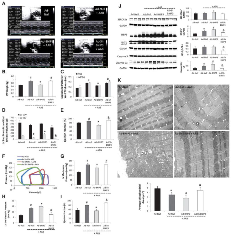
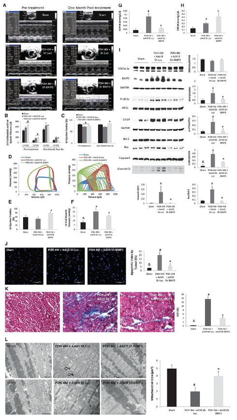
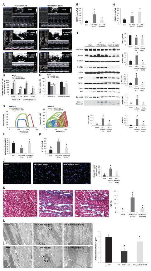
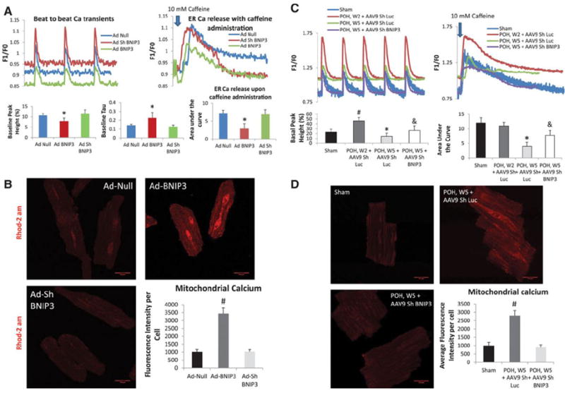
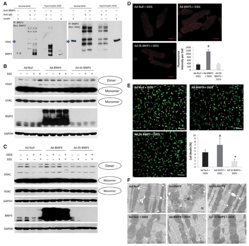
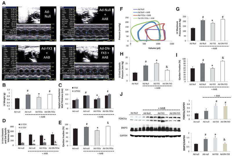
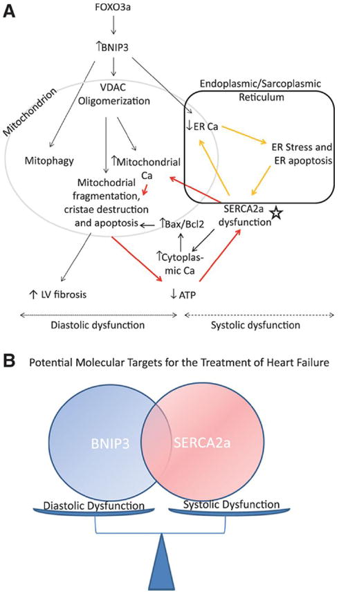
Methods and results: In a rat model of pressure overload HF, BNIP3 knockdown significantly decreased left ventricular (LV) volumes with significant improvement in LV diastolic and systolic function. There were significant decreases in myocardial apoptosis and LV interstitial fibrosis. Ultrastructurally, BNIP3 knockdown attenuated mitochondrial fragmentation and restored mitochondrial morphology and integrity. On the molecular level, there were significant decreases in endoplasmic reticulum (ER) stress and mitochondrial apoptotic markers. One of the mechanisms by which BNIP3 mediates mitochondrial dysfunction is via the oligomerization of the voltage-dependent anion channels causing a shift of calcium from the ER to mitochondrial compartments, leading to the decrease in ER calcium content, mitochondrial damage, apoptosis, and LV interstitial fibrosis, and hence contributes to both systolic and diastolic myocardial dysfunction, respectively. In systolic HF, the downregulation of SERCA2a (sarcoplasmic-endoplasmic reticulum calcium ATPase), along with an increased BNIP3 expression, further worsen myocardial diastolic and systolic function and contribute to the major remodeling seen in systolic HF as compared with diastolic HF with normal SERCA2a expression.

Conclusions: The increase in BNIP3 expression contributes mainly to myocardial diastolic dysfunction through mitochondrial apoptosis, LV interstitial fibrosis, and to some extent to myocardial systolic dysfunction attributable to the shift of calcium from the ER to the mitochondria and to the decrease in ER calcium content. However, SERCA2a downregulation remains a prerequisite for the major LV remodeling seen in systolic HF.

Keywords: apoptosis; gene therapy; heart failure; hypertrophy; remodeling.


Figures

Figure 1
Figure 1. BNIP3 overexpression increased cardiomyocyte death in vitro and impaired LV systolic and diastolic function in a rat model of early POH
1 A-E: M-mode images two weeks after gene delivery via a cross clamp technique and AAB. There were significant decreases in LV weight, IVSd, LVPWd and LVEF and significant increase in LV end systolic volume in the Ad-BNIP3 + AAB group, *P<0.05 vs Ad-Null + AAB and Ad-Sh BNIP3 + AAB, #P<0.05 vs Ad-Null. There was slight but significant decrease in LV end systolic volume and increase in LVEF in the Ad-Sh BNIP3 + AAB group, &P<0.05 vs Ad-Null + AAB. 1 F-I: Pressure-volume loop measurements showed significant decreases in LV maximum pressure and LVEF and significant increases in LVEDP in the Ad-BNIP3 + AAB group, *P<0.05 vs Ad-Null + AAB and Ad-Sh BNIP3 + AAB, #P<0.05 vs Ad-Null. There was significant decrease in LVEDP and a slight but significant increase in LVEF in the Ad-Sh BNIP3 + AAB group, &P<0.05 vs Ad-Null + AAB. 1-J: BNIP3 expression as well as cleaved caspase 3 significantly increased in the Ad-Null + AAB and was the highest in Ad-BNIP3 + AAB, *P<0.05 vs Ad-Null, #P<0.05 vs all other groups. BNIP3 knockdown significantly attenuated the increase in BNIP3 expression, cleaved caspase 3 and the conversion of LC3-1 to LC3-2 in response to PO, &P<0.05 vs Ad-Null + AAB and Ad-BNIP3 + AAB. There was no difference in SERCA2a expression in all groups. 1-K: Ultrastructurally, there was significant decrease in mitochondrial area two weeks after PO, *P<0.05 vs Ad-Null and was the worst in Ad-BNIP3 group with significant mitochondrial fragmentation, cristae destruction and myofibrillar damage, #P<0.05 vs all other groups. BNIP3 knockdown prevented mitochondrial damage in PO, &P<0.05 vs Ad-Null + AAB and Ad-BNIP3 + AAB. Arrows are showing autophagosomes. Images 5,000X magnified, scale bar 2μm.
Figure 2
Figure 2. Tail vein delivery of 5E10 vg/ml of AAV9 Sh BNIP3 reversed cardiac remodeling and improved LV diastolic function and contractility in a rat model of diastolic HF with preserved EF
2 A-C: M-mode images of the above group of animals before and one month after treatment with AAV9 Sh Luc vs AAV9 Sh BNIP3. LV volumes were significantly decreased and the LVEF was significantly increased one month after treatment with AAV9 Sh BNIP3, *P<0.05 vs POH 4M + AAV9 Sh Luc, #P<0.05 vs sham. 2-D: P-V loop tracings in the different groups at baseline and during inferior vena cava occlusion. 2-E: LVEF significantly improved one month after Sh BNIP3 treatment, *P<0.05 vs other two groups. 2 F-G: LVEDP and EDPVR were significantly increased in the Sh Luc group, #P<0.05 vs sham, &P<0.05 vs other two groups. Those parameters were significantly decreased one month after Sh BNIP3 treatment, *P<0.05 vs POH 4M + AAV9 Sh Luc. 2-H: LV contractility significantly increased in the Sh BNIP3 group, *P<0.05 vs POH 4M + AAV9 Sh Luc. Note that the Sh Luc group has a falsely increased ESPVR compared to sham animals as their LVESP are higher, but their V0 is significantly shifted to the right as compared to the sham group. 2-I: Western blot analysis of LV tissue lysate showed robust decrease in BNIP3 expression, ER stress (p-eIF2a) and ER stress apoptotic marker (CHOP) as well as in Bax to Bcl-2 ratio and in cleaved caspase 3 in the Sh BNIP3 group, P<0.05 vs POH 4M + AAV9 Sh Luc, #P<0.05 vs sham and &P<0.05 vs other two groups. There was no difference in SERCA2a expression between all groups. 2 J-K: There were robust decreases in myocardial apoptosis and in LV interstitial fibrosis in the Sh BNIP3 group, *P<0.05 vs POH 4M + AAV9 Sh Luc, #P<0.05 vs sham and &P<0.05 vs other two groups. 2-L: Ultrastructurally, there were significant mitochondrial fragmentation and cristae destruction with dilated T tubules and damaged myofibrils. BNIP3 knockdown robustly attenuated mitochondrial fragmentation and cristae destruction with the mitochondrial area almost back to control level and attenuated myofibrillar damage, #P<0.05 vs sham and *P<0.05 vs POH 4M + AAV9 Sh Luc. Arrows showing the presence of autophagosomes. Above images are 5,000X magnified, scale bar 2μm. Lower images are 12,000X magnified, scale bar 1μm.
Figure 3
Figure 3. Tail vein delivery of 5E10 vg/ml of AAV9 Sh BNIP3 reversed cardiac remodeling and improved LV diastolic function and contractility in a rat model of systolic HF
3 A-C: M-mode images of the above group of animals before and one month after treatment with AAV9 Sh Luc vs AAV9 Sh BNIP3. LV volumes were significantly decreased and the LVEF was significantly increased one month after treatment with AAV9 Sh BNIP3, *P<0.05 vs HF + AAV9 Sh Luc, #P<0.05 vs sham. 3-D: P-V loop tracings in the different groups at baseline and during inferior vena cava occlusion. 3-E: LVEF significantly improved one month after Sh BNIP3 treatment, *P<0.05 vs HF + AAV9 Sh Luc and #P<0.05 vs sham. 3 F-G: LVEDP and EDPVR were significantly increased in the Sh Luc group, #P<0.05 vs sham, &P<0.05 vs other two groups. Those parameters were significantly decreased one month after Sh BNIP3 treatment, *P<0.05 vs HF + AAV9 Sh Luc. 3-H: LV contractility significantly increased in the Sh BNIP3 group, *P<0.05 vs HF + AAV9 Sh Luc. Note that the Sh Luc group has a falsely increased ESPVR compared to sham animals as their LVESP are higher, but their V0 is significantly shifted to the right as compared to the sham group. 3-I: Western blot analysis of LV tissue lysate showed robust decrease in BNIP3 expression, ER stress (p-eIF2a) and ER stress apoptotic marker (CHOP) as well as in Bax to Bcl-2 ratio and in cleaved caspase 3 in the Sh BNIP3 group, P<0.05 vs HF + AAV9 Sh Luc, #P<0.05 vs sham and &P<0.05 vs other two groups. There was significant decrease in SERCA2a expression in the HF groups, #P<0.05 vs sham. 3 J-K: There were robust decreases in myocardial apoptosis and in LV interstitial fibrosis in the Sh BNIP3 group, *P<0.05 vs HF + AAV9 Sh Luc, #P<0.05 vs sham and &P<0.05 vs other two groups. 3-L: Ultrastructurally, there was significant mitochondrial fragmentation and cristae destruction with dilated T tubules and damaged myofibrils. BNIP3 knockdown robustly attenuated mitochondrial fragmentation and cristae destruction with the mitochondrial area almost back to control level and attenuated myofibrillar damage, #P<0.05 vs sham and *P<0.05 vs HF + AAV9 Sh Luc. Arrows showing the presence of autophagosomes. Above images are 5,000X magnified, scale bar 2μm. Lower images are 12,000X magnified, scale bar 1μm.
Figure 4
Figure 4. BNIP3 regulates ER and mitochondrial calcium homeostasis
4-A: BNIP3 overexpression in normal cardiomyocytes significantly decreased beat-to-beat Ca2+ release and ER Ca2+ content with significant increase in Tau, *P<0.05 vs Ad-Null and Ad-Sh BNIP3. There were no differences between the Ad-Null and Ad-Sh BNIP3 treated cardiomyocytes. 4-B: There was threefold increase in mitochondrial Ca2+ in the Ad-BNIP3 group, *P<0.05 vs Ad-Null and Ad-Sh BNIP3. 4-C: In hypertrophic cardiomyocytes, there is robust increase in beat-to-beat Ca2+ release with no change in ER Ca2+ content two weeks after PO, #P<0.05 vs sham. However, five weeks after PO beat-to-beat Ca2+ transients are significantly decreased due to a significant decrease in ER Ca2+ content, *P<0.05 vs all other groups. BNIP3 knockdown significantly increased beat to beat Ca2+ transients and ER Ca2+ content in hypertrophic cardiomyocytes five weeks after PO, &P<0.05 vs POH, W5 + AAV9 Sh Luc 4-D: Mitochondrial Ca2+ significantly increased in hypertrophic cardiomyocytes five weeks after PO, #P<0.05 vs other two groups.
Figure 5
Figure 5. BNIP3 modulates the VDAC channels and shifts the Ca from the ER into the mitochondria
5-A: Co-immunoprecipitation did not show that BNIP3 and VDAC channels form a complex. 5-B: Rather there is modulation of the VDAC channels by BNIP3. The increase in BNIP3 expression causes oligomerization of the VDAC channels, mainly in the form of a dimer, with and without the presence of a cross linking reagent (EGS) leading to the increase in mitochondrial Ca2+. 5 C-D: DIDS, an anion channel inhibitor, inhibited the oligomerization of the VDAC channels in Ad-BNIP3 infected cardiomyocytes. There was threefold decrease in mitochondrial Ca2+ in all groups with significant decrease in the Ad-BNIP3 infected cardiomyocytes treated with DIDS, #P<0.05 vs Ad-Null + DIDS and Ad-Sh BNIP3 + DIDS. 5-E DIDS significantly attenuated cell death in Ad-BNIP3 infected cardiomyocytes, #P<0.05 vs Ad-Null + DIDS and *P<0.05 vs other two groups. 5-F: Ultrastructurally, DIDS significantly attenuated mitochondrial fragmentation and autophagosome formation in Ad-BNIP3 infected cardiomyocytes. The mitochondrial area was significantly higher in cardiomyocytes treated with DIDS compared to no DIDS treatment. Images 12,000X magnified, scale bar 1 μm.
Figure 6
Figure 6. The expression of a constitutively active FOXO3a impairs cardiac diastolic and systolic function in a rat model of early POH
6-A: M-mode images two weeks after gene delivery via a cross clamp technique and AAB. 6 B-E: There were significant decreases in LV weight, IVSd, LVPWd and LV EF and significant increase in LV end systolic volume in the Ad-FX3a + AAB group, *P<0.05 vs Ad-Null + AAB and Ad-DN-FX3a + AAB, #P<0.05 vs Ad-Null. There was slight but significant decrease in LV end systolic volume and increase in LVEF in the Ad-DN-FX3 + AAB group, &P<0.05 vs Ad-Null + AAB. 6 F-I: Pressure-volume loop measurements showed significant decreases in LV maximum pressure and EF and significant increases in LVEDP in the Ad-FX3a + AAB group, *P<0.05 vs Ad-Null + AAB and Ad-DN-FX3a + AAB, #P<0.05 vs Ad-Null. There was significant decrease in LVEDP and a slight but significant increase in LVEF in the Ad-DN-FX3a + AAB group, &P<0.05 vs Ad-Null + AAB. 6-J: FOXO3a expression increased in the Ad-FX3a and Ad-DN-FX3a groups, #P<0.05 vs Ad-Null and Ad-Null + AAB. BNIP3 expression increased in the Ad-Null + AAB and in the Ad-FX3a + AAB groups, #P<0.05 vs Ad-Null and *P<0.05 vs all other groups. The delivery of Ad-DN-FX3a significantly attenuated the increase in BNIP3 expression in response to PO, &P<0.05 vs Ad-Null + AAB and Ad-FX3a + AAB.
Figure 7
Figure 7. Schematic illustration of the mechanism by which BNIP3 induces mitochondrial damage and cell death
7-A: The increase in BNIP3 expression leads to the oligomerization of the VDAC channels causing the shift in ER Ca2+ towards the mitochondria. This shift of ER Ca2+ has two sequelae, the first being the increase in mitochondrial Ca2+ and the second is the decrease in ER Ca2+ content. The increase in mitochondrial Ca2+ leads to mitochondrial fragmentation, mitophagy and apoptosis, decline in cardiac energetics and LV interstitial fibrosis (diastolic dysfunction). Whereas, the decrease in ER Ca2+ leads to ER stress and ER stress induced apoptosis (systolic dysfunction). In systolic HF, SERCA2a downregulation (star), which is an independent process from the increase in BNIP3 expression, contributes to the further decline in ER Ca2+ and to the further increase in mitochondrial Ca2+ leading to two vicious cycles and hence becomes the core of these two cycles as shown in the figure above (red and yellow arrows). 7-B: Cartoon highlighting the interplay between BNIP3 and SERCA2a in modulating diastolic and systolic function of the cardiomyocyte, respectively.

Similar articles

Cited by

References

    1. Diwan A, Krenz M, Syed FM, Wansapura J, Ren X, Koesters AG, Li H, Kirshenbaum LA, Hahn HS, Robbins J, Jones WK, Dorn GW. Inhibition of ischemic cardiomyocyte apoptosis through targeted ablation of bnip3 restrains postinfarction remodeling in mice. J Clin Invest. 2007;117:2825–2833. - PMC - PubMed
    1. Dorn GW., 2nd Mitochondrial pruning by nix and bnip3: An essential function for cardiac-expressed death factors. J Cardiovasc Transl Res. 2010;3:374–383. - PMC - PubMed
    1. Gang H, Hai Y, Dhingra R, Gordon JW, Yurkova N, Aviv Y, Li H, Aguilar F, Marshall A, Leygue E, Kirshenbaum LA. A novel hypoxia-inducible spliced variant of mitochondrial death gene bnip3 promotes survival of ventricular myocytes. Circ Res. 2011;108:1084–1092. - PubMed
    1. Kubli DA, Quinsay MN, Huang C, Lee Y, Gustafsson AB. Bnip3 functions as a mitochondrial sensor of oxidative stress during myocardial ischemia and reperfusion. Am J Physiol Heart Circ Physiol. 2008;295:H2025–2031. - PMC - PubMed
    1. Landes T, Emorine LJ, Courilleau D, Rojo M, Belenguer P, Arnaune-Pelloquin L. The bh3-only bnip3 binds to the dynamin opa1 to promote mitochondrial fragmentation and apoptosis by distinct mechanisms. EMBO Rep. 2010;11:459–465. - PMC - PubMed

Publication types

MeSH terms