

Review
doi: 10.1007/s00018-013-1307-3.
Epub 2013 Mar 19.
Checkpoint kinase 1 in DNA damage response and cell cycle regulation
Affiliations
- PMID: 23508805
- PMCID: PMC3731415
- DOI: 10.1007/s00018-013-1307-3
Item in Clipboard
Review
Checkpoint kinase 1 in DNA damage response and cell cycle regulation
Cell Mol Life Sci.
2013 Nov.
Abstract
Originally identified as a mediator of DNA damage response (DDR), checkpoint kinase 1 (Chk1) has a broader role in checkpoint activation in DDR and normal cell cycle regulation. Chk1 activation involves phosphorylation at conserved sites. However, recent work has identified a splice variant of Chk1, which may regulate Chk1 in both DDR and normal cell cycle via molecular interaction. Upon activation, Chk1 phosphorylates a variety of substrate proteins, resulting in the activation of DNA damage checkpoints, cell cycle arrest, DNA repair, and/or cell death. Chk1 and its related signaling may be an effective therapeutic target in diseases such as cancer.
Figures
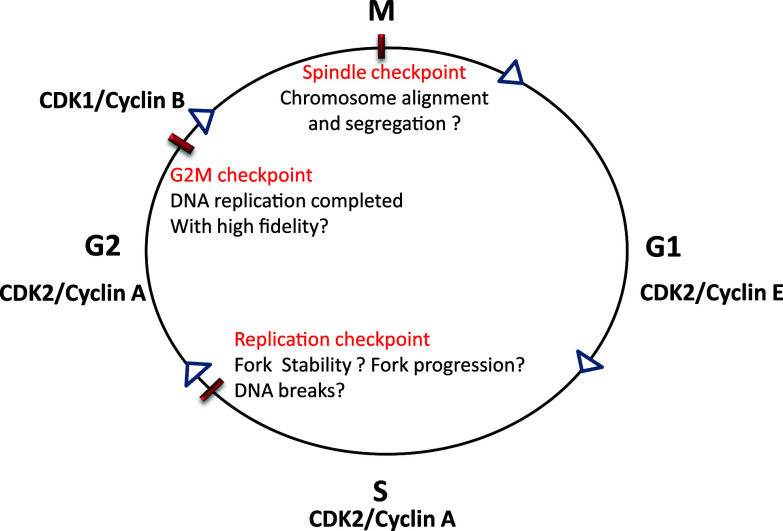
Cell cycle checkpoints. The cell cycle consists of G1, S, G2, and M phases, which are driven by various cyclin/CDK complexes. Progression from one phase to the next phase in the cell cycle is monitored by different checkpoints. S phase is regulated by replication checkpoint that monitors the initiation of replication, replication fork stability, fork progression, and DNA lesions. G2/M checkpoint monitors the completion of DNA replication with high fidelity. Spindle checkpoint makes sure that chromosomes are aligned and segregated for even distribution into two daughter cells
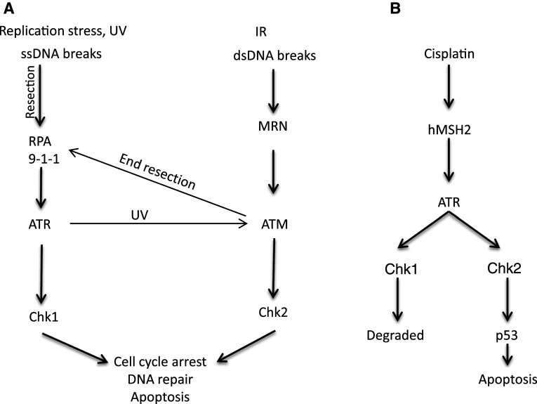
Chk1 and Chk2 activation in response to DNA damage. a Double-strand DNA (dsDNA) breaks are sensed by the MRN complex, which recruits and activates ATM. MRN-mediated nuclease activity generates SS DNA, which can activate ATR. ATM is also activated in UV-induced DNA damage in ATR-dependent manner. Activated ATM phosphorylates several substrates including Chk2. RPA senses and binds single-strand DNA (ssDNA) lesions, which recruit ATR through ATRIP. 9-1-1 (Rad9-Rad1-Hus1) complex binds to ssDNA and dsDNA junction, which recruits TOPBP1 to activate ATR, which phosphorylates Chk1. b ATR is also activated by DNA damage induced by cisplatin. Cisplatin-induced DNA lesion recruits MSH2 (mismatch repair protein 2), which in turn helps in the recruitment and activation of ATR. Activated ATR phosphorylates both Chk1 and Chk2, however, Chk1 is degraded and Chk2 remains active, which phosphorylates and activates p53 leading to apoptosis
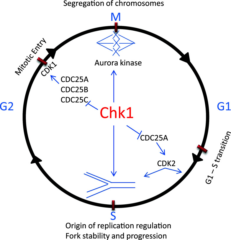
Cell cycle checkpoints regulated by Chk1. Chk1 helps maintain genomic integrity by regulating DNA replication, G2/M, and spindle checkpoints. Chk1 monitors DNA replication, slows down the replication to favor fork stability, prevent stalling of forks and DNA breaks. Chk1 prevents premature entry into mitosis before DNA replication is completed with high fidelity by inhibiting Cdc25 family phosphatases. Chk1 also regulates segregation of chromosomes through activation of aurora kinase to prevent genomic instability
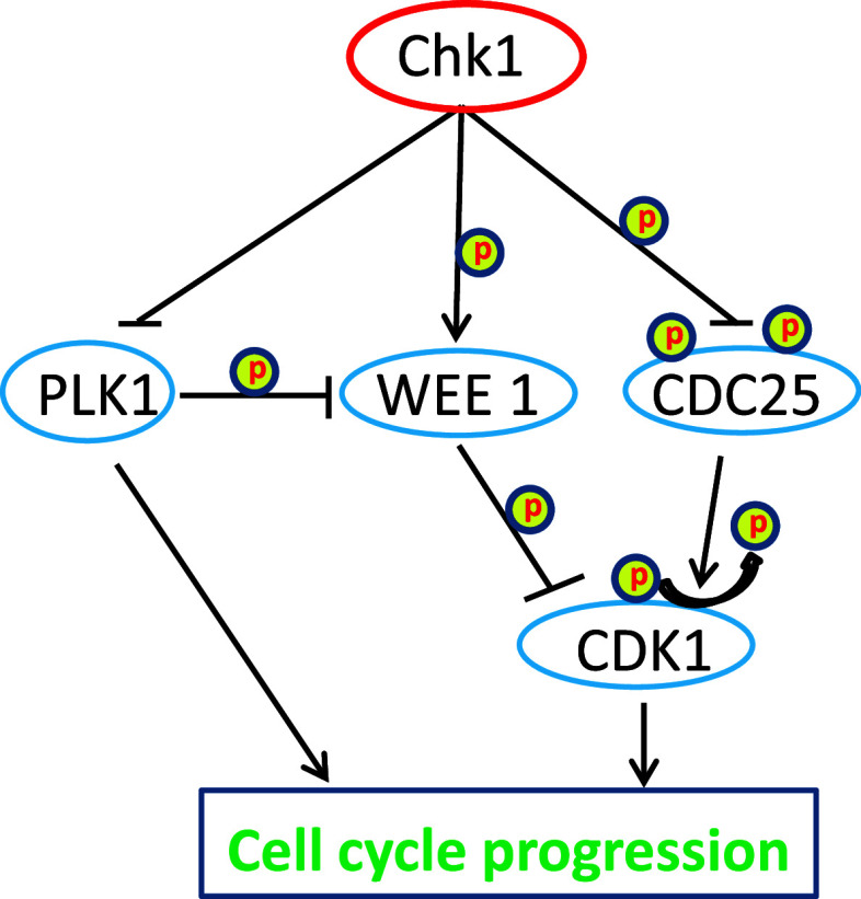
Chk1 regulation of cell cycle progression. Chk1 regulates progression of cell cycle by inhibiting Cdc25 family phosphatases and polo-like kinase 1(PLK1), and activating WEE 1 kinase. Cdc25 is a phosphatase and activator of CDK1, whereas WEE1 inhibits CDK1 by phosphorylation. PLK1 can activate CDK1 by inhibiting WEE1. PLK1 can also directly promote cell cycle progression
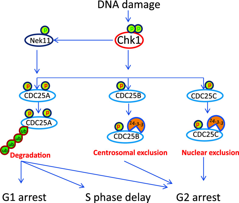
Chk1-mediated cell cycle arrest in response to DNA damage. Chk1 phosphorylates Cdc25 family resulting in their inhibition. Chk1 can also activate Nek11, which further phosphorylates Cdc25A on multiple sites to target it for degradation. Cdc25A is crucial for G1-S transition, S phase progression, and mitotic entry; as a result, Cdc25A inhibition leads to G1 arrest, slows down replication, and G2 arrest. Chk1 phosphorylates Cdc25B to promote its binding to 14-3-3 protein and sequestration from centrosome. Chk1 also phosphorylates Cdc25C to induce its binding to 14-3-3 protein and nuclear exclusion. Cdc25B and Cdc25C inhibition mainly causes G2 arrest
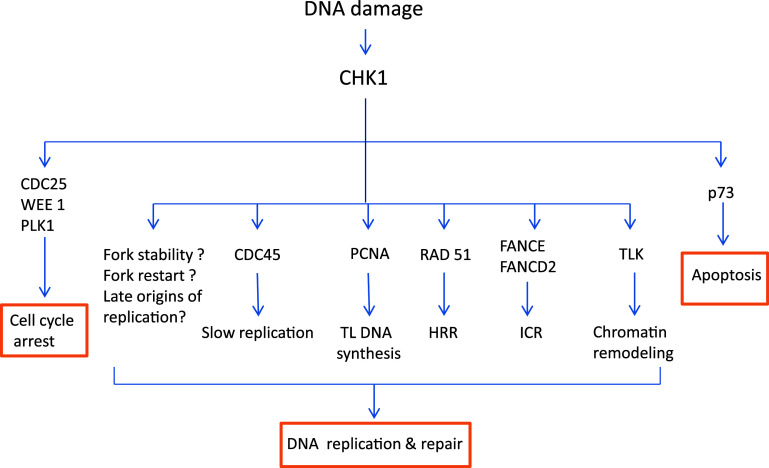
Chk1-mediated DNA repair. Activated Chk1 phosphorylates several substrates to mediate DNA repair and apoptosis. Chk1 slows down DNA replication directly by affecting CDC45 loading on replication complex to allow time for DNA repair. Chk1 promotes monoubiquitination of PCNA, which recruits translesion polymerases to help translesion DNA synthesis. Chk1 phosphorylates TLK-tousled like kinase to promote chromatin assembly. Chk1 phosphorylates Rad51 to target it to double-strand breaks to promote HRR-homologous recombination and repair. Chk1 phosphorylates FANCE and monoubiquitinates FANCD2, which help resolve ICR-interstrand crosslink DNA lesion. Chk1 also regulates replication fork stability, fork restart, and late origins of replication, the mechanistic details of which are currently unclear. In addition to DNA repair, Chk1 may also induce cell cycle arrest and apoptosis by regulating different substrates
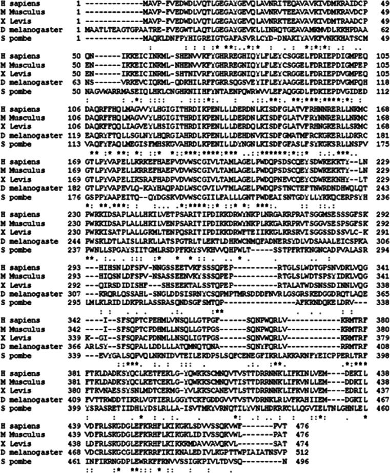
CHk1 sequence alignment between human, mice, fruit fly, Xenopus and yeast. Chk1 sequences of human, mice, Drosophila, Xenopus, and yeast were analyzed using T-coffee, a Web server for multiple sequence alignment tool. The N-terminal kinase domain and regulatory SQ/TQ domain of Chk1 are highly conserved, while the C-terminal domain is less conserved. Asterisk indicates highly conserved sequence, Colon indicates conserved substitution, Dot indicate semi conserved substitutions
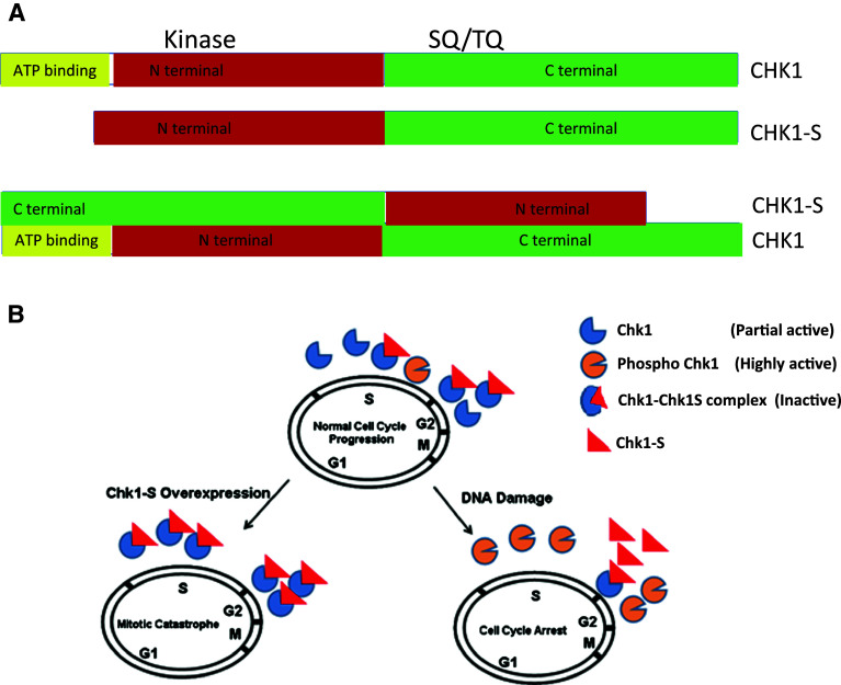
Physical and functional interactions between Chk1 and Chk1-S. a Schematic representation of Chk1 and Chk1S domains and their interaction. Checkpoint kinase 1 has ATP binding domain, N-terminal kinase domain, SQ/TQ domain, and C-terminal domain. Chk1S lacks ATP binding domain. C-terminal domain of Chk1 interacts with N-terminal domain of CHK1-S. b Regulation of Chk1 by Chk1-S. Chk1-S interacts with Chk1 to inhibit its kinase activity. In unperturbed cell cycles, Chk1-S is expressed in late S to G2 phase to inhibit Chk1 and promote mitotic entry. In DNA damage, Chk1 is phosphorylated to prevent Chk1-S binding and inhibition, resulting in Chk1 activation. When Chk1-S is overexpressed, it antagonizes Chk1, leading to premature mitotic entry and consequent cell death via mitotic catastrophe. Figure 8b is adopted from Pabla et al. PNAS 109, 197-202, 2012
References
-
- Walworth N, Davey S, Beach D. Fission yeast Chk1-protein kinase links the rad checkpoint pathway to Cdc2. Nature. 1993;363:368–371. - PubMed
-
- Fogarty P, Kalpin RF, Sullivan W. The Drosophila maternal-effect mutation grapes causes a metaphase arrest at nuclear cycle 13. Development. 1994;120:2131–2142. - PubMed
-
- Peng CY, Graves PR, Thoma RS, Wu Z, Shaw AS, Piwnica-Worms H. Mitotic and G2 checkpoint control: regulation of 14–3-3 protein binding by phosphorylation of Cdc25C on serine-216. Science. 1997;277:1501–1505. - PubMed
-
- Elledge SJ. Cell cycle checkpoints: preventing an identity crisis. Science. 1996;274:1664–1672. - PubMed
Publication types
MeSH terms
Substances
Grants and funding
LinkOut - more resources
Full Text Sources
Other Literature Sources
Miscellaneous
