

Interobserver agreement for the detection of Barrett's esophagus with optical frequency domain imaging
- PMID: 23508980
- PMCID: PMC3732518
- DOI: 10.1007/s10620-013-2625-x
Interobserver agreement for the detection of Barrett's esophagus with optical frequency domain imaging
Abstract
Background: Optical frequency domain imaging (OFDI) is a second-generation form of optical coherence tomography (OCT) providing comprehensive cross-sectional views of the distal esophagus at a resolution of ~7 μm.
Aim: Using validated OCT criteria for squamous mucosa, gastric cardia mucosa, and Barrett's esophagus (BE), the objective of this study was to determine the inter- and intra-observer agreements by a large number of OFDI readers for differentiating these tissues.
Methods: OFDI images were obtained from nine subjects undergoing screening and surveillance for BE. Sixty-four OFDI image regions of interest were randomly selected for review. A training set of 19 images was compiled distinguishing squamous mucosa from gastric cardia and BE using previously validated OCT criteria. The ten readers then interpreted images in a test set of 45 different images of squamous mucosa (n = 15), gastric cardia (n = 15), or BE (n = 15). Interobserver agreement differentiating the three tissue types and BE versus non-BE mucosa was determined using multi-rater Fleiss's κ value. The images were later randomized again and four readers repeated the test 3 weeks later to assess intraobserver reliability.
Results: All ten readers showed excellent agreement for the differentiation of BE versus non-BE mucosa (κ = 0.811 p < 0.0001) and for differentiating BE versus gastric cardia versus squamous mucosa (κ = 0.866, p < 0.0001). For the four readers who repeated the test, the median intraobserver agreement (BE vs. non-BE) was high (κ = 0.975, IQR: 0.94, 1.0).
Conclusions: Trained readers have a high interobserver agreement for differentiating BE, squamous, and gastric cardia mucosa using OFDI.
Figures
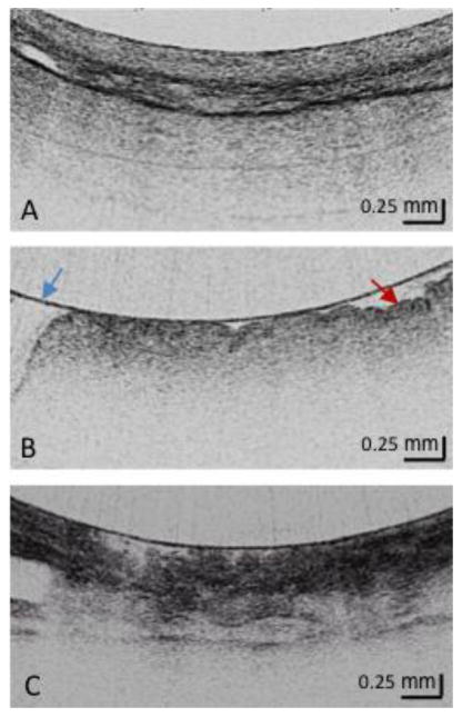
Squamous Mucosa Layered structure No glands in the epithelial layer Squamous epithelium is homogeneous
Gastric Cardia Mucosa (at least 2) Vertical pit architecture Highly reflective surface (red arrow) Relatively poor image penetration Broad regular foveolar region Rugae Blue arrow is the balloon wall
Barrett's Metaplasia (at least 2) Loss of layered or vertical pit and crypt architecture Irregular mucosal surface Heterogeneous scattering
References
-
- Lagergren J, Bergstrom R, Lindgren A, et al. Barrett's esophagus. A prevalent, occult complication of gastroesophageal reflux disease. Gastroenterology. 1987;92:118–24. - PubMed
-
- Pohl H, Sirovich B, Welch HG. Esophageal adenocarcinoma incidence: Are we reaching the peak? Cancer Epidemiol Biomarkers Prev. 2010;19:1468–70. - PubMed
-
- Siegel R, Niashadham D, Jemal A. Cancer Statistics, 2012. Ca Cancer J Clin. 2012;62(1):10–29. - PubMed
-
- Fitzgerald RC, Lascar R, Triadafilopoulos G. Review article: Barrett's oesophagus, dysplasia and pharmacologic acid suppression. Aliment Pharmacol Ther. 2001;15(3):269–276. - PubMed
Publication types
MeSH terms
Grants and funding
LinkOut - more resources
Full Text Sources
Other Literature Sources
