. 2013 Apr 2;110(14):E1263-72.
doi: 10.1073/pnas.1303309110. Epub 2013 Mar 18.

Activity-enhancing mutations in an E3 ubiquitin ligase identified by high-throughput mutagenesis

Affiliations

Activity-enhancing mutations in an E3 ubiquitin ligase identified by high-throughput mutagenesis

Lea M Starita et al. Proc Natl Acad Sci U S A. .

Abstract

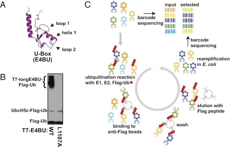
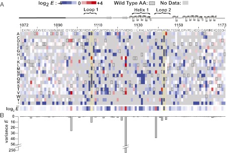
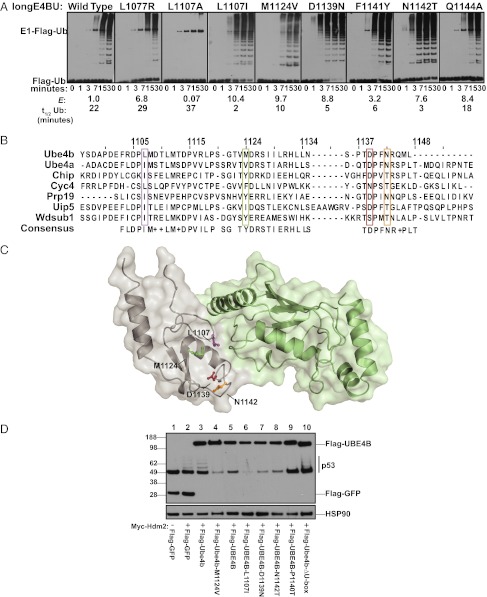
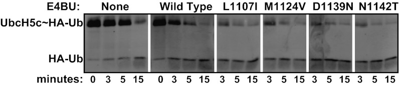
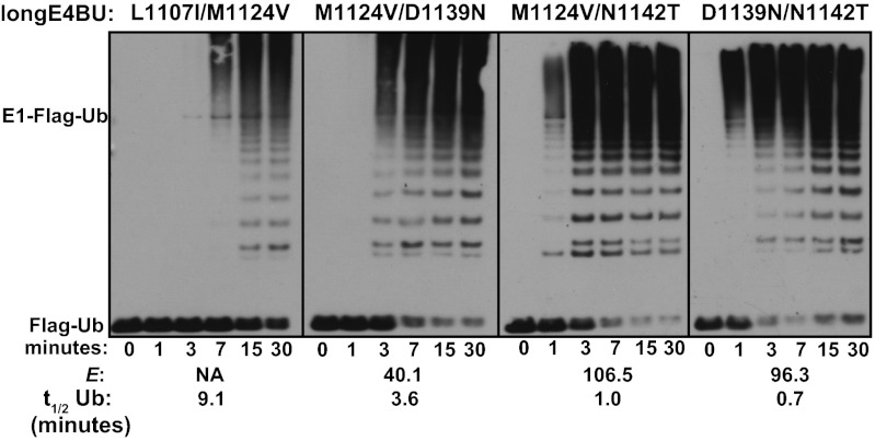
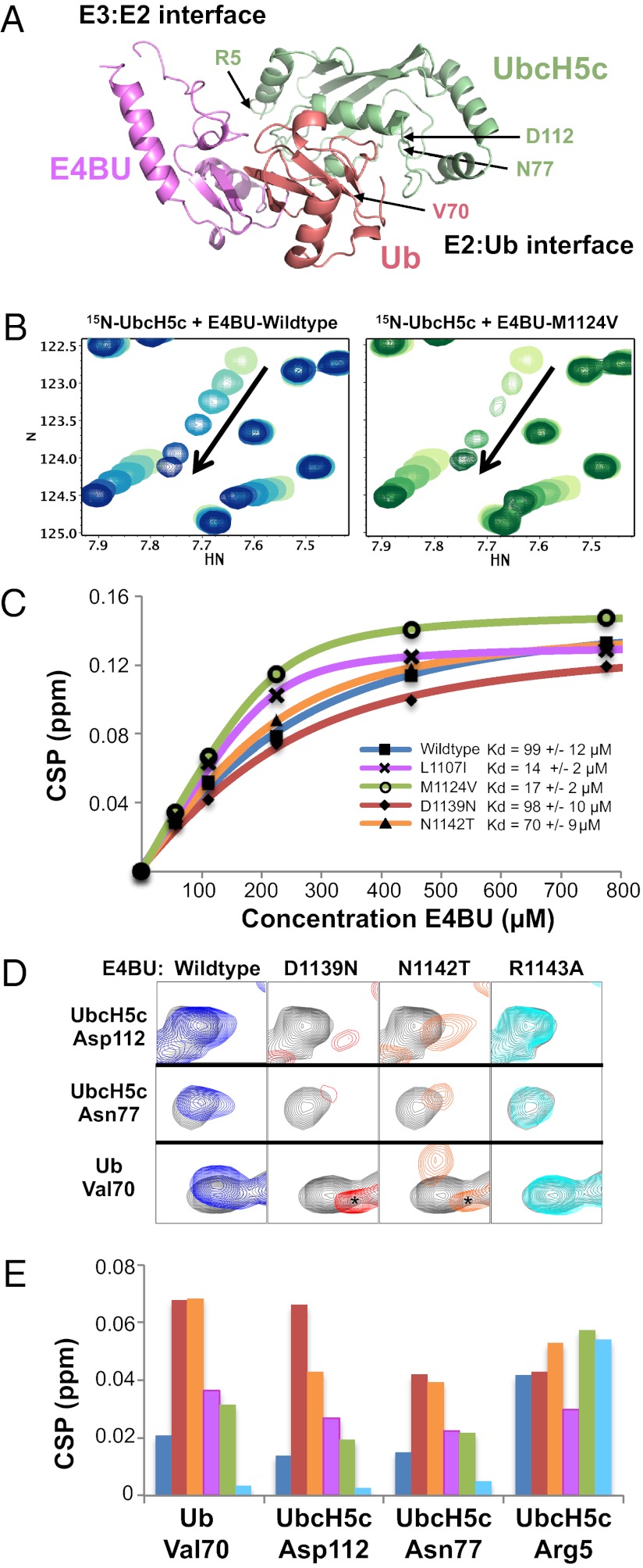
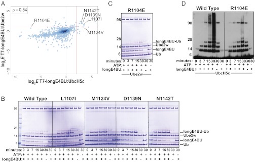
Although ubiquitination plays a critical role in virtually all cellular processes, mechanistic details of ubiquitin (Ub) transfer are still being defined. To identify the molecular determinants within E3 ligases that modulate activity, we scored each member of a library of nearly 100,000 protein variants of the murine ubiquitination factor E4B (Ube4b) U-box domain for auto-ubiquitination activity in the presence of the E2 UbcH5c. This assay identified mutations that enhance activity both in vitro and in cellular p53 degradation assays. The activity-enhancing mutations fall into two distinct mechanistic classes: One increases the U-box:E2-binding affinity, and the other allosterically stimulates the formation of catalytically active conformations of the E2∼Ub conjugate. The same mutations enhance E3 activity in the presence of another E2, Ube2w, implying a common allosteric mechanism, and therefore the general applicability of our observations to other E3s. A comparison of the E3 activity with the two different E2s identified an additional variant that exhibits E3:E2 specificity. Our results highlight the general utility of high-throughput mutagenesis in delineating the molecular basis of enzyme activity.


Conflict of interest statement

The authors declare no conflict of interest.

Figures

Fig. 1.
Fig. 1.
Highly parallel method for examining Ub ligase activity. (A) Cartoon representation of the 82-aa E4BU solution structure (Protein Data Bank ID code 2kr4). Residues within loop 1, loop 2, and helix 1 form the E3:E2 interface. The longE4BU domain contains an extra 20 amino-terminal segment required for auto-ubiquitination activity. (B) Western blot for Flag-Ub shows Ub ligase activity of longE4BU expressed on the phage surface. LongE4BU-WT (lane 1) or longE4BU-L1107A (lane 2) was fused to the coat protein of T7 bacteriophage. Amplified phage lysate was incubated with recombinant E1, UbcH5c, ATP/Mg, and Flag-Ub. The reaction was analyzed by Western blot with anti-Flag to follow Flag-Ub. (C) Library of longE4BU sequence variants was generated from doped oligonucleotides, with a degenerate 18-base barcode inserted 3′ of the stop codon. The variable region and barcode were cloned into the phage genome and displayed as a carboxyl-terminal fusion with the T7 coat protein. The library of T7-longE4BU sequence variants was subjected to multiple rounds of selection for functional Ub ligase activity by incubation with recombinant 6× His-E1, UbcH5c, ATP/Mg, and Flag-Ub. The ubiquitinated phages were selected on anti-Flag agarose, and unbound phages were washed away. Bound Flag-ubiquitinated T7-longE4BU variants were eluted by competition with 3× Flag peptide. Eluted phages were reamplified and subjected to additional rounds of selection. DNA was purified from the input and selected T7-longE4BU populations, Illumina libraries were constructed, and the barcodes were sequenced by 36-base single end reads.
Fig. 2.
Fig. 2.
Effects of known inactivating mutations and >900 unique mutations on E3 function are uncovered by deep mutational scanning of longE4BU. (A) Sequence–function map of log2 E scores for 932 T7-longE4BU variants with a single amino acid change. Blue, white, and red boxes represent T7-longE4BU variants that were depleted, neutral, or enriched, respectively, during the selection process; gray represents no data that passed quality filters; and boxed rectangles represent the WT residue. A schematic of the E4BU secondary structure is shown above. The amino-terminal 20 amino acids that were not included in either deposited structure are represented by a black line. Loop 1, loop 2, and helix 1 are indicated. The longE4BU sequence is represented on the x axis, and the possible amino acid substitutions are represented on the y axis. Below are the position-averaged E scores for each position in T7-longE4BU. (B) Variance of E scores for each position represented in a bar graph is shown.
Fig. 3.
Fig. 3.
Enriched longE4BU variants are more active Ub ligases than the WT protein. (A) Ubiquitination assays. Recombinant WT longE4BU and the indicated variants were incubated with recombinant E1, UbcH5c, ATP/Mg, and Flag-Ub at 37 °C for the indicated time. Ubiquitination products were monitored by Western blot to follow Flag-Ub. E scores and the approximate t1/2 of unmodified Ub calculated from densitometry of Coomassie-stained reactions are indicated below (see also Fig. S3 B and C). (B) Multiple sequence alignment of the U-box domains for the seven murine U-box–containing proteins. Positions corresponding to E4BU L1107 (purple), M1124 (green), D1139 (red), and N1142 (orange) are indicated. (C) Sites of mutation in highly enriched T7-longE4BU variants mapped to the E4BU:UbcH5c crystal structure (Protein Data Bank ID code 3L1Z). L1107 (purple), M1124 (green), D1139 (red), and N1142 (orange) are depicted on gray E4BU. UbcH5c is pale green. (D) H1299 cells were transfected with pCMV-neo-p53, and cells in lanes 2–8 were transfected with pCMV-Myc-Hdm2. Flag-tagged, full-length mouse Ube4b or human UBE4B constructs were transfected as indicated. Blots were probed for p53 and the Flag epitope. Endogenous heat shock protein 90 (HSP90) was used as a protein loading control (see also Fig. S4).
Fig. 4.
Fig. 4.
Activating mutations enhance the general reactivity of an E2∼Ub conjugate lysine reactivity assay. Recombinant minimal U-box domains of WT E4BU (1,092–1,173) and the indicated variants were incubated with purified UbcH5c∼HA-Ub and free lysine at 37 °C for the times indicated. Breakdown of the UbcH5c∼HA-Ub thioester linkage was monitored by Western blot with anti-HA antibodies.
Fig. 5.
Fig. 5.
Effects of some activating mutations are synergistic and create hyperactive U-box domains. Ubiquitination assays. The indicated purified variants of longE4BU were incubated with recombinant E1, UbcH5c, ATP/Mg, and Flag-Ub at 37 °C for the indicated times. Ubiquitination products were monitored by Western blot analysis to follow Flag-Ub. E scores and the approximate t1/2 of unmodified Ub calculated from densitometry of Coomassie-stained reactions are indicated below (see also Fig. S3).
Fig. 6.
Fig. 6.
NMR analysis reveals that E4BU activating mutations fall into two classes. (A) One member from the E4BU:UbcH5c∼Ub model (3) is shown for reference with the E3:E2 and E2:Ub interfaces annotated. (B) Representative portion of the UbcH5c HSQC spectrum shows CSPs on addition of 0.25, 0.5, 1, 2, and 3.4 mol eq of E4BU WT (Left, blue) or M1124V (Right, green) to 225 μM 15N-UbcH5c. The M1124V mutant exhibits binding phenomena that approach intermediate exchange, as illustrated by line-width broadening at early titration points. (C) CSP values fit with quadratic binding equations using NMRView. Kd values were determined for WT (blue, 99 ± 12 μM), L1107I (magenta, 14 ± 2 μM), M1124V (green, 17 ± 2 μM), D1139N (red, 98 ± 10 μM), and N1142T (orange, 70 ± 9 μM) E4BU titrated into 15N-UbcH5c. (D) Resonances corresponding to D112 in UbcH5c helix 2, N77 near the UbcH5c active site, and V70 within the Ub hydrophobic surface act as indicators for closed, active UbcH5c∼Ub conformations promoted by E4BU binding. Compared with the addition of 0.25 molar equivalences of WT E4BU (blue) to 225 μM 15N-UbcH5c-O∼15N-Ub, both the D1139N (red) and N1142T (orange) mutants induce further population of closed UbcH5c∼Ub conformations. We could not collect NMR data at higher additions of E4BU due to the catalyzed hydrolysis of the oxyester effected by the E3. Addition of the catalytically inactive E4BU mutant R1143A (cyan) is shown for reference. Signal arising from small quantities of free, hydrolyzed Ub is marked by asterisks. (E) CSPs shown in D are quantified and shown in histogram format for the addition of 0.25 mol eq of WT (blue), D1139N (red), N1142T (orange), L1107I (magenta), M1124V (green), and R1143A (cyan) E4BU to 225 μM 15N-UbcH5c-O∼15N-Ub. Perturbations that reflect E3:E2 binding directly are largely unaffected, as shown by UbcH5c residue R5.
Fig. 7.
Fig. 7.
E4BU activating mutation behaves differently when paired with a different E2, Ube2w. (A) Log2-transformed E scores calculated from the deep mutational scan of T7-longE4BU performed with Ube2w as the E2 (y axis) compared with log2-transformed E scores from the deep mutational scan of T7-longE4BU performed with UbcH5c (x axis). Only E scores from variants with a single amino acid change are compared. The red lines signify a twofold increase in variant E scores over WT for Ube2w and a fourfold increase for UbcH5c. A Spearman’s rank correlation coefficient of 0.54 was calculated. (B and C) Ubiquitination assays. The indicated purified variants of longE4BU were incubated with recombinant E1, UbcH5c or Ube2w ATP/Mg, and Ub at 37 °C for the indicated time. Ubiquitination products were monitored by Coomassie staining. (D) Ubiquitination assays. The indicated purified variants of longE4BU were incubated with recombinant E1, UbcH5c or Ube2w ATP/Mg, and Ub at 37 °C for the indicated time. Ubiquitination products were monitored by Western blot analysis to follow Ub.

References

    1. Pickart CM, Eddins MJ. Ubiquitin: Structures, functions, mechanisms. Biochim Biophys Acta. 2004;1695(1-3):55–72. - PubMed
    1. Dou H, Buetow L, Sibbet GJ, Cameron K, Huang DT. BIRC7-E2 ubiquitin conjugate structure reveals the mechanism of ubiquitin transfer by a RING dimer. Nat Struct Mol Biol. 2012;19(9):876–883. - PMC - PubMed
    1. Pruneda JN, et al. Structure of an E2:E3∼Ub complex reveals an allosteric mechanism shared among RING/U-box ligases. Mol Cell. 2012;47(6):933–942. - PMC - PubMed
    1. Ozkan E, Yu H, Deisenhofer J. Mechanistic insight into the allosteric activation of a ubiquitin-conjugating enzyme by RING-type ubiquitin ligases. Proc Natl Acad Sci USA. 2005;102(52):18890–18895. - PMC - PubMed
    1. Reverter D, Lima CD. Insights into E3 ligase activity revealed by a SUMO-RanGAP1-Ubc9-Nup358 complex. Nature. 2005;435(7042):687–692. - PMC - PubMed

Publication types