. 2013 Apr 2;110(14):5624-9.
doi: 10.1073/pnas.1219662110. Epub 2013 Mar 18.

Bacterial flagella explore microscale hummocks and hollows to increase adhesion

Affiliations

Bacterial flagella explore microscale hummocks and hollows to increase adhesion

Ronn S Friedlander et al. Proc Natl Acad Sci U S A. .

Abstract

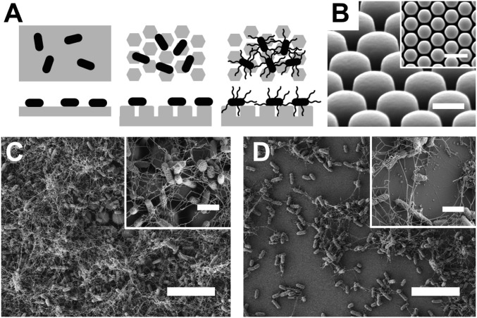
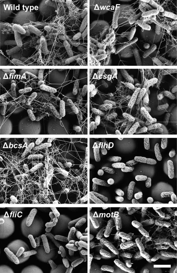
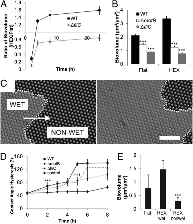
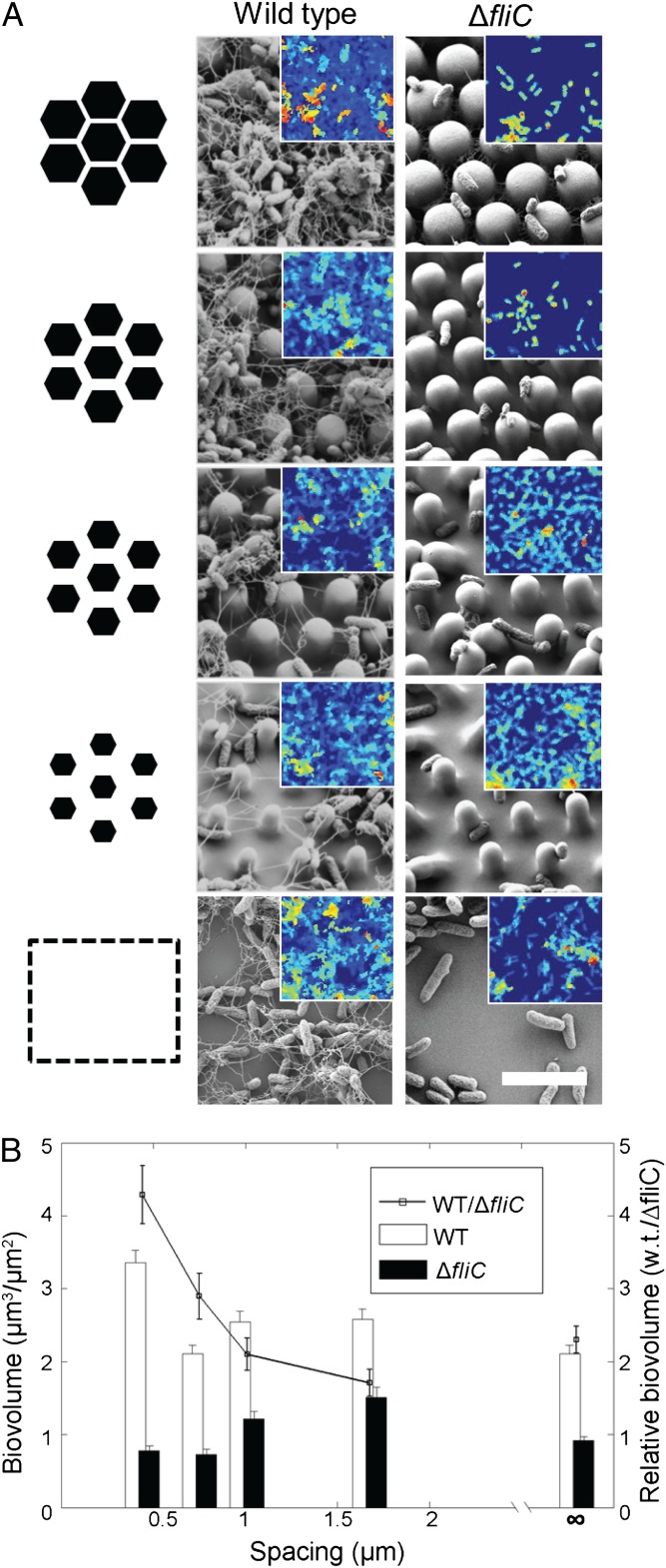
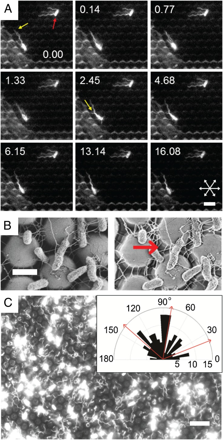
Biofilms, surface-bound communities of microbes, are economically and medically important due to their pathogenic and obstructive properties. Among the numerous strategies to prevent bacterial adhesion and subsequent biofilm formation, surface topography was recently proposed as a highly nonspecific method that does not rely on small-molecule antibacterial compounds, which promote resistance. Here, we provide a detailed investigation of how the introduction of submicrometer crevices to a surface affects attachment of Escherichia coli. These crevices reduce substrate surface area available to the cell body but increase overall surface area. We have found that, during the first 2 h, adhesion to topographic surfaces is significantly reduced compared with flat controls, but this behavior abruptly reverses to significantly increased adhesion at longer exposures. We show that this reversal coincides with bacterially induced wetting transitions and that flagellar filaments aid in adhesion to these wetted topographic surfaces. We demonstrate that flagella are able to reach into crevices, access additional surface area, and produce a dense, fibrous network. Mutants lacking flagella show comparatively reduced adhesion. By varying substrate crevice sizes, we determine the conditions under which having flagella is most advantageous for adhesion. These findings strongly indicate that, in addition to their role in swimming motility, flagella are involved in attachment and can furthermore act as structural elements, enabling bacteria to overcome unfavorable surface topographies. This work contributes insights for the future design of antifouling surfaces and for improved understanding of bacterial behavior in native, structured environments.


Conflict of interest statement

The authors declare no conflict of interest.

Figures

Fig. 1.
Fig. 1.
Bacterial surface adhesion. (A) Schematics of en face (Upper) and cross-sectional (Lower) views of rod-like bacteria adhering to flat (Left) or patterned (Center) substrates and attachment of bacteria possessing surface appendages to a patterned substrate (Right), when the length scale of surface topography is on the order of the bacterial diameter. (B) Scanning EM of a HEX PDMS substrate. (Scale bar, 2 µm.) Inset is orthogonal view at lower magnification. (Scale bar: Inset, 5 μm.) (C) Scanning EM of wild-type E. coli grown for 24 h at 37 °C in M63+ on a HEX-patterned PDMS substrate. Inset is higher magnification. (D) E. coli grown on flat PDMS substrate. Inset is higher magnification. (Scale bars: in C and D, 10 μm; Inset in C and D, 2 µm.)
Fig. 2.
Fig. 2.
Phenotypes of biofilm-associated knockouts on patterned PDMS. Wild-type (ZK2686) and mutant derivatives (as labeled) were grown on topographically patterned PDMS substrates for 24 or 48 h at 37 °C in M63+. Scanning electron micrographs depict the morphological properties of each strain. (Scale bar, 2 μm.)
Fig. 3.
Fig. 3.
Colonization of patterned substrates by wild-type and nonmotile bacteria and its relationship to surface wetting. (A) Biovolume on patterned (HEX) relative to flat substrates for wild-type and ΔfliC cells at various times. (B) Biovolume of cells adherent to submerged flat or HEX-patterned PDMS coupons after 24 h. Error bars indicate standard error of the mean of at least five independent experiments (five z stacks per experiment). ***P < 0.001 by Student’s two-tailed t test, compared with WT. (C) Phase-contrast images of the advancement of the wetting front during culture, at 4 h. The meniscus (dotted line) advances through the patterned substrate, exposing channels between surface features, thus increasing available surface area for bacterial attachment. The area to the Left of the dotted line is fully wetted, whereas the area to the Right of the line contains air pockets. The white arrow indicates the direction of the wetting front progression. Thirty minutes have elapsed between the images on the Left and Right. (Scale bar, 20 µm.) For full movie, see Movie S1. (D) Contact angle hysteresis measurements of substrates that have been exposed to growing E. coli cells for increasing incubation periods or M63+ medium only, followed by sonication. Error bars represent SD. ***P < 0.001 by Student’s two-tailed t test, comparing WT to control at 2 h. †††P < 0.001 by Student’s two-tailed t test, comparing WT to ∆fliC or ∆motB. See also Fig. S1. (E) Biovolume of cells adherent to submerged substrates after 2 h of culture. HEX substrates were either force-wet using ethanol followed by rinsing (HEX-wet), or left in their nonwetting state (HEX-nonwet). Error bars represent SD. ***P < 0.001 by Student’s two-tailed t test, comparing HEX-wet to HEX-nonwet.
Fig. 4.
Fig. 4.
Differential response of wild-type and ΔfliC cells to changes in surface feature spacing. (A) Schematic of underlying surface topography, illustrating increasing spacing with constant pitch (left column) and the scanning EM and confocal images of wild type and ΔfliC cells (center and right columns) grown for 24 h on corresponding PDMS substrates and then fixed. Samples were imaged in the hydrated state using confocal microscopy, and then dehydrated and imaged using scanning EM. Scanning EM images are shown, with representative thickness maps derived from confocal z stacks shown in the corresponding Inset (color mapping is for clarity and has arbitrary scale). (Scale bar, 5 µm.) (B) Biovolume was quantified for each topographical pattern and normalized to projected surface area. Biovolumes are shown for wild type and ΔfliC mutants (plotted as bars), as well as their ratios (black squares connected by lines). Error bars indicate SEM of ≥26 data points.
Fig. 5.
Fig. 5.
Flagellar appendages “reach” and “grasp” to improve surface adhesion on patterned surfaces. (A) Selected frames from a video of Alexa 594-stained cells taken at 15 frames per second. Times are given (in seconds) in each panel. In frame 1, note that the upper cell (red arrow) is fully adherent (it remains stationary throughout the frames). Notably, its middle flagellum is nestled between the surface features slightly out of focus, because it is below the imaging plane. The other two flagella are resting atop the surface features in the focal plane. The lower bacterium is in the early steps of adhesion, tethered by one flagellum (yellow arrow), also nestled between the surface features. Its remaining free flagella continue to rotate rapidly until 2.45 s, at which time another (short) flagellum makes surface contact (yellow arrow). The cell body continues to slowly reorient as it makes more intimate contact (via other flagella and/or pili) and settles in its final position at 13.14 s. (Scale bar, 5 μm.) Axes of symmetry of the substrate are indicated by arrows in the bottom-right image. For full movie, see Movie S2. (B) Scanning EM images of the same field of wild-type E. coli grown on HEX PDMS posts. Images were acquired with Everhart-Thornley (Left) and in-lens detectors (Right). The difference in shadowing between the two images highlights the depth of penetration of the flagellar filaments into the channels between the surface features. Note specifically the region indicated by the red arrow. (Scale bar, 2 μm.) (C) Image of Alexa 594-stained cells after initial adhesion to HEX substrate. (Scale bar, 10 μm.) Inset shows angular histogram of filament orientations of adherent E. coli. The histogram illustrates preferential alignment of filaments along the two of the three planes of symmetry of the hexagonal surface pattern. Axes of symmetry of the substrate are indicated with red arrows.

References

    1. Bos R, van der Mei HC, Busscher HJ. Physico-chemistry of initial microbial adhesive interactions—its mechanisms and methods for study. FEMS Microbiol Rev. 1999;23(2):179–230. - PubMed
    1. Costerton JW, Stewart PS, Greenberg EP. Bacterial biofilms: A common cause of persistent infections. Science. 1999;284(5418):1318–1322. - PubMed
    1. Aslam S, Darouiche RO. Role of antibiofilm-antimicrobial agents in controlling device-related infections. Int J Artif Organs. 2011;34(9):752–758. - PMC - PubMed
    1. Regev-Shoshani G, Ko M, Miller C, Av-Gay Y. Slow release of nitric oxide from charged catheters and its effect on biofilm formation by Escherichia coli. Antimicrob Agents Chemother. 2010;54(1):273–279. - PMC - PubMed
    1. Park KD, et al. Bacterial adhesion on PEG modified polyurethane surfaces. Biomaterials. 1998;19(7–9):851–859. - PubMed

Publication types