. 2013 Apr 2;110(14):E1301-10.
doi: 10.1073/pnas.1210353110. Epub 2013 Mar 18.

Met synergizes with p53 loss to induce mammary tumors that possess features of claudin-low breast cancer

Affiliations

Met synergizes with p53 loss to induce mammary tumors that possess features of claudin-low breast cancer

Jennifer F Knight et al. Proc Natl Acad Sci U S A. .

Abstract

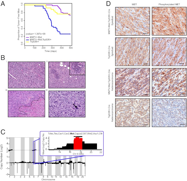
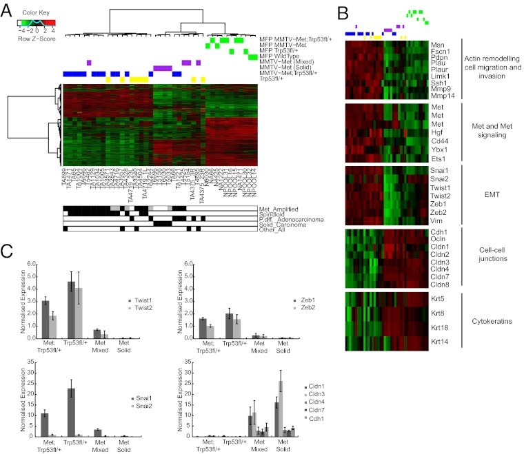
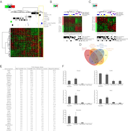
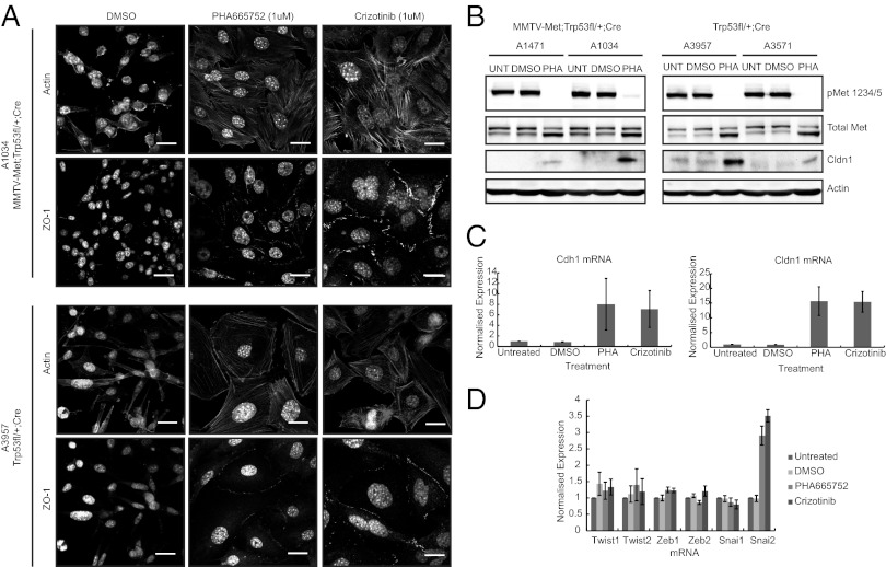
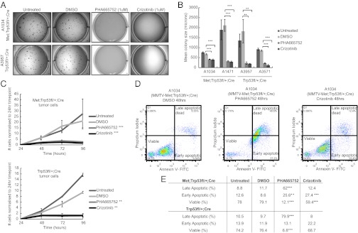
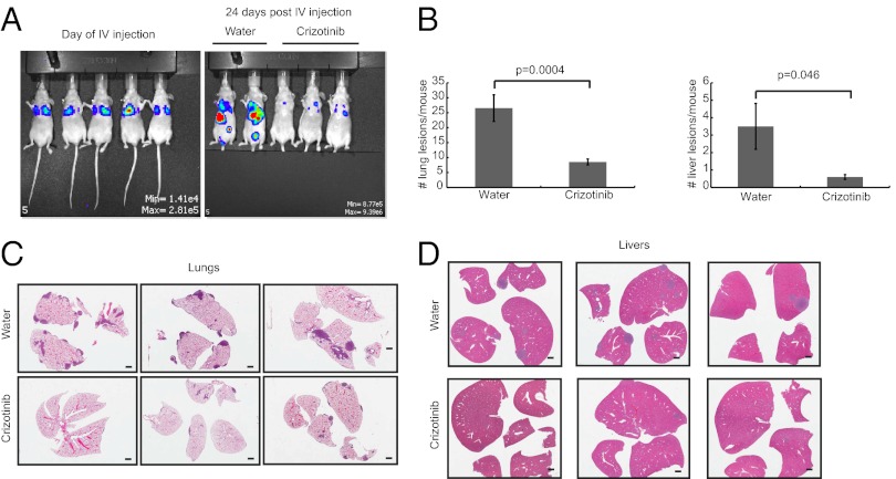
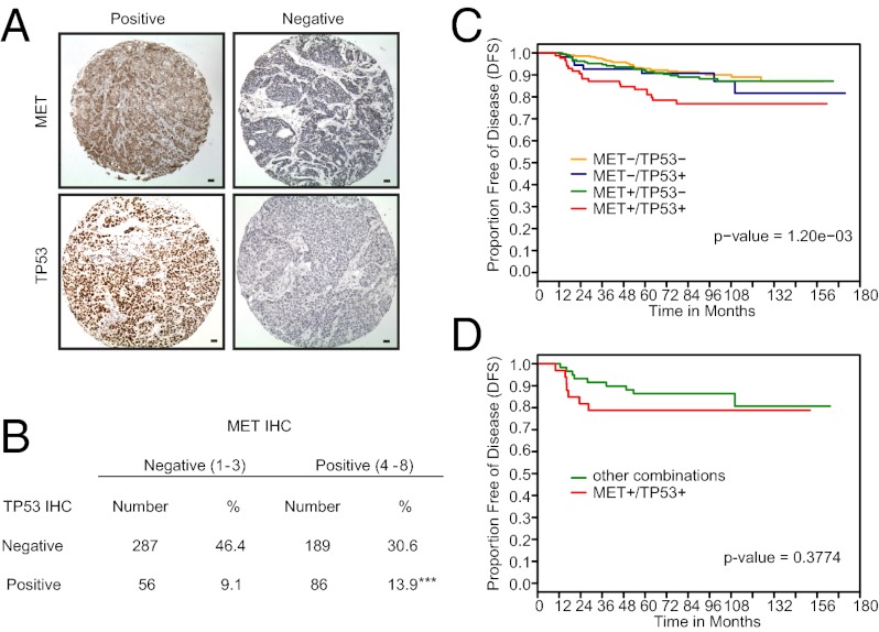
Triple-negative breast cancer (TNBC) accounts for ∼20% of cases and contributes to basal and claudin-low molecular subclasses of the disease. TNBCs have poor prognosis, display frequent mutations in tumor suppressor gene p53 (TP53), and lack targeted therapies. The MET receptor tyrosine kinase is elevated in TNBC and transgenic Met models (Met(mt)) develop basal-like tumors. To investigate collaborating events in the genesis of TNBC, we generated Met(mt) mice with conditional loss of murine p53 (Trp53) in mammary epithelia. Somatic Trp53 loss, in combination with Met(mt), significantly increased tumor penetrance over Met(mt) or Trp53 loss alone. Unlike Met(mt) tumors, which are histologically diverse and enriched in a basal-like molecular signature, the majority of Met(mt) tumors with Trp53 loss displayed a spindloid pathology with a distinct molecular signature that resembles the human claudin-low subtype of TNBC, including diminished claudins, an epithelial-to-mesenchymal transition signature, and decreased expression of the microRNA-200 family. Moreover, although mammary specific loss of Trp53 promotes tumors with diverse pathologies, those with spindloid pathology and claudin-low signature display genomic Met amplification. In both models, MET activity is required for maintenance of the claudin-low morphological phenotype, in which MET inhibitors restore cell-cell junctions, rescue claudin 1 expression, and abrogate growth and dissemination of cells in vivo. Among human breast cancers, elevated levels of MET and stabilized TP53, indicative of mutation, correlate with highly proliferative TNBCs of poor outcome. This work shows synergy between MET and TP53 loss for claudin-low breast cancer, identifies a restricted claudin-low gene signature, and provides a rationale for anti-MET therapies in TNBC.


Conflict of interest statement

The authors declare no conflict of interest.

Figures

Fig. 1.
Fig. 1.
MMTV-Metmt;Trp53fl/+;Cre mammary tumors are highly penetrant, have a spindloid pathology, and selectively amplify the endogenous Met locus. A Kaplan-Meier plot illustrates that MMTV-Metmt;Trp53fl/+;Cre (n = 35) and Trp53fl/+;Cre mice (n = 25) have similar tumor onsets (∼300 d), occurring earlier than tumors in MMTV-Metmt mice (n = 52) (∼400 d) (A). However, MMTV-Metmt;Trp53;Cre mice are associated with a significantly higher tumor penetrance (∼70%) compared with Trp53fl/+;Cre mice (∼24%), resulting in a steeper curve (A). Tumor pathology was similar between MMTV-Metmt;Trp53fl/+;Cre and Trp53fl/+;Cre mice, ranging from spindloid to poorly differentiated adenocarcinomas (B). Cells with enlarged nuclei (arrow in B, iv) and large areas of necrosis (outlined in B, iii) were common. Spindloid tumors often contained ducts with atypical morphology (Inset, B, ii). All MMTV-Metmt;Trp53fl/+;Cre tumors contained genomic amplification of Met and adjacent loci, as determined by array-CGH (C), a phenomenon also observed in Trp53fl/+Cre tumors of spindloid pathology but not in Trp53fl/+;Cre adenocarcinomas (Fig. S2). High expression and activation (phosphorylation) of endogenous MET in MMTV-Metmt;Trp53fl/+;Cre and Trp53fl/+;Cre tumors was confirmed by immunostaining (D). A Trp53fl/+;Cre adenocarcinoma without amplification of Met and little activated MET is shown as a comparison (D). (Scale bars, 50 µm.)
Fig. 2.
Fig. 2.
MMTV-Metmt;Trp53fl/+;Cre spindloid tumors display elevated expression of genes associated with a mesenchymal, migratory phenotype and are distinct from MMTV-Metmt mammary tumors. Unsupervised hierarchical clustering identifies three distinct groups. In the first group, 12 MMTV-Metmt;Trp53fl/+;Cre tumors (blue) form a cluster with six Trp53fl/+;Cre tumors (yellow) and one MMTV-Metmt tumor (purple); this cluster represents tumors of predominantly spindloid pathology and with genomic amplification of Met. In the next cluster, poorly differentiated adenocarcinomas (two MMTV-Metmt;Trp53fl/+;Cre and two Trp53fl/+;Cre tumors) cluster with tumors of the MMTV-Metmt model. MMTV-Metmt tumors further segregate into solid and mixed subtypes in accordance with their pathology (14). Normal mammary gland controls (green) form the third cluster. Tumor characterizations below the heat map are represented in white for negative, black for positive, and gray for unknown. “Other_All” refers to tumors of various pathology types; for example, tumor A899 contained regions of spindloid and adenocarcinoma-type pathologies. Genes highly expressed in MMTV-Metmt;Trp53fl/+;Cre and Trp53fl/+;Cre spindloid tumors are associated with cell migration and invasion, signaling through the MET receptor, and EMT (B). Low expression of cell-cell junction markers and moderate expression of epithelial cytokeratins is also observed (B). A number of these genes were validated by qRT-PCR (n = 5 MMTV-Metmt;Trp53fl/+;Cre, 5 Trp53f/+;Cre tumors, 3 MMTV-Metmt mixed tumors, and 3 MMTV-Metmt solid tumors); expression relative to wild-type mammary gland is shown. Error bars, SEM (C).
Fig. 3.
Fig. 3.
Gene and miRNA expression profiles of MMTV-Metmt;Trp53fl/+;Cre and Trp53fl/+;Cre spindloid tumors correlate with those of human claudin-low breast cancer. A cross-species comparison with human breast cancer subtypes reveals that a large proportion of MMTV-Metmt;Trp53fl/+;Cre tumors and Trp53fl/+;Cre tumors cluster with the claudin-low molecular subclass at the level of gene expression (A). Application of a published claudin-low breast cancer gene expression signature to the mouse model data confirmed this association (P < 0.0001) (B) and showed that tumors of spindloid pathology were those that correlated with the signature. Similarly, a significant association in miRNA expression was identified through the application of a human claudin-low miRNA signature to MMTV-Metmt;Trp53fl/+;Cre and Trp53fl/+;Cre tumor data (P = 4 × 10⁻⁴) (C). To further identify genes associated with claudin-low tumor cell biology and to remove genes expressed by cells in the tumor microenvironment, an intersect of genes highly expressed in human claudin-low breast cancers, MMTV-Metmt;Trp53fl/+;Cre and Trp53fl/+;Cre spindloid tumors (compared with MMTV-Metmt tumors) and human basal B (claudin-low) breast cancer cell lines, was generated (D). This comprised 36 genes (E), a selection of which was validated by qRT-PCR (n = 5 MMTV-Metmt;Trp53fl/+;Cre, 5 Trp53f/+;Cre tumors, 3 MMTV-Metmt mixed tumors, and 3 MMTV-Metmt solid tumors), data were normalized to wild-type mammary gland. Error bars, SEM (F).
Fig. 4.
Fig. 4.
Treatment of spindloid tumor cells with pharmacological MET inhibitors leads to reversal of the claudin-low phenotype. MMTV-Metmt;Trp53fl/+;Cre and Trp53fl/+;Cre spindloid tumor cells were treated in vitro with small-molecule inhibitors of MET kinase (PHA665752 [1 μM] or Crizotinib [1 μM]) for 48–72 h. On treatment, cells underwent a distinct morphological change from a mesenchymal to an epithelial-like state (A), which included the formation of cell-cell junctions, as demonstrated by the appearance of cortical actin and localization of ZO-1 at sites of cell-cell contact (A). (Scale bars, 20 μm.) This was also accompanied by elevated levels of Claudin1 protein, as shown by Western blotting (B). Although we also observed an increase in mRNA levels of Claudin1 (Cldn1) and E-cadherin (Cdh1) on Met inhibition (C), there was no corresponding decrease in genes that are well-established as transcriptional drivers of EMT (Twist1/2, Zeb1/2, and Snai1/2) (D). Averaged PCR data for four spindloid tumor cell lines (two MMTV-Metmt;Trp53fl/+;Cre and two Trp53fl/+;Cre lines) are presented. Error bars, SEM.
Fig. 5.
Fig. 5.
Inactivation of MET kinase inhibits the proliferation and survival of Met-amplified spindloid tumor cells. Tumor cells isolated from two MMTV-Metmt;Trp53fl/+;Cre and two Trp53fl/+;Cre spindloid mammary tumors formed smaller colonies in soft agar during a 10-d assay in the presence of MET kinase inhibitors (PHA665752 [1 μM] and Crizotinib [1 μM]); representative images for two cell lines are shown (A). (Scale bars, 1,000 μm.) Reduction in colony size was highly significant in all four cell lines (B). Error bars, SEM. Significantly impaired proliferation resulting from MET inhibition was also demonstrated in a 4-d proliferation assay in which the same cell lines were grown on tissue culture plastic and counted every 24 h (C). Error bars, SEM. To assess any effect on cell viability, cells treated with MET inhibitors for 48 h were stained with Annexin-V and propidium iodide and analyzed by flow cytometry. Representative plots for one MMTV-Metmt;Trp53fl/+;Cre cell line are shown (D), and averaged data for two MMTV-Metmt;Trp53fl/+;Cre and two Trp53fl/+ cell lines are tabulated (E). All four cell lines responded similarly and showed a dramatic increase in the proportion of cells in late-stage apoptosis after treatment with PHA665752 (e.g., 11.7% of MMTV-Metmt;Trp53fl/+;Cre cells were in late apoptosis in the DMSO control vs. 62% in the PHA667572 treatment). The effect of Crizotinib on cell viability was more moderate (only 12.4% of MMTV-Metmt;Trp53fl/+;Cre cells treated with Crizotinib were in late apoptosis). ***P < 0.01; **P < 0.05 (E).
Fig. 6.
Fig. 6.
MET inhibition impairs the metastatic potential of spindloid mammary tumor cells. An MMTV-Metmt;Trp53fl/+;Cre spindloid tumor cell line expressing firefly luciferase was injected i.v. by the tail vein into 35 nude mice (0.5 × 10⁶ cells/mouse). Mice were imaged on the day of injection (A) and twice per week thereafter to monitor the development of metastases. A control group of 15 mice was gavaged daily with water and compared with 20 mice receiving a daily gavage of Crizotinib (45 mg/kg/d). By day 24, control mice showed extensive metastatic burden compared with Crizotinib-treated mice (A). Lungs and livers were harvested from all animals at day 24 and scored histologically for metastatic lesions. Mice treated with Crizotinib showed a significant reduction in the number of lesions detected in both the lungs and liver (B). Representative histology from three control and three Crizotinib-treated mice is shown (C and D).
Fig. 7.
Fig. 7.
Elevated MET expression in human breast cancer is associated with TP53 mutation and combining MET with TP53 positive IHC identifies patients with poor prognosis. A human breast cancer tissue microarray comprising 618 node-negative patients was stained for MET and TP53 (A). Analysis showed that MET-positive tumors were more likely to stain positively for TP53 (indicative of mutated TP53) than MET-negative tumors (B) and that patients with MET-positive–TP53-positive tumors had a significantly worse outcome than patients with either MET or TP53 positivity alone (P = 0.0012) (C). Within TN patients specifically (n = 93), there was a trend toward MET-TP53 copositivity correlating with a poorer outcome (P = 0.3774), with a clear separation from patients with other combinations of MET and TP53 IHC within the first 36 mo after diagnosis.

References

    1. Ferlay, et al. Globocan 2008 v2.0, Cancer Incidence and Mortality Worldwide. IARC CancerBase 10. 2008. Available at https://globocan.iarc.fr.
    1. Sørlie T, et al. Gene expression patterns of breast carcinomas distinguish tumor subclasses with clinical implications. Proc Natl Acad Sci USA. 2001;98(19):10869–10874. - PMC - PubMed
    1. Herschkowitz JI, et al. Identification of conserved gene expression features between murine mammary carcinoma models and human breast tumors. Genome Biol. 2007;8(5):R76. - PMC - PubMed
    1. Brenton JD, Carey LA, Ahmed AA, Caldas C. Molecular classification and molecular forecasting of breast cancer: Ready for clinical application? J Clin Oncol. 2005;23(29):7350–7360. - PubMed
    1. Sorlie T, et al. Repeated observation of breast tumor subtypes in independent gene expression data sets. Proc Natl Acad Sci USA. 2003;100(14):8418–8423. - PMC - PubMed

Publication types

Associated data