. 2013 Apr 2;110(14):5492-7.
doi: 10.1073/pnas.1217745110. Epub 2013 Mar 18.

Conformational plasticity of the type I maltose ABC importer

Affiliations

Conformational plasticity of the type I maltose ABC importer

Simon Böhm et al. Proc Natl Acad Sci U S A. .

Abstract

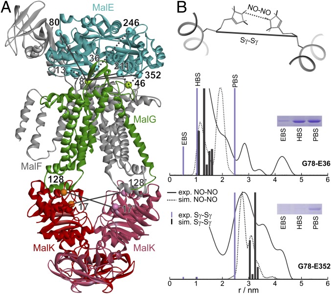
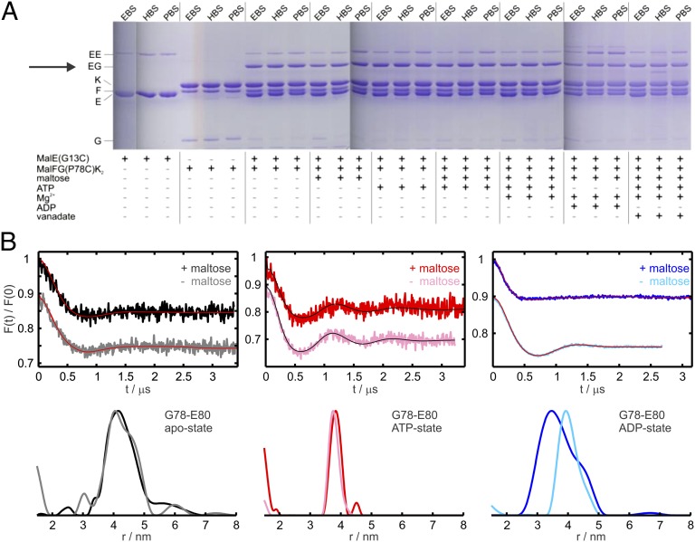
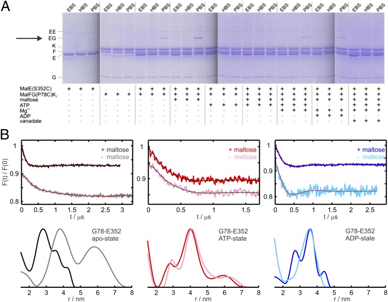
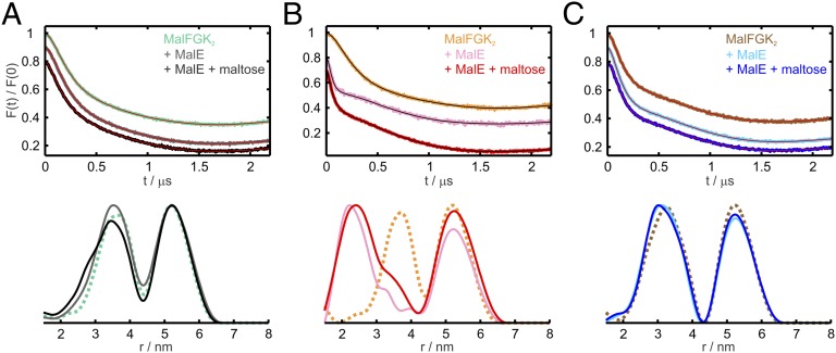
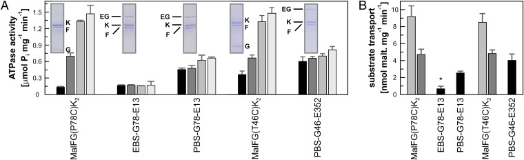
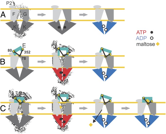
ATP-binding cassette (ABC) transporters couple the translocation of solutes across membranes to ATP hydrolysis. Crystal structures of the Escherichia coli maltose importer (MalFGK2) in complex with its substrate binding protein (MalE) provided unprecedented insights in the mechanism of substrate translocation, leaving the MalE-transporter interactions still poorly understood. Using pulsed EPR and cross-linking methods we investigated the effects of maltose and MalE on complex formation and correlated motions of the MalK2 nucleotide-binding domains (NBDs). We found that both substrate-free (open) and liganded (closed) MalE interact with the transporter with similar affinity in all nucleotide states. In the apo-state, binding of open MalE occurs via the N-lobe, leaving the C-lobe disordered, but upon maltose binding, closed MalE associates tighter to the transporter. In both cases the NBDs remain open. In the presence of ATP, the transporter binds both substrate-free and liganded MalE, both inducing the outward-facing conformation trapped in the crystal with open MalE at the periplasmic side and NBDs tightly closed. In contrast to ATP, ADP-Mg(2+) alone is sufficient to induce a semiopen conformation in the NBDs. In this nucleotide-driven state, the transporter binds both open and closed MalE with slightly different periplasmic configurations. We also found that dissociation of MalE is not a required step for substrate translocation since a supercomplex with MalE cross-linked to MalG retains the ability to hydrolyze ATP and to transport maltose. These features of MalE-MalFGK2 interactions highlight the conformational plasticity of the maltose importer, providing insights into the ATPase stimulation by unliganded MalE.


Conflict of interest statement

The authors declare no conflict of interest.

Figures

Fig. 1.
Fig. 1.
DEER and cross-linking distance restraints vs. MalFGK2-E structure. (A) Apo-state crystal structure [Protein Data Bank (PDB) 3PV0]. The Cα atoms of the residues replaced by cysteines in this study are shown in ball representation. Some pairs were chosen for both cross-linking and DEER measurements (solid lines), others only for cross-linking (dotted lines). (B) Experimental DEER (straight black line) and cross-linking (violet bar) distances obtained with the pairs G78-E36 and G78-E352 (the letters denote the subunit of the site) compared with the simulated nitroxide–nitroxide (NO–NO, dotted line) and sulfur–sulfur (Sγ–Sγ, black bar) distances simulated with MMM using spin-labeled rotamers (MTSL library at 175 K) on the structure 3PV0. The insets show the corresponding bands in the gel.
Fig. 2.
Fig. 2.
Cross-linking and DEER data between MalFG(P78C)K2 and sites in the N-lobe of MalE. (A) MalFG(P78C)K2 in DDM micelles (2.5 µM) was incubated with MalE(G13C) (5 µM) and homobifunctional thiosulfonate cross-linkers (1 mM) in the presence of different cofactors as indicated. The cross-linked products were subsequently analyzed by SDS/PAGE. The cross-linkers EBS, HBS, and PBS have approximate spacer lengths of 5.2 Å, 10.4 Å, and 24.7 Å (see SI Materials and Methods for more details). (B) DEER analysis on MalFGK2 transporters solubilized in n-Dodecyl β-D-maltoside carrying the spin label at position 78 in MalG incubated with MalE spin-labeled at the N-lobe (position 80) in different nucleotide states in the presence and absence of maltose, as indicated. (Upper) Normalized DEER Form factors F(t). (Lower) Distance distributions obtained with Tikhonov regularization using the software DeerAnalysis2011 (35). The mutants are denoted by the name of the relative subunit (e.g., G or E) and the corresponding residue number.
Fig. 3.
Fig. 3.
Cross-linking and DEER data between MalFG(P78C)K2 and sites in the C-lobe of MalE. (A) MalFG(P78C)K2 in DDM micelles (2.5 µM) was incubated with MalE(S352C) (5 µM) and homobifunctional thiosulfonate cross-linkers (1 mM) in the presence of different cofactors as indicated. (B) DEER analysis on MalFGK2 transporters solubilized in DDM carrying the spin label at position 78 in MalG incubated with MalE spin-labeled at the C-lobe (position 352) in different nucleotide states in the presence and absence of maltose, as indicated. (Upper) Normalized DEER Form factors F(t). (Lower) Distance distributions obtained with Tikhonov regularization using the software DeerAnalysis2011 (35). The mutants are denoted by the name of the relative subunit (e.g., G or E) and the corresponding residue number.
Fig. 4.
Fig. 4.
MalE and maltose effects on MalK2 interspin distances in different nucleotide states. (Upper) Normalized DEER Form factors F(t). (Lower) Distance distributions obtained with Tikhonov regularization using the software DeerAnalysis2011 (35) on MalFGK2 transporters solubilized in DDM spin-labeled at positions 17 and 128 in MalK. The short distance peak (< 4.5 nm) represents the 17–128′ and 128–17′ contributions. (A) Apo-state of the MalFGK2 transporters alone (turquoise) and after incubation with wild-type MalE in the absence (gray) and in the presence (black) of maltose. (B) Analogous DEER analysis performed in the presence of 10 mM AMP–PNP and 10 mM MgCl2. MalFGK2 transporters without MalE/maltose (orange) and incubated with WT–MalE in the absence (pink) and presence (red) of maltose. (C) Analogous DEER analysis performed in the presence of 10 mM ADP, 10 mM MgCl2. MalFGK2 transporters without MalE/maltose (brown) and incubated with WT–MalE in the absence (cyan) and in the presence (blue) of maltose. For clarity, the distance distribution obtained with the MalFGK2 transporters alone are presented as dotted lines. The primary data and the distances simulated with MMM on the corresponding crystal structures are shown in Fig. S5.
Fig. 5.
Fig. 5.
ATPase and transport activities of cross-linked MalE–FGK2 supercomplexes. (A) ATPase activity of MalFG(P78C), EBS-, or PBS-linked MalFG(P78C)–MalE(G13C) (named EBS-G78-E13, PBS-G78-E13) in the presence of increasing concentration of freely diffusible MalE(G13C) and of MalFG(T46C) and PBS-linked MalFG(T46C)–MalE(S352C) (named PBS-G46-E13) in the presence of increasing concentration of freely diffusible MalE(S352C). (B) [14C]maltose transport of reconstituted MalFG(P78C) and MalFG(T46C) with added MalE(G13C) and MalE(S352C), respectively, at molar ratios 10:1 (gray) or 1:1 (dark gray) and of the supercomplexes EBS-G78-E13, PBS-G78-E13, and PBS-G46-E352. The asterisks in the EBS-linked supercomplex denote the observed basal maltose incorporation, with no time-dependent increase. Each value is the mean of three separate determinations with SD shown as error bars.
Fig. 6.
Fig. 6.
Model for MalE–MalFGK2 interaction during the nucleotide cycle. (A) MalFGK2 alone during nucleotide cycle. Only ADP–Mg2+ is shown to affect the relative displacement of the NBDs. (B) MalFGK2-E conformations during nucleotide cycle in the absence of maltose. The spin-labeled positions 78 in MalG, 80 in MalE (N-lobe), and 352 in MalE (C-lobe) are highlighted by yellow circles. (C) Nucleotide cycle in MalFGK2-E associated with maltose translocation. The two experimentally detected ADP-states are presented as posthydrolytic states after translocation of maltose (open MalE) and upon loading of a new maltose-loaded closed MalE. The available X-ray structures of MalFGK2 alone in the apo-state (PDB 3FH6) and MalFGK2-E in the pretranslocation state (3PV0) and in the AMP–PNP-state (3PUY) are presented behind the corresponding schematic models.

References

    1. Davidson AL, Dassa E, Orelle C, Chen J. Structure, function, and evolution of bacterial ATP-binding cassette systems. Microbiol Mol Biol Rev. 2008;72(2):317–364. - PMC - PubMed
    1. Moussatova A, Kandt C, O’Mara ML, Tieleman DP. ATP-binding cassette transporters in Escherichia coli. Biochim Biophys Acta. 2008;1778(9):1757–1771. - PubMed
    1. Vasiliou V, Vasiliou K, Nebert DW. Human ATP-binding cassette (ABC) transporter family. Hum Genomics. 2009;3(3):281–290. - PMC - PubMed
    1. Eitinger T, Rodionov DA, Grote M, Schneider E. Canonical and ECF-type ATP-binding cassette importers in prokaryotes: Diversity in modular organization and cellular functions. FEMS Microbiol Rev. 2011;35(1):3–67. - PubMed
    1. Rees DC, Johnson E, Lewinson O. ABC transporters: The power to change. Nat Rev Mol Cell Biol. 2009;10(3):218–227. - PMC - PubMed

Publication types

MeSH terms

LinkOut - more resources