. 2013 Apr 9;110(15):E1407-15.
doi: 10.1073/pnas.1219217110. Epub 2013 Mar 18.

Identification of a previously undescribed divergent virus from the Flaviviridae family in an outbreak of equine serum hepatitis

Affiliations

Identification of a previously undescribed divergent virus from the Flaviviridae family in an outbreak of equine serum hepatitis

Sanjay Chandriani et al. Proc Natl Acad Sci U S A. .

Abstract

Theiler's disease is an acute hepatitis in horses that is associated with the administration of equine blood products; its etiologic agent has remained unknown for nearly a century. Here, we used massively parallel sequencing to explore samples from a recent Theiler's disease outbreak. Metatranscriptomic analysis of the short sequence reads identified a 10.5-kb sequence from a previously undescribed virus of the Flaviviridae family, which we designate "Theiler's disease-associated virus" (TDAV). Phylogenetic analysis clusters TDAV with GB viruses of the recently proposed Pegivirus genus, although it shares only 35.3% amino acid identity with its closest relative, GB virus D. An epidemiological survey of additional horses from three separate locations supports an association between TDAV infection and acute serum hepatitis. Experimental inoculation of horses with TDAV-positive plasma provides evidence that several weeks of viremia preceded liver injury and that liver disease may not be directly related to the level of viremia. Like hepatitis C virus, the best characterized Flaviviridae species known to cause hepatitis, we find TDAV is capable of efficient parenteral transmission, engendering acute and chronic infections associated with a diversity of clinical presentations ranging from subclinical infection to clinical hepatitis.


Conflict of interest statement

Conflict of interest statement: A.L.K., S.C., P.S.-C., T.J.D., and B.C.T. are coinventors on a patent application relating to the results herein. A.L.K., S.C., P.S.-C., D.E.G., and W.Z. are employees of the Novartis Institutes for BioMedical Research.

Figures

Fig. 1.
Fig. 1.
Overview of a Theiler's disease outbreak. Twenty-two horses on Farm A suspected of exposure to botulinum toxin were prophylactically treated with i.v. equine antibotulinum toxin hyperimmune plasma. Five horses received antitoxin from one source (gray horses, antitoxin 1), whereas 17 horses received an independently sourced batch (black horses, antitoxin 2). Fifteen horses followed in this study went untreated (white horses, untreated). Within 8 wk of antitoxin administration, 8 horses treated with antitoxin 2 showed signs of acute hepatitis (yellow boxes). All other horses were clinically asymptomatic (no boxes).
Fig. 2.
Fig. 2.
Genome of Theiler's disease-associated virus (TDAV). (A) Schematic of the TDAV genome. Protein cleavage sites are putative and were annotated on the basis of homologous inference from HCV (Fig. S1). The “*” gene corresponds to HCV p7, GBV-B p13, GBV-D X, and GBV-A 21 kDa protein (19). (B) Coverage map of sequencing reads from horse A1, horse A2, and antitoxin 2. (C) Median calculated amplicon size based on the distance between the 5′ ends of paired-end sequencing reads mapping to each nucleotide. (D) Overlapping clones recovered and sequenced to confirm the genome assembly (black) and the location of amplicons used in the TDAV detection assay (green; primer pairs labeled below). (E) Pairwise amino acid percentage of identity plot (100-aa windows) of TDAV compared with HCV-gt1, NPHV, and GBV-D. Genome position scale (Lower) refers to A–E.
Fig. 3.
Fig. 3.
Phylogenetic analysis of Flaviviridae based on polyprotein sequences. Multiple-sequence alignments (available upon request) were generated using MUSCLE (39) and a neighbor-joining phylogenetic tree with 100 bootstrap replicates was generated in MEGA5 (40). TDAV is in boldface type for emphasis. All branches are numbered starting at “*” and incremented clockwise (see Table S1 for key). Closer branch groups are labeled with a range of sequence numbers; a representative member of a group is listed in parentheses with a thicker line illustrating the branch for the representative member. WNV, West Nile virus; DENV, Dengue virus 1; DV, Donggang virus; YFV, Yellow fever virus; MV, Modoc virus; TBEV, Tick-borne encephalitis virus; KRV, Kamiti River virus; TBV, Tamana bat virus; BVDV, Bovine viral diarrhea virus 1; TDAV, Theiler's disease-associated virus; GBV-(A-D), GB virus (A-D); HCV, Hepatitis C virus genotype 1; NPHV, NPHV #1 (AFJ20709.1). “” indicates nearly identical branches 8 and 9, corresponding to Tembusu virus and Duck flavivirus TA, respectively. Species text color denotes genus classification: Pegivirus (blue), Hepacivirus (orange), Pestivirus (green), and Flavivirus (black). The NPHVs and TDAV [gray and black (in boldface type), respectively] have yet to be classified into a genus.
Fig. 4.
Fig. 4.
Quantitative PCR-based assay to detect TDAV in different cohorts. Cycles to threshold (Ct) from the qRT-PCR TDAV assay (primers EVT-146/147) are plotted on an inverted y axis; a lower Ct value represents a greater viral load. (A) Serum/plasma from horses on Farms A, B, and D with antitoxin treatment status indicated. All samples were analyzed in parallel with a second qRT-PCR TDAV assay (primers EVT-162/163). Those samples in which a qPCR signal was absent by the second assay are denoted as “confirmed negative” by gray circles. (B) Antitoxin 2-treated horses from Farm A were categorized as “clinical” when they had characteristic signs of acute hepatitis including icterus, lethargy, and anorexia and when there were elevations in the serum activity of at least three of the tested liver enzymes (AST, GGT, SDH, and GLDH). Horses classified as “subclinical” had no overt clinical manifestations of acute hepatitis but had significant elevations in the serum activity of at least three of the tested liver enzymes. Asymptomatic cases had neither clinical signs of acute hepatitis nor abnormalities in the serum activities of the tested liver enzymes.
Fig. 5.
Fig. 5.
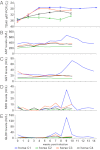
TDAV viral load and liver enzyme function in animals experimentally inoculated with antitoxin 2. (A) qRT-PCR quantification of TDAV RNA. Cycles to threshold (Ct) values from the qRT-PCR assay [average values and SD (n = 4) with primers EVT-146/147] are plotted on an inverted y axis; a lower Ct value represents a greater viral load. (B–E) Biochemical tests for AST, GGT, SDH, and GLDH. Samples were taken at time 0 and weekly for 10–14 wk after inoculation with 500 mL of the same lot of antitoxin 2 linked to the serum hepatitis outbreak. One horse became symptomatic (horse C1, blue), whereas the other three did not (horse C2, green; horse C3, orange; and horse C4, black).
Fig. 6.
Fig. 6.
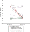
TDAV can cause chronic infection. TDAV was monitored in paired samples harvested during the outbreak and approximately 1 y later. Cycles to threshold (Ct) from the qRT-PCR TDAV assay (primers EVT-146/147) are plotted on an inverted y axis; a lower Ct value represents a greater viral load. The qRT-PCR assays were performed in parallel on paired samples, permitting direct comparison of Ct values.
Fig. P1.
Fig. P1.
Discovery of Theiler’s disease-associated virus (TDAV). Within 8 wk of prophylactic treatment with a batch of antibotulinum toxin, 8 of 17 horses showed signs of acute hepatitis (yellow boxes). RNA was extracted from 2 of the most clinically ill horses and an aliquot of the antitoxin was then analyzed by high-throughput sequencing. Horse-derived reads (black sequences) were filtered from the dataset of more than 60 million reads from these samples, and virus-derived reads (red sequences) were found among the remaining nonhorse sequences in the dataset. De novo assembly of the detected viral reads revealed that all three samples shared a nearly identical 10.5-kb viral genome sequence, Theiler’s disease-associated virus (TDAV), corresponding to a highly divergent member of the Flaviviridae viral family.

References

    1. Kahn CM, Line S, editors. 2011. Merck Veterinary Manual (Merck, Whitehouse Station, NJ), 9th Ed. Available at www.merckvetmanual.com/mvm/index.jsp?cfile=htm/bc/22802.htm.
    1. Theiler A. 1919. Acute liver-atrophy and parenchymatous hepatitis in horses. The Fifth and Sixth Reports of the Director of Veterinary Research, April, 1918. Department of Agriculture, Union of South Africa (The Government Printing and Stationery Office, Pretoria, Union of South Africa), pp. 7–164.
    1. Panciera RJ. Serum hepatitis in the horse. J Am Vet Med Assoc. 1969;155(2):408–410. - PubMed
    1. Hjerpe CA. Serum hepatitis in the horse. J Am Vet Med Assoc. 1964;144:734–740. - PubMed
    1. Aleman M, Nieto JE, Carr EA, Carlson GP. Serum hepatitis associated with commercial plasma transfusion in horses. J Vet Intern Med. 2005;19(1):120–122. - PubMed

Publication types

Associated data