

Uncoupling RARA transcriptional activation and degradation clarifies the bases for APL response to therapies
- PMID: 23509325
- PMCID: PMC3620357
- DOI: 10.1084/jem.20122337
Uncoupling RARA transcriptional activation and degradation clarifies the bases for APL response to therapies
Abstract
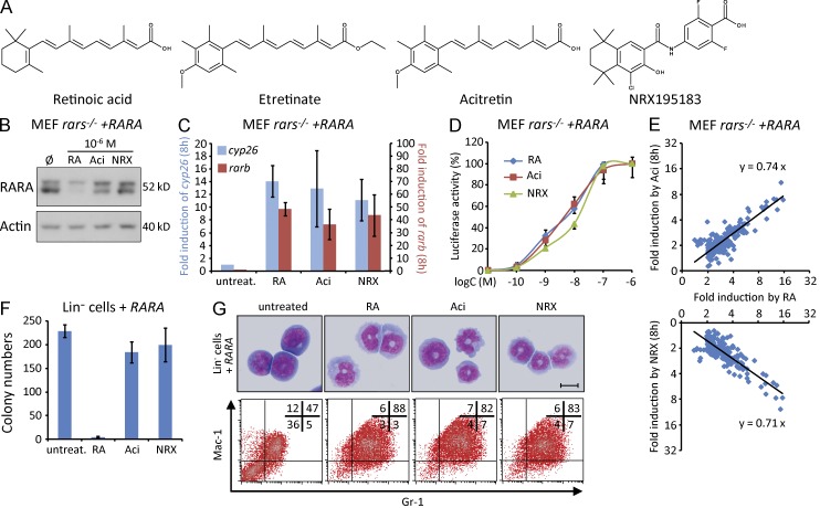
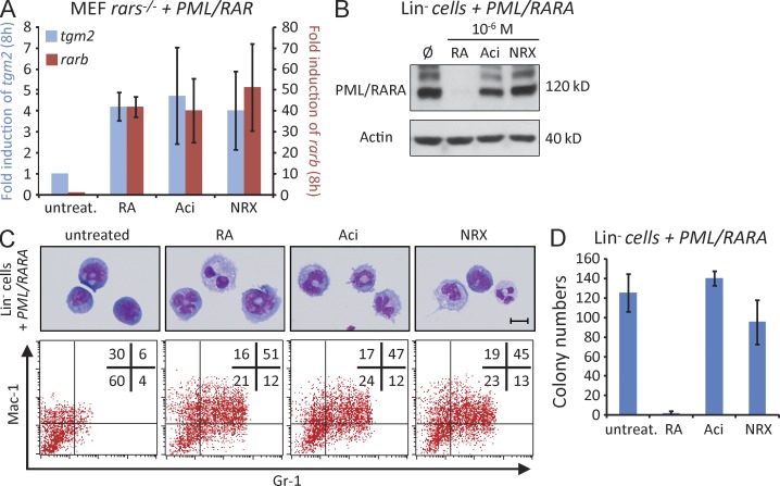
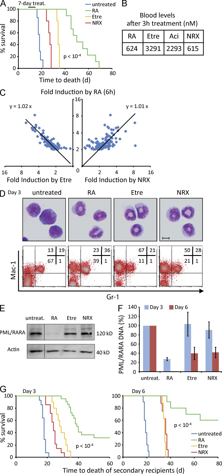
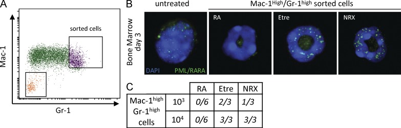
In PML/RARA-driven acute promyelocytic leukemia (APL), retinoic acid (RA) induces leukemia cell differentiation and transiently clears the disease. Molecularly, RA activates PML/RARA-dependent transcription and also initiates its proteasome-mediated degradation. In contrast, arsenic, the other potent anti-APL therapy, only induces PML/RARA degradation by specifically targeting its PML moiety. The respective contributions of RA-triggered transcriptional activation and proteolysis to clinical response remain disputed. Here, we identify synthetic retinoids that potently activate RARA- or PML/RARA-dependent transcription, but fail to down-regulate RARA or PML/RARA protein levels. Similar to RA, these uncoupled retinoids elicit terminal differentiation, but unexpectedly fail to impair leukemia-initiating activity of PML/RARA-transformed cells ex vivo or in vivo. Accordingly, the survival benefit conferred by uncoupled retinoids in APL mice is dramatically lower than the one provided by RA. Differentiated APL blasts sorted from uncoupled retinoid-treated mice retain PML/RARA expression and reinitiate APL in secondary transplants. Thus, differentiation is insufficient for APL eradication, whereas PML/RARA loss is essential. These observations unify the modes of action of RA and arsenic and shed light on the potency of their combination in mice or patients.
Figures
Similar articles
-
Activation of a promyelocytic leukemia-tumor protein 53 axis underlies acute promyelocytic leukemia cure.Nat Med. 2014 Feb;20(2):167-74. doi: 10.1038/nm.3441. Epub 2014 Jan 12. Nat Med. 2014. PMID: 24412926
-
Novel treatment of acute promyelocytic leukemia: As₂O₃, retinoic acid and retinoid pharmacology.Curr Pharm Biotechnol. 2013;14(9):849-58. doi: 10.2174/1389201015666140113095812. Curr Pharm Biotechnol. 2013. PMID: 24433507 Review.
-
PML-RARA-RXR oligomers mediate retinoid and rexinoid/cAMP cross-talk in acute promyelocytic leukemia cell differentiation.J Exp Med. 2004 Apr 19;199(8):1163-74. doi: 10.1084/jem.20032226. J Exp Med. 2004. PMID: 15096541 Free PMC article.
-
Clearance of PML/RARA-bound promoters suffice to initiate APL differentiation.Blood. 2014 Dec 11;124(25):3772-80. doi: 10.1182/blood-2014-03-561852. Epub 2014 Sep 25. Blood. 2014. PMID: 25258343
-
Function of PML-RARA in Acute Promyelocytic Leukemia.Adv Exp Med Biol. 2024;1459:321-339. doi: 10.1007/978-3-031-62731-6_14. Adv Exp Med Biol. 2024. PMID: 39017850 Review.
Cited by
-
Active Pin1 is a key target of all-trans retinoic acid in acute promyelocytic leukemia and breast cancer.Nat Med. 2015 May;21(5):457-66. doi: 10.1038/nm.3839. Epub 2015 Apr 13. Nat Med. 2015. PMID: 25849135 Free PMC article.
-
The Promise of Retinoids in the Treatment of Cancer: Neither Burnt Out Nor Fading Away.Cancers (Basel). 2023 Jul 8;15(14):3535. doi: 10.3390/cancers15143535. Cancers (Basel). 2023. PMID: 37509198 Free PMC article. Review.
-
Synergy against PML-RARa: targeting transcription, proteolysis, differentiation, and self-renewal in acute promyelocytic leukemia.J Exp Med. 2013 Dec 16;210(13):2793-802. doi: 10.1084/jem.20131121. J Exp Med. 2013. PMID: 24344243 Free PMC article. Review.
-
Pin1 inhibition exerts potent activity against acute myeloid leukemia through blocking multiple cancer-driving pathways.J Hematol Oncol. 2018 May 30;11(1):73. doi: 10.1186/s13045-018-0611-7. J Hematol Oncol. 2018. PMID: 29848341 Free PMC article.
-
Non-genetic heterogeneity, altered cell fate and differentiation therapy.EMBO Mol Med. 2021 Mar 5;13(3):e12670. doi: 10.15252/emmm.202012670. Epub 2021 Feb 8. EMBO Mol Med. 2021. PMID: 33555144 Free PMC article. Review.
References
-
- Ablain J., Nasr R., Bazarbachi A., de Thé H. 2011. The drug-induced degradation of oncoproteins: an unexpected Achilles’ heel of cancer cells? Cancer Discovery. 1:117–127 10.1158/2159-8290.CD-11-0087 - DOI - PubMed
-
- Altucci L., Rossin A., Hirsch O., Nebbioso A., Vitoux D., Wilhelm E., Guidez F., Schiavone E.M., Grimwade D., Zelent A., et al. 2005. Rexinoid-triggered differentiation and tumor-selective apoptosis of acute myeloid leukemia by protein kinase A-mediated desubordination of retinoid X receptor. Cancer Res. 65:8754–8765 10.1158/0008-5472.CAN-04-3569 - DOI - PubMed
-
- Breitman T.R., Collins S.J., Keene B.R. 1981. Terminal differentiation of human promyelocytic leukemic cells in primary culture in response to retinoic acid. Blood. 57:1000–1004 - PubMed
Publication types
MeSH terms
Substances
LinkOut - more resources
Full Text Sources
Other Literature Sources
