. 2013 Feb;10(1):1-9.
doi: 10.1513/AnnalsATS.201206-029OC.

Fasting 2-deoxy-2-[18F]fluoro-D-glucose positron emission tomography to detect metabolic changes in pulmonary arterial hypertension hearts over 1 year

Affiliations

Fasting 2-deoxy-2-[18F]fluoro-D-glucose positron emission tomography to detect metabolic changes in pulmonary arterial hypertension hearts over 1 year

Erika L Lundgrin et al. Ann Am Thorac Soc. 2013 Feb.

Abstract

Background: The development of tools to monitor the right ventricle in pulmonary arterial hypertension (PAH) is of clinical importance. PAH is associated with pathologic expression of the transcription factor hypoxia-inducible factor (HIF)-1α, which induces glycolytic metabolism and mobilization of proangiogenic progenitor (CD34(+)CD133(+)) cells. We hypothesized that PAH cardiac myocytes have a HIF-related switch to glycolytic metabolism that can be detected with fasting 2-deoxy-2-[(18)F]fluoro-d-glucose positron emission tomography (FDG-PET) and that glucose uptake is informative for cardiac function.

Methods: Six healthy control subjects and 14 patients with PAH underwent fasting FDG-PET and echocardiogram. Blood CD34(+)CD133(+) cells and erythropoietin were measured as indicators of HIF activation. Twelve subjects in the PAH cohort underwent repeat studies 1 year later to determine if changes in FDG uptake were related to changes in echocardiographic parameters or to measures of HIF activation.

Measurements and results: FDG uptake in the right ventricle was higher in patients with PAH than in healthy control subjects and correlated with echocardiographic measures of cardiac dysfunction and circulating CD34(+)CD133(+) cells but not erythropoietin. Among patients with PAH, FDG uptake was lower in those receiving β-adrenergic receptor blockers. Changes in FDG uptake over time were related to changes in echocardiographic parameters and CD34(+)CD133(+) cell numbers. Immunohistochemistry of explanted PAH hearts of patients undergoing transplantation revealed that HIF-1α was present in myocyte nuclei but was weakly detectable in control hearts.

Conclusions: PAH hearts have pathologic glycolytic metabolism that is quantitatively related to cardiac dysfunction over time, suggesting that metabolic imaging may be useful in therapeutic monitoring of patients.


Figures
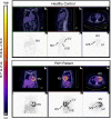
Figure 1.
Figure 1.
Fasting 2-deoxy-2-[18F]fluoro-d-glucose positron emission tomography (FDG-PET) imaging of control participant and patient with pulmonary arterial hypertension (PAH). Fasting FDG-PET imaging reveals generalized enhanced FDG standardized uptake value in the PAH heart. IVS = interventricular septum; LV = left ventricle; RV = right ventricle.
Figure 2.
Figure 2.
Baseline fasting relative 2-deoxy-2-[18F]fluoro-d-glucose standardized uptake value (FDG-SUV) in pulmonary arterial hypertension (PAH) and control hearts. Box plot representation of the relative FDG-SUV at baseline in four heart regions: right atrium (RA), right ventricle (RV), left ventricle (LV), and interventricular septum (IVS). P values reported are from the two-sided rank-sums test. Fasting relative FDG-SUV is increased in PAH RV and RA compared with control subjects.
Figure 3.
Figure 3.
Changes in fasting relative 2-deoxy-2-[18F]fluoro-d-glucose standardized uptake value (FDG-SUV) over 1 year by clinical status classification. Fasting relative FDG-SUV of patients with pulmonary arterial hypertension (PAH) is shown at baseline and at 1 year in four heart regions: (right atrium [RA], right ventricle [RV], left ventricle [LV], and interventricular septum [IVS]), by clinical status classification (improved, stable, or worsened). P values reported are from the signed rank test. Each dashed line represents an independent PAH patient. *WHO functional class III-IV patient who was transplanted a month after the 1-year visit.
Figure 4.
Figure 4.
Fasting relative 2-deoxy-2-[18F]fluoro-d-glucose standardized uptake value (FDG-SUV) relates to circulating CD34+CD133+ cells in pulmonary arterial hypertension (PAH). Each open circle represents an independent patient with PAH. At baseline, there is a positive relationship between right atrium (RA) and right ventricle (RV) FDG-SUV and % circulating CD34+CD133+ cells. However, percent change in circulating progenitors over 1 year negatively correlates to percent change in relative FDG-SUV of the RV and LV. IVS = interventricular septum.
Figure 5.
Figure 5.
Hypoxia-inducible factor (HIF)-1α protein expression in pulmonary arterial hypertension (PAH) plexiform lesions and cardiac myocytes. Arrows point to a few of the many brown-staining nuclei of PAH pulmonary endothelial cells (B) and cardiac myocytes (D). Nuclear localization was confirmed by sequential sections of heart in which nuclei were identified by DAPI (blue) immunofluorescence (E, F). Scale bar: 25 μm. Ctrl = control; RV = right ventricle.
Figure 6.
Figure 6.
Relative 2-deoxy-2-[18F]fluoro-d-glucose (FDG) uptake of right ventricle (RV) and right atrium (RA) relates to echocardiographic parameters in pulmonary arterial hypertension (PAH). Each open circle represents an independent patient with PAH at the baseline visit.
Figure 7.
Figure 7.
Percent changes in fasting relative 2-deoxy-2-[18F]fluoro-d-glucose standardized uptake value (FDG-SUV) over 1 year correlates with changes in echocardiographic disease severity measures. Each open circle represents an independent patient with pulmonary arterial hypertension. LV = left ventricle; PR = pulmonary regurgitation; RA = right atrium; RV = right ventricle.

References

    1. Firth AL, Mandel J, Yuan J X-J. Idiopathic pulmonary arterial hypertension. Dis Model Mech. 2010;3:268–273. - PMC - PubMed
    1. Humbert M, Sitbon O, Chaouat A, Bertocchi M, Habib G, Gressin V, Yaïci A, Weitzenblum E, Cordier JF, Chabot F, et al. Survival in patients with idiopathic, familial, and anorexigen-associated pulmonary arterial hypertension in the modern management era. Circulation. 2010;122:156–163. - PubMed
    1. Toblli JE, Lombraña A, Duarte P, Di Gennaro F. Intravenous iron reduces NT-pro-brain natriuretic peptide in anemic patients with chronic heart failure and renal insufficiency. J Am Coll Cardiol. 2007;50:1657–1665. - PubMed
    1. Sztrymf B, Souza R, Bertoletti L, Jaïs X, Sitbon O, Price LC, Simonneau G, Humbert M. Prognostic factors of acute heart failure in patients with pulmonary arterial hypertension. Eur Respir J. 2010;35:1286–1293. - PubMed
    1. WXisneski JA, Gertz EW, Neese RA, Mayr M. Myocardial metabolism of free fatty acids: studies with 14C-labeled substrates in humans. J Clin Invest. 1987;79:359–366. - PMC - PubMed

Publication types

MeSH terms