

Gastric duplication cyst: two case reports and review of the literature
- PMID: 23509656
- PMCID: PMC3590563
- DOI: 10.1155/2013/605059
Gastric duplication cyst: two case reports and review of the literature
Abstract
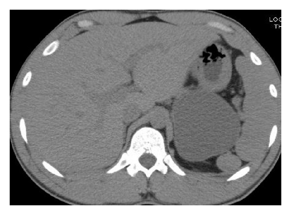
Background. Duplication of the alimentary tract is a rare congenital anomaly. Gastric duplication cysts (GDCs) represent 4% of all alimentary tract duplications, and approximately 67% manifest within the first year of life. Duplication cysts in adults are generally encountered as incidental findings at endoscopy or laparotomy. Herein, we report two rare cases of symptomatic GDC presenting in adults. Case 1. A 27-year-old male presented with a five-month history of back pain. Exam revealed mild epigastric tenderness with a vague palpable mass in left upper abdomen. CT scan showed 8 × 7.4 × 6 cm homogenous, nonseptated cystic mass posterosuperior to pancreatic tail. On laparotomy, a cystic mass measuring 11 × 8 cm was found, which was densely adherent to posterior wall of stomach suggestive of GDC. Case 2. A 28-year-old woman presented with epigastric pain associated with vomiting for 2 months. Exam revealed mild epigastric tenderness. CT scan showed four cystic lesions in the medial wall of distal stomach measuring approximately one cm each suggestive of duplication cysts. Exploratory laparotomy with antrectomy and truncal vagotomy with Billroth II reconstruction were performed. Pathology in both patients was diagnostic of GDC. Conclusion. GDC is a rare anomaly, and its presentation in adults is even rarer.
Figures


References
-
- Maeda H, Okabayashi T, Nishimori I, et al. Diagnostic challenge to distinguish gastric duplication cyst from pancreatic cystic lesions in adult. Internal Medicine. 2007;46(14):1101–1104. - PubMed
-
- Johnston J, Wheatley GH, El Sayed HF, Marsh WB, Ellison EC, Bloomston M. Gastric duplication cysts expressing carcinoembryonic antigen mimicking cystic pancreatic neoplasms in two adults. American Surgeon. 2008;74(1):91–94. - PubMed
-
- Kim DH, Kim JS, Nam ES, Shin HS. Foregut duplication cyst of the stomach. Pathology International. 2000;50(2):142–145. - PubMed
LinkOut - more resources
Full Text Sources
Other Literature Sources
