. 2013:2013:509714.
doi: 10.1155/2013/509714. Epub 2012 Dec 27.

RNase P-associated external guide sequence effectively reduces the expression of human CC-chemokine receptor 5 and inhibits the infection of human immunodeficiency virus 1

Affiliations

RNase P-associated external guide sequence effectively reduces the expression of human CC-chemokine receptor 5 and inhibits the infection of human immunodeficiency virus 1

Wenbo Zeng et al. Biomed Res Int. 2013.

Abstract

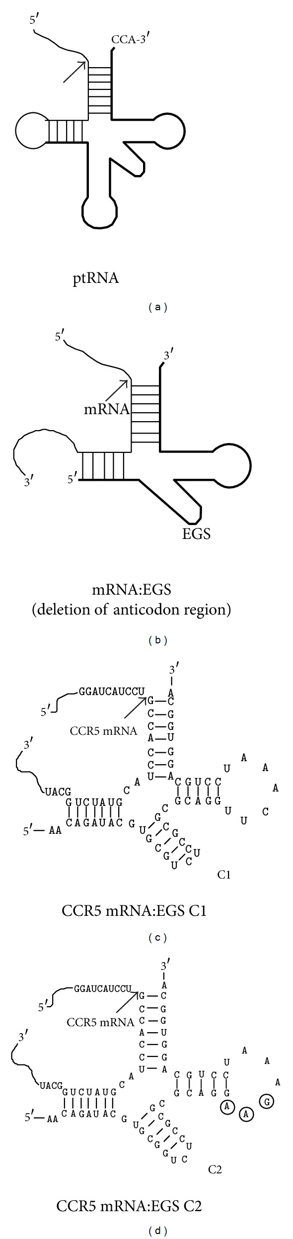
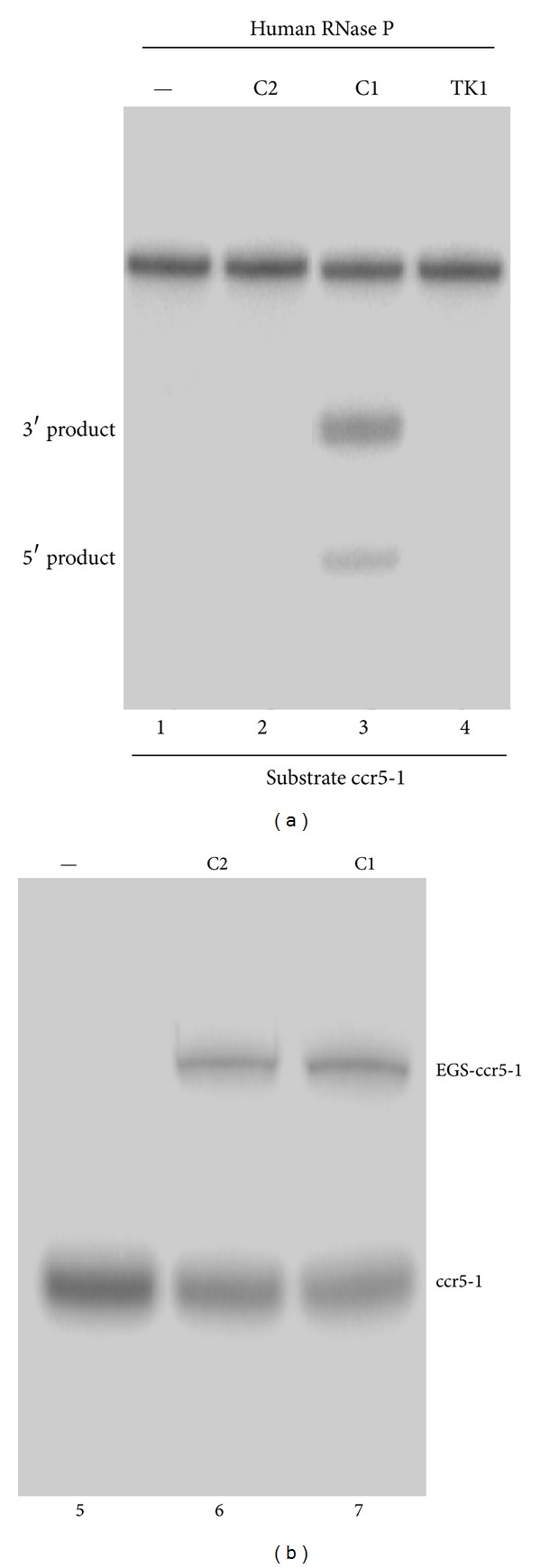
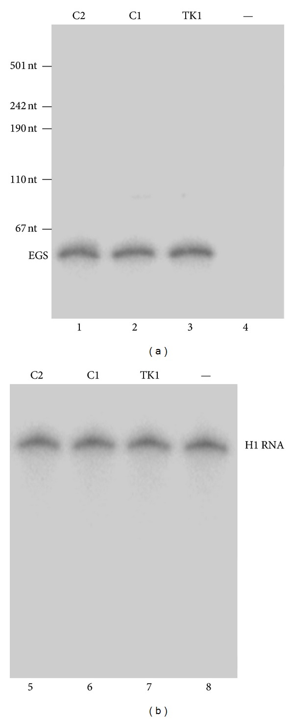
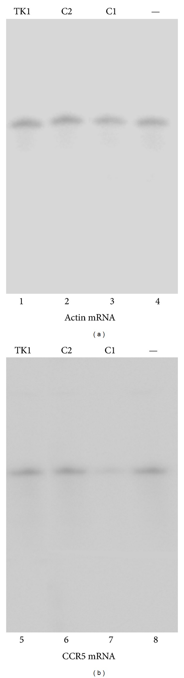
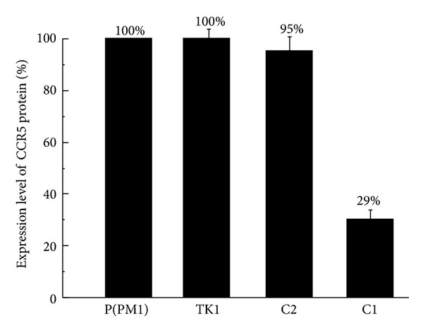
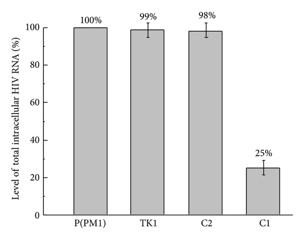
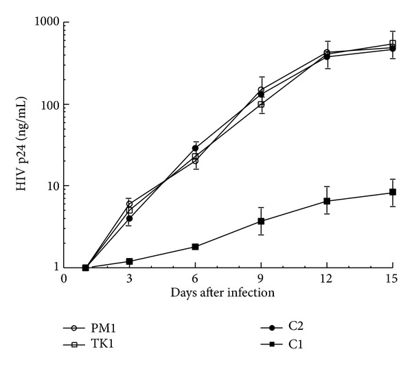
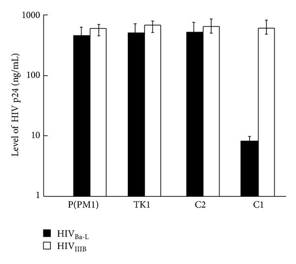
External guide sequences (EGSs) represent a new class of RNA-based gene-targeting agents, consist of a sequence complementary to a target mRNA, and render the target RNA susceptible to degradation by ribonuclease P (RNase P). In this study, EGSs were constructed to target the mRNA encoding human CC-chemokine receptor 5 (CCR5), one of the primary coreceptors for HIV. An EGS RNA, C1, efficiently directed human RNase P to cleave the CCR5 mRNA sequence in vitro. A reduction of about 70% in the expression level of both CCR5 mRNA and protein and an inhibition of more than 50-fold in HIV (R5 strain Ba-L) p24 production were observed in cells that expressed C1. In comparison, a reduction of about 10% in the expression of CCR5 and viral growth was found in cells that either did not express the EGS or produced a "disabled" EGS which carried nucleotide mutations that precluded RNase P recognition. Furthermore, the same C1-expressing cells that were protected from R5 strain Ba-L retained susceptibility to X4 strain IIIB, which uses CXCR4 as the coreceptor instead of CCR5, suggesting that the RNase P-mediated cleavage induced by the EGS is specific for the target CCR5 but not the closely related CXCR4. Our results provide direct evidence that EGS RNAs against CCR5 are effective and specific in blocking HIV infection and growth. These results also demonstrate the feasibility to develop highly effective EGSs for anti-HIV therapy.


Figures

Figure 1
Figure 1
Schematic representation of substrates for RNase P. (a) A natural substrate (ptRNA). (b) A hybridized complex of a target RNA (e.g., mRNA) and a EGS that resembles the structure of a tRNA. (c) and (d) Complexes between CCR5 mRNA sequence and EGS C1 and C2, respectively. The sequences of C1 and C2 that were equivalent to the T-stem and loop and variable region of a tRNA molecule were derived from tRNAser [19]. Only the exact sequence of the CCR5 mRNA around the targeting site was shown. The EGS sequence is shown in bold. The site of cleavage by RNase P is marked with an arrowhead. The three nucleotides that are mutated in C2 are in circles.
Figure 2
Figure 2
(a) Cleavage of CCR5 mRNA sequence (substrate ccr5-1) by human RNase P in the presence of different EGSs. No EGS was added to the reaction mixture in lane 1. 1 nM of the EGS C2 (lane 2), C1 (lane 3), and TK1 (lane 4) was incubated with [32P]-labeled CCR5 mRNA substrate (20 nM) and human RNase P (2 units) at 37°C in a volume of 10 μL for 15 minutes in buffer A (50 mM Tris, pH 7.4, 100 mM NH4Cl, and 10 mM MgCl2). (b) In vitro binding of [32P]-labeled substrate ccr5-1 and EGSs. Substrate ccr5-1 at a concentration of 0.1 nM was incubated either alone (lane 5) or in the presence of 2 nM EGS C1 (lane 7) and C2 (lane 6) in buffer B for 15 min to allow binding and then loaded on a nondenaturing polyacrylamide gel. Experimental details can be found in Section 2.
Figure 3
Figure 3
The expression of EGS RNAs in cultured cells. Northern analyses were carried out using nuclear RNA fractions isolated from parental PM1 cells (—, lanes 4 and 8) and a cloned cell line that expressed C2 (lanes 1 and 5), C1 (lanes 2 and 6), and TK1 (lanes 3 and 7). Equal amounts of each RNA sample (30 μg) were separated on 2% agarose gels that contained formaldehyde, transferred to nitrocellulose membranes, and hybridized to a [32P]-radiolabeled probe that contained the DNA sequence coding for EGS C1 (lanes 1–4) or H1 RNA (lanes 5–8), the RNA subunit of human RNase P and a nuclear RNA [15]. The hybridized products corresponding to the full-length retroviral transcripts (~6 kb), transcribed from the LTR promoter, are at the top of the gel and are not shown.
Figure 4
Figure 4
Levels of CCR5 mRNA. Northern analyses were carried out using RNA isolated from parental PM1 cells (—, lanes 4 and 8) and cell lines that expressed TK1 (lanes 1 and 5), C2 (lanes 2 and 6), and C1 (lanes 3 and 7). Equal amounts of each RNA sample (30 μg) were separated on agarose gels that contained formaldehyde, transferred to nitrocellulose membranes, and hybridized to a [32P]-radiolabeled probe that contained the actin (lanes 1–4) and CCR5 mRNA sequences (lanes 5–8).
Figure 5
Figure 5
Fluorescence-activated cell sorting (FACS) analysis of CCR5 expression at the surface of the parental PM1 cells [P(PM1)] and cells that expressed EGS TK1 (TK1), C2 (C2), and C1 (C1). Quantitation was carried out by labeling the cells with a PE-conjugated anti-CCR5 antibody. The results are expressed in percent of fluorescence compared with the parental PM1 cells, which serve as the control. The values are the means from triplicate experiments. The standard deviation is indicated by the error bars.
Figure 6
Figure 6
Schematic representation of the levels of total (unspliced and spliced) intracellular HIV RNA in HIVBa-L-infected PM1 cells that did not express an EGS [P(PM1)] or expressed EGS C1 (C1), C2 (C2), and TK1 (TK1). The RNA samples were isolated from cells at 48 hours after infection, and the levels of HIV RNA were determined using a real-time PCR assay. The values shown are the averages from three independent experiments.
Figure 7
Figure 7
Growth of HIV-1 in PM1 cells and cell lines that expressed EGS RNAs. 5 × 10⁵ cells were infected with HIV-1 at a MOI of 0.02–0.1. Viral production was determined by a p24 antigen assay as a function of time postinfection. The values are the means from triplicate experiments. The standard deviation is indicated by the error bars.
Figure 8
Figure 8
Schematic representation of the expression levels of supernatant HIV-1 p24 protein in viral infected cells that did not express an EGS [P(PM1)] or expressed EGS C1 (C1), C2 (C2), and TK1 (TK1). The cells were either infected with X4 strain HIVBa-L or R5 strain HIVIIIB. The protein samples were isolated from culture media at 15 days after infection. The levels of p24 protein were measured with an HIV p24 ELISA kit. The values shown are the averages from three independent experiments. Solid bars: HIVBa-L; open bars: HIVIIIB.

Similar articles

Cited by

References

    1. Scherer LJ, Rossi JJ. Approaches for the sequence-specific knockdown of mRNA. Nature Biotechnology. 2003;21(12):1457–1465. - PubMed
    1. Santoro SW, Joyce GF. A general purpose RNA-cleaving DNA enzyme. Proceedings of the National Academy of Sciences of the United States of America. 1997;94(9):4262–4266. - PMC - PubMed
    1. Stein CA, Cheng YC. Antisense oligonucleotides as therapeutic agents—is the bullet really magical? Science. 1993;261(5124):1004–1012. - PubMed
    1. Wong-Staal F, Poeschla EM, Looney DJ. A controlled, Phase 1 clinical trial to evaluate the safety and effects in HIV-1 infected humans of autologous lymphocytes transduced with a ribozyme that cleaves HIV-1 RNA. Human Gene Therapy. 1998;9(16):2407–2425. - PubMed
    1. Zhou J, Rossi JJ. Current progress in the development of RNAi-based therapeutics for HIV-1. Gene Therapy. 2011;18(12):1134–1138. - PMC - PubMed

Publication types