. 2013:2013:801214.
doi: 10.1155/2013/801214. Epub 2012 Dec 23.

Effect of nanoparticles and environmental particles on a cocultures model of the air-blood barrier

Affiliations

Effect of nanoparticles and environmental particles on a cocultures model of the air-blood barrier

Rossella Bengalli et al. Biomed Res Int. 2013.

Abstract

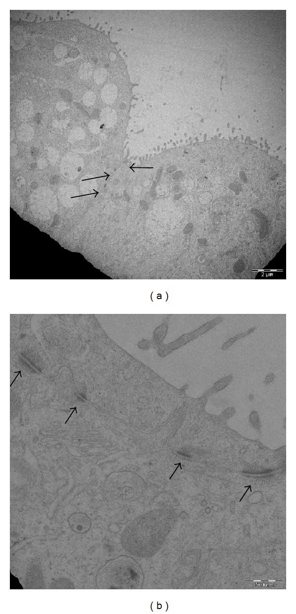
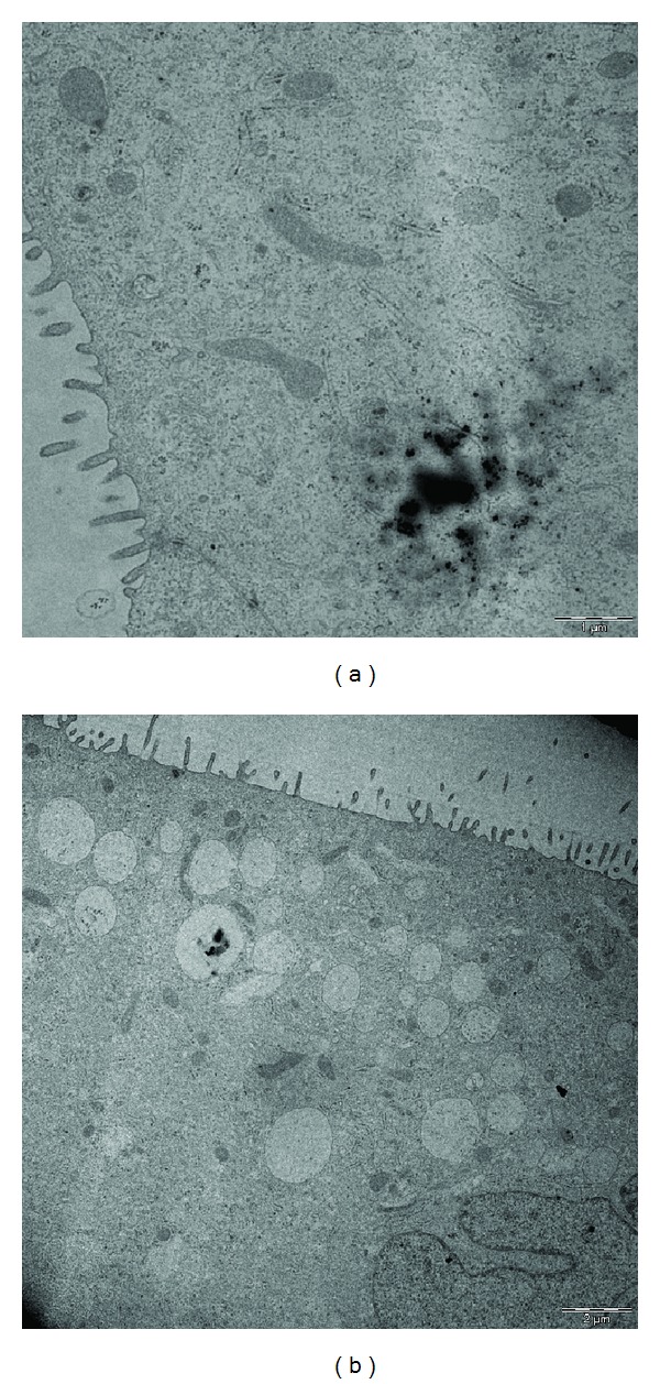
Exposure to engineered nanoparticles (NPs) and to ambient particles (PM) has increased significantly. During the last decades the application of nano-objects to daily-life goods and the emissions produced in highly urbanized cities have considerably augmented. As a consequence, the understanding of the possible effects of NPs and PM on human respiratory system and particularly on the air-blood barrier (ABB) has become of primary interest. The crosstalk between lung epithelial cells and underlying endothelial cells is indeed essential in determining the effects of inhaled particles. Here we report the effects of metal oxides NPs (CuO and TiO2) and of PM on an in vitro model of the ABB constituted by the type II epithelial cell line (NCI-H441) and the endothelial one (HPMEC-ST1.6R). The results demonstrate that apical exposure of alveolar cells induces significant modulation of proinflammatory proteins also in endothelial cells.


Figures

Figure 1
Figure 1
Immunofluorescent staining of the TJ cytoplasmic plaque protein ZO-1 (green) of the NCI-H441 monolayer on day 12, performed as described in Section 2. NCI-h441 differentiated with 1 μM Dexamethasone after day 3 of culture were positively stained for ZO-1 at the cell-cell interface (a) confirming the formation of functional TJs while in cells without Dexamethasone treatment the ZO-1 staining clearly demonstrates the absence of TJs formation; (b) nuclei were stained with DAPI (blue), ZO-1 (green), and actin (red).
Figure 2
Figure 2
(a) and (b) Transmission electron microscope of NCI-H441 cells after 12 days of coculture. The TEM pictures show the formation of tight (↑) and adherens () junctions.
Figure 3
Figure 3
Transepithelial electric resistance (TEER) values of the ABB coculture of NCI-H441 (treated at day 3 with Dexamethasone 1 μM) and HPMEC-ST1.6R. TEER values are expressed as Ω∗cm2. The highest values of TEER were reached after 11–13 days in culture (703 ± 118 Ω∗cm2). Gelatine-coated inserts without cells were used as blank. Data are expressed as means ± S.E. of seven different experiments.
Figure 4
Figure 4
Transepithelial electric resistance (TEER) values of the ABB coculture of NCI-H441 (treated at day 3 with Dexamethasone 1 μM) and HPMEC-ST1.6R. The ABB has been apically treated with PM10 (Figure 4(a)) and metal oxide NPs (Figure 4(b)). TEER values are expressed as Ω∗cm2 and show no significant differences between control and PM10 and TiO2-treated cells. Cells treated with CuO NPs showed a significant reduction in TEER values. ↑ indicates the day of apical treatment with particles. Gelatine-coated inserts without cells were used as blanks. Data are expressed as means ± SE of 3 different experiments. *Statistically different from control P < 0.05, ANOVA.
Figure 5
Figure 5
IL-1β release from the ABB model exposed to Milan summer PM10 for 24 h at day 12 of culture. The release of the interleukin is significantly increased in the basolateral compartment (endothelial cells) after apical exposure of the system (NCI-H441 compartment). *Statistically different from control P < 0.05, ANOVA.
Figure 6
Figure 6
IL-6 and IL-8 release from the ABB model exposed to metal oxides NPs (25 μg/mL) at day 12 of culture. The release of IL-6 is significantly increased by CuO and TiO2 NPs in the apical compartment (a) while IL-8 is significantly increased in the apical compartment only after treatment with CuO NPs. (b) *Statistically different from control P < 0.05, ANOVA.
Figure 7
Figure 7
Transmission electron microscope of NCI-H441 cells after 12 days of cocultures treated with NPs. (a) NCI-H441 cells treated with CuO (25 μg/mL). (b) NCI-H441 cells treated with TiO2 (25 μg/mL). The presence of CuO NPs free in the cytoplasm can be related to the oxidative potential of these particles which determines the rupture of endosomal vesicles as well as to a different route of entry into the cells.

References

    1. Maniatis NA, Kotanidou A, Catravas JD, Orfanos SE. Endothelial pathomechanisms in acute lung injury. Vascular Pharmacology. 2008;49(4–6):119–133. - PMC - PubMed
    1. Crandall ED, Matthay MA, Garfinkel S. Alveolar epithelial transport: basic science to clinical medicine. American Journal of Respiratory and Critical Care Medicine. 2001;163(4):1021–1029. - PubMed
    1. Rogaczewska T, Matczak W. Evaluation of occupational exposure to cadmium based on air analysis of the work area. I. Cadmium oxide level in the air of work areas in a cadmium and nickel cumulator factory. Medycyna Pracy. 1985;36(4):273–279. - PubMed
    1. Weichenthal S, Dufresne A, Infante-Rivard C. Indoor ultrafine particles and childhood asthma: exploring a potential public health concern. Indoor Air. 2007;17(2):81–91. - PubMed
    1. Lanone S, Rogerieux F, Geys J, et al. Comparative toxicity of 24 manufactured nanoparticles in human alveolar epithelial and macrophage cell lines. Particle and Fibre Toxicology. 2009;6, article 14 - PMC - PubMed

LinkOut - more resources