

Positron emission tomography imaging of fibrillar parenchymal and vascular amyloid-β in TgCRND8 mice
- PMID: 23509918
- PMCID: PMC3629748
- DOI: 10.1021/cn300226q
Positron emission tomography imaging of fibrillar parenchymal and vascular amyloid-β in TgCRND8 mice
Abstract
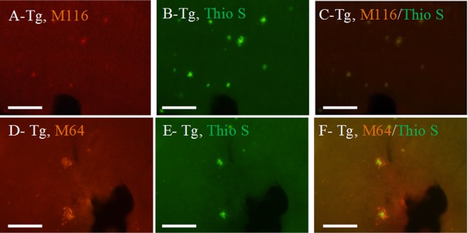
Few quantitative diagnostic and monitoring, tools are available to clinicians treating patients with Alzheimer's disease. Further, many of the promising quantitative imaging tools under development lack clear specificity toward different types of Amyloid-β (Aβ) pathology such as vascular or oligomeric species. Antibodies offer an opportunity to image specific types of Aβ pathology because of their excellent specificity. In this study, we developed a method to translate a panel of anti-Aβ antibodies, which show excellent histological performance, into live animal imaging contrast agents. In the TgCRND8 mouse model of Alzheimer's disease, we tested two antibodies, M64 and M116, that target parenchyma aggregated Aβ plaques and one antibody, M31, that targets vascular Aβ. All three antibodies were administered intravenously after labeling with both poly(ethylene glycol) to enhance circulation and (64)Cu to allow detection via positron emission tomography (PET) imaging. We were clearly able to differentiate TgCRND8 mice from wild type controls by PET imaging using either M116, the anti-Aβ antibody targeting parenchymal Aβ or M31, the antivascular Aβ antibody. To confirm the validity of the noninvasive imaging of specific Aβ pathology, brains were examined after imaging and showed clear evidence of binding to Aβ plaques.
Figures


Similar articles
-
[124I]IBETA: A New Aβ Plaque Positron Emission Tomography Imaging Agent for Alzheimer's Disease.Molecules. 2022 Jul 17;27(14):4552. doi: 10.3390/molecules27144552. Molecules. 2022. PMID: 35889425 Free PMC article.
-
Butyrylcholinesterase-knockout reduces fibrillar β-amyloid and conserves 18FDG retention in 5XFAD mouse model of Alzheimer's disease.Brain Res. 2017 Sep 15;1671:102-110. doi: 10.1016/j.brainres.2017.07.009. Epub 2017 Jul 17. Brain Res. 2017. PMID: 28729192
-
Characterization of IMPY as a potential imaging agent for beta-amyloid plaques in double transgenic PSAPP mice.Eur J Nucl Med Mol Imaging. 2004 Aug;31(8):1136-45. doi: 10.1007/s00259-004-1487-z. Epub 2004 Mar 9. Eur J Nucl Med Mol Imaging. 2004. PMID: 15007564
-
Compounds for imaging amyloid-β deposits in an Alzheimer's brain: a patent review.Expert Opin Ther Pat. 2015 Apr;25(4):413-23. doi: 10.1517/13543776.2015.1007953. Epub 2015 Mar 8. Expert Opin Ther Pat. 2015. PMID: 25746836 Review.
-
Positron emission tomography radiopharmaceuticals for imaging brain Beta-amyloid.Semin Nucl Med. 2011 Jul;41(4):283-99. doi: 10.1053/j.semnuclmed.2011.02.005. Semin Nucl Med. 2011. PMID: 21624562 Review.
Cited by
-
What amyloid ligands can tell us about molecular polymorphism and disease.Neurobiol Aging. 2016 Jun;42:205-12. doi: 10.1016/j.neurobiolaging.2016.03.019. Epub 2016 Mar 24. Neurobiol Aging. 2016. PMID: 27143437 Free PMC article. Review.
-
Synaptic Amyloid-β Oligomers Precede p-Tau and Differentiate High Pathology Control Cases.Am J Pathol. 2016 Jan;186(1):185-98. doi: 10.1016/j.ajpath.2015.09.018. Am J Pathol. 2016. PMID: 26718979 Free PMC article.
-
ImmunoPET Directed to the Brain: A New Tool for Preclinical and Clinical Neuroscience.Biomolecules. 2023 Jan 13;13(1):164. doi: 10.3390/biom13010164. Biomolecules. 2023. PMID: 36671549 Free PMC article. Review.
-
Monoclonal antibodies against Aβ42 fibrils distinguish multiple aggregation state polymorphisms in vitro and in Alzheimer disease brain.J Biol Chem. 2014 Nov 14;289(46):32131-32143. doi: 10.1074/jbc.M114.594846. Epub 2014 Oct 3. J Biol Chem. 2014. PMID: 25281743 Free PMC article.
-
Recent progress in the development of metal complexes as β-amyloid imaging probes in the brain.Medchemcomm. 2017 May 16;8(7):1393-1407. doi: 10.1039/c7md00064b. eCollection 2017 Jul 1. Medchemcomm. 2017. PMID: 30108850 Free PMC article. Review.
References
-
- Hampel H.; Prvulovic D.; Teipel S.; Jessen F.; Luckhaus C.; Froliche L.; Riepe M. W.; Dodel R.; Leyhe T.; Bertram L.; Hoffmann W.; Faltraco F.; German Task Force Alzheimer’s D. (2011) The future of Alzheimer’s disease: The next 10 years. Prog. Neurobiol. 95, 718–728. - PubMed
-
- Brookmeyer R.; Johnson E.; Ziegler-Graham K.; Arrighi H. M. (2007) Forecasting the global burden of Alzheimer’s disease. Alzheimer’s Dementia 3, 186–191. - PubMed
-
- Holtzman J. L. (2010) Are We Prepared to Deal With the Alzheimer’s Disease Pandemic?. Clin. Pharmacol. Ther. 88, 563–565. - PubMed
-
- McKhann G. M.; Knopman D. S.; Chertkow H.; Hyman B. T.; Jack C. R. Jr; Kawas C. H.; Klunk W. E.; Koroshetz W. J.; Manly J. J.; Mayeux R.; Mohs R. C.; Morris J. C.; Rossor M. N.; Scheltens P.; Carrillo M. C.; Thies B.; Weintraub S.; Phelps C. H. (2011) The diagnosis of dementia due to Alzheimer’s disease: Recommendations from the National Institute on Aging-Alzheimer’s Association workgroups on diagnostic guidelines for Alzheimer’s disease. Alzheimer’s Dementia 7, 263–269. - PMC - PubMed
-
- Tanzi R. E.; Bertram L. (2005) Twenty Years of the Alzheimer’s Disease Amyloid Hypothesis: A Genetic Perspective. Cell 120, 545–555. - PubMed
Publication types
MeSH terms
Substances
Grants and funding
LinkOut - more resources
Full Text Sources
Other Literature Sources
