. 2013 Mar 20;4(3):393-407.
doi: 10.1021/cn3001203. Epub 2012 Dec 17.

Curcumin modulates α-synuclein aggregation and toxicity

Affiliations

Curcumin modulates α-synuclein aggregation and toxicity

Pradeep K Singh et al. ACS Chem Neurosci. .

Abstract

In human beings, Parkinson's disease (PD) is associated with the oligomerization and amyloid formation of α-synuclein (α-Syn). The polyphenolic Asian food ingredient curcumin has proven to be effective against a wide range of human diseases including cancers and neurological disorders. While curcumin has been shown to significantly reduce cell toxicity of α-Syn aggregates, its mechanism of action remains unexplored. Here, using a series of biophysical techniques, we demonstrate that curcumin reduces toxicity by binding to preformed oligomers and fibrils and altering their hydrophobic surface exposure. Further, our fluorescence and two-dimensional nuclear magnetic resonance (2D-NMR) data indicate that curcumin does not bind to monomeric α-Syn but binds specifically to oligomeric intermediates. The degree of curcumin binding correlates with the extent of α-Syn oligomerization, suggesting that the ordered structure of protein is required for effective curcumin binding. The acceleration of aggregation by curcumin may decrease the population of toxic oligomeric intermediates of α-Syn. Collectively; our results suggest that curcumin and related polyphenolic compounds can be pursued as candidate drug targets for treatment of PD and other neurological diseases.


Figures

Figure 1
Figure 1
Curcumin binds α-Syn oligomers and reduces their toxicity. (a) SEC profiles of α-Syn incubated with and without curcumin at room temperature (RT) and 37 °C. (b) Curcumin fluorescence emission spectra of oligomer–Cur complex and preformed oligomers incubated with curcumin (Oligo+Cur). Only curcumin (Cur) and oligomers (Oligo) were used as control. (c) MTT reduction by SH-SY5Y neuronal cell in the presence of 5 μM preformed oligomers in presence and absence of 3 μM curcumin. 20 mM MES buffer, pH 6.0 was used as a control. (d) LDH release assay in SH-SY5Y neuronal cells using 5 μM preformed oligomers in presence and absence of 3 μM curcumin. 0.5% triton X-100 was used as positive control, and the buffer was used as negative control. Differentiated SH-SY5Y cells were treated with buffer, oligo and oligo+Cur for 40 h. (e) Oligo treated cells showed more neurite damage and reduced synaptophysin staining compared to oligo+Cur. Scale bars are 50 μm. (f) DAPI staining indicates that the nuclear morphology of oligo+ Cur treated samples were more intact compared to oligo treated cells. Scale bars are 50 μm. (g) The fluorescence intensity of the oxidized 2-hydroethidium showing significantly high intensity in oligomers treated cells compared to control and cells treated with oligomers+curcumin. Scale bars are 50 μm. (h) Flow cytometry analysis showing that reduction of oligomers toxicity in presence of curcumin. Quadrants Q1, Q2, Q3, and Q4 represent dead cells, late apoptotic cells/necrosis, live cells, and early apoptotic cells, respectively. Statistical significance: *P < 0.05, **P < 0.01.
Figure 2
Figure 2
Curcumin modulates morphology of α-Syn oligomers. (a) AFM images of preformed oligomers isolated from SEC. Top left panel showing oligomers directly isolated from SEC. Top right panel showing oligomers morphology of α-Syn–curcumin complex isolated from SEC. Bottom panels showing 30 min incubated α-Syn oligomers in the presence (bottom right) and absence (bottom left) of curcumin. Scale bars are 500 nm. (b) Proteinase K digestion profile of oligomers in the presence and absence of curcumin showing similar extent of proteinase K digestion.
Figure 3
Figure 3
Curcumin binds to α-Syn fibrils and reduces their toxicity. 100 μM preformed α-Syn fibrils were incubated with and without 100 μM curcumin for 20 h, and these samples were used for all the assays. (a) Significant increase in curcumin fluorescence at ∼500 nm was observed after binding to fibrils when excited at 426 nm. (b) CD spectra of preformed α-Syn fibrils without (red) and with curcumin (blue). Both the spectra showed mostly β-sheet conformation without any significant change. (c) CR absorbance at 540 nm after binding to α-Syn fibrils in the presence and absence of curcumin. (d) CR fluorescence at 595 nm showing decreased in CR binding when preformed α-Syn fibrils were incubated in the presence of curcumin. (e) MTT reduction assay using SH-SY5Y cell line by preformed α-Syn fibrils in the presence and absence of curcumin. (f) LDH release assay using SH-SY5Y cells showing less LDH release by fibrils incubated in the presence of curcumin. 0.5% Triton X-100 used as a positive control and showed 100% cell death. (g) AFM morphology of α-Syn fibrils in the presence and absence of curcumin. Left panel shows the distinct fibrillar morphology of α-Syn amyloids, whereas the right panel shows clustered and clumped fibrillar aggregates along with some amorphous species when α-Syn fibrils were incubated with curcumin. Scale bars are 500 nm. (h) Altered proteinase K digestion profile evident from SDS-PAGE analysis showing α-Syn fibrils in the presence of curcumin is more resistant to proteolytic degradation. Statistical significance *P < 0.05; **P < 0.01; NS P > 0.05.
Figure 4
Figure 4
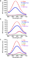
Curcumin reduces the exposed hydrophobic surfaces of oligomeric and fibrilar α-Syn. NR binding to oligomers and fibrils in presence and absence of curcumin was measured by fluorescence. (a) NR binding of α-Syn oligomers obtained from SEC and incubated in presence (Oligo+Cur) and absence (Oligo) of curcumin. The NR fluorescence of curcumin (Cur), NR, and NR+Cur was taken as control. (b) NR binding of Oligomer–curcumin complex (Oligo-Cur) isolated from SEC. Only NR was used as a control. (c) NR binding of α-Syn fibrils in presence and absence curcumin. Curcumin and NR alone showed insignificant fluorescence. All spectra were measured by exciting the solution at 550 nm and emission in the range of 560–720 nm.
Figure 5
Figure 5
Relative binding of curcumin to different α-Syn species. (a) Curcumin fluorescence spectra of varying concentrations of curcumin (1–20 μM) in the presence of 5 μM preformed α-Syn fibrils. (b) CD spectra of 5 μM α-Syn monomers and fibrils in the presence and absence of 20 μM curcumin. (c) Curcumin fluorescence value at λmax (500 nm) in the presence of different α-Syn species with varying concentrations of curcumin showing maximum curcumin binding for fibrils. (d) Comparative increase in curcumin fluorescence at 500 nm in the presence of 5 μM each of the LMW 50 kDa, LMW 100 kDa and monomer showing increase in curcumin binding according to the oligomer order. (e) Double-reciprocal plots of various α-Syn species. (f) Scatchard plot of the α-Syn fibrils–curcumin complex. Curb and Curf indicate bound and free curcumin, respectively.
Figure 6
Figure 6
Probing interaction of curcumin with monomer and LMW 100 kDa α-Syn by NMR spectroscopy. (a) Overlay of 1H–15N HSQC spectra of 150 μM monomeric α-Syn in the absence (red spectrum) and in the presence of 75 μM curcumin (yellow spectrum, top left). Top right panel shows the overlay of the 1H–15N HSQC spectra of 150 μM 100 kDa LMW α-Syn in the absence (black spectrum) and in the presence of 75 μM curcumin (red spectrum, top right). In the HSQC spectrum of 100 kDa LMW, many additional peaks were observed compared to the HSQC spectrum of monomers and were numbered arbitrarily. (b) Difference in chemical shifts (Δδ) of individual amino acids in monomer (red) and 100 kDa LMW (blue) of α-Syn in the presence and absence of curcumin were calculated. The Δδ (in ppm) was plotted against individual amino acid residues. (c) Intensities of all extra peaks in HSQC spectrum of oligomers in presence (I) and absence (I0) of curcumin were calculated, and relative intensities I/I0 were plotted against oligomer numbers (assigned arbitrary).
Figure 7
Figure 7
Curcumin accelerates α-Syn aggregation. (a) Time dependent CD spectra of α-Syn in the presence and absence of curcumin. α-Syn in the presence of curcumin showing accelerated conversion of random coil to β-sheet conformation during aggregation. (b) Electron micrographs of the aggregates after 3 days of incubation showing fibrillar morphology of α-Syn in the absence (left) and presence (right) of curcumin. Altered morphology of α-Syn aggregates were seen in the presence of curcumin. Scale bars are 500 nm. (c) SDS-PAGE analysis showing different proteinase K digestion pattern by α-Syn fibrils formed in the presence and absence of curcumin. (d) Time dependent AFM analysis of α-Syn aggregation in the presence and absence of curcumin showing accelerated conversion of oligomers to fibrils in the presence of curcumin. Scale bars are 700 nm.
Figure 8
Figure 8
Schematic representation showing the effect of curcumin on α-Syn aggregation and toxicity. Curcumin may interact with various oligomeric intermediates of α-Syn and accelerate their conversion to fibrils. Curcumin also binds to preformed oligomers and fibrils and reduces their toxicity.

Similar articles

Cited by

References

    1. Cookson M. R. (2005) The biochemistry of Parkinson’s disease. Annu. Rev. Biochem. 74, 29–52. - PubMed
    1. Lansbury P. T.; Brice A. (2002) Genetics of Parkinson’s disease and biochemical studies of implicated gene products - Commentary. Curr. Opin Cell. Biol. 14, 653–660. - PubMed
    1. Feany M. B. (2000) Studying human neurodegenerative diseases in flies and worms. J. Neuropathol. Exp. Neurol. 59, 847–856. - PubMed
    1. Conway K. A.; Harper J. D.; Lansbury P. T. (1998) Accelerated in vitro fibril formation by a mutant α-synuclein linked to early-onset Parkinson disease. Nat. Med. 4, 1318–1320. - PubMed
    1. Conway K. A.; Lee S. J.; Rochet J. C.; Ding T. T.; Williamson R. E.; Lansbury P. T. (2000) Acceleration of oligomerization, not fibrillization, is a shared property of both a-synuclein mutations linked to early-onset Parkinson’s disease: Implications for pathogenesis and therapy. Proc. Natl. Acad. Sci. U.S.A. 97, 571–576. - PMC - PubMed

Publication types

MeSH terms