. 2013 Jul;104(7):865-70.
doi: 10.1111/cas.12154. Epub 2013 Apr 19.

Suppression of autophagy by CUB domain-containing protein 1 signaling is essential for anchorage-independent survival of lung cancer cells

Affiliations

Suppression of autophagy by CUB domain-containing protein 1 signaling is essential for anchorage-independent survival of lung cancer cells

Takamasa Uekita et al. Cancer Sci. 2013 Jul.

Abstract

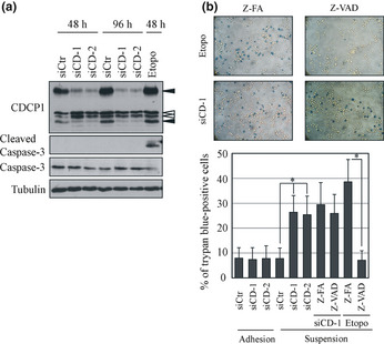
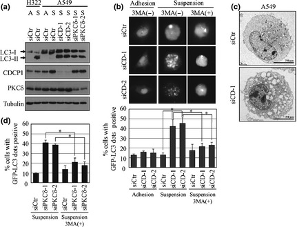
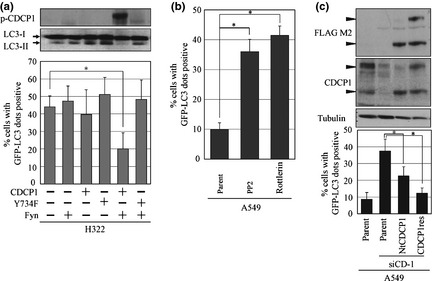
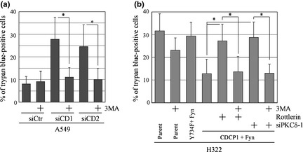
CUB (C1r/C1s, urchin embryonic growth factor, BMP1) domain-containing protein 1 (CDCP1) has been implicated in promoting metastasis of cancer cells through several mechanisms, including the inhibition of anoikis, which is cell death triggered by the loss of extracellular matrix interactions. However, the mechanism inhibiting cell death regulated by CDCP1 remains elusive. Inhibition of CDCP1 expression using small interfering RNA (siRNA) induced the cell death of suspended cancer cells without cleaving caspase-3, a marker of apoptosis; cell death was not inhibited by a general caspase inhibitor, suggesting that the loss of CDCP1 induces caspase-independent cell death. In contrast, knockdown of CDCP1 as well as protein kinase Cδ (PKCδ), a downstream effector of CDCP1, in a suspension culture of lung cancer cells resulted in marked induction of membranous microtubule-associated protein 1 light chain 3 (LC3)-II protein, a hallmark of autophagy, and caused the formation of an autophagosome structure visualized using green fluorescent protein-tagged LC3-II. Expression and phosphorylation of exogenous CDCP1 by Fyn kinase reduced the formation of autophagosomes and inhibited phosphorylation of CDCP1 by PP2, a Src kinase inhibitor or inhibited PKCδ by rottlerin, stimulating autophagosome formation. Moreover, death of suspended lung cancer cells induced by CDCP1 siRNA or by PKCδ siRNA was reduced by the autophagy inhibitor 3-methyladenine. These results indicate that CDCP1-PKCδ signaling plays a critical role in inhibiting autophagy, which is responsible for anoikis resistance of lung cancer cells.


Figures

Figure 1
Figure 1
Cell death induced by CUB (C1r/C1s, urchin embryonic growth factor, BMP1) domain‐containing protein 1 (CDCP1) in lung cancer cells cultured under suspension condition is caspase independent. (a) A549 cells were treated with control (siCtr) or CDCP1 siRNA (siCD‐1 and siCD‐2) and cultured in a low cell‐binding plate (2‐methacryloyloxyethyl phosphorylcholine plate) for 48 or 96 h, respectively. The cell lysate was subjected to western blot analysis with the indicated antibodies. Black arrowheads indicate CDCP1. White arrowheads indicate non‐specific bands. (b) The effect of CDCP1 siRNA, Z‐VADFMK (Z‐VAD) and etoposide (Etopo) on cell death in suspension culture was determined using trypan blue staining and expressed as a percentage. Upper panels represent trypan blue staining of A549 cells treated with siCD‐1 and Z‐FAFMK (Z‐FA) or Z‐VAD and treated with etoposide and Z‐FA or Z‐VAD. Data are the means ± SD from three experiments. *P‐values are based on Student's t‐test and were considered significant for < 0.01.
Figure 2
Figure 2
CUB (C1r/C1s, urchin embryonic growth factor, BMP1) domain‐containing protein 1 (CDCP1) and protein kinase Cδ (PKCδ) inhibit autophagosome formation in lung cancer cells cultured under suspension conditions. (a) A549 cells and H322 cells were treated with control (siCtr), CDCP1 (siCD‐1 and siCD‐2), or PKCδ siRNA (siPKCδ‐1 and siPKCδ‐2) and cultured in a normal cell culture plate or 2‐methacryloyloxyethyl phosphorylcholine plate for 48 h. The cell lysate was subjected to western blot analysis with the indicated antibodies. A, adhesion culture; S, suspension culture. (b) The effect of CDCP1 siRNA with or without 3‐methyladenine (3MA) treatment on autophagosome formation in adhesion or suspension cultures was determined by measuring the green fluorescent protein (GFP)‐LC3 dot‐positive cells as a percentage. The upper panel represents the GFPLC3 dot‐positive A549 cells treated with siCD‐1 and siCD‐2 with or without 3MA treatment. The lower panel represents the percentage of GFPLC3 dot‐positive cells. (c) Electron micrographs showing the structure of A549 cells treated with control siRNA (siCtr) or CDCP1 siRNA‐1 (siCD‐1). (d) The effect of PKCδ siRNA with or without 3MA treatment on autophagosome formation in suspension culture of A549 cells was determined by measuring GFPLC3 dot‐positive cells as a percentage. Data are the means ± SD from three experiments. *P‐values based on Student's t‐test were considered significant for < 0.01.
Figure 3
Figure 3
Phosphorylation of CUB (C1r/C1s, urchin embryonic growth factor, BMP1) domain‐containing protein 1 (CDCP1) inhibits autophagy in suspension culture. (a) H322 cells overexpressing CDCP1 or CDCP1 mutant (Y734F) and Fyn tagged with HA (Fyn) were used to measure autophagosome formation. Note that phosphorylated CDCP1, which is present following overexpression of CDCP1 and Fyn, reduced the number of LC3‐II protein and green fluorescent protein (GFP)‐LC3 dot‐positive cells. (b) A549 cells were treated with or without PP2 or rottlerin in suspension culture and autophagy was detected by GFPLC3 dot‐positive cells. (c) A549 cells stably expressed N‐terminus‐truncated CDCP1 (NtCDCP1) or full‐length CDCP1 rescue mutant (CDCP1res) were treated with CDCP1 siRNA and autophagy was detected by the number of GFPLC3 dot‐positive cells. Black arrowheads indicate CDCP1. Data are the means ± SD from three experiments. *P‐values were determined using the Student's t‐test and considered significant for < 0.01.
Figure 4
Figure 4
CUB (C1r/C1s, urchin embryonic growth factor, BMP1) domain‐containing protein 1 (CDCP1)‐protein kinase Cδ (PKCδ) signaling regulates cancer cell survival by inhibiting autophagy. (a) The effect of the control or CDCP1 siRNA with or without 3‐methyladenine (3MA) treatment on cell death in suspension culture was determined by measuring cells staining positive for trypan blue as a percentage. (b) H322 cells overexpressing CDCP1 or CDCP1 mutant (Y734F) and Fyn tagged with HA (Fyn) were used to measure cell death in suspension culture. The effect of rottlerin and PKCδ siRNA (siPKCδ‐1) with or without 3MA treatment on cell death of H322 cells overexpressing CDCP1 and Fyn in suspension culture was determined by measuring trypan blue‐positive cells as a percentage. Data are the means ± SD from three experiments. *P‐values were determined using Student's t‐test and were considered significant for < 0.01.

References

    1. Hooper JD, Zijlstra A, Aimes RT et al Subtractive immunization using highly metastatic human tumor cells identifies SIMA135/CDCP1, a 135 kDa cell surface phosphorylated glycoprotein antigen. Oncogene 2003; 22: 1783–94. - PubMed
    1. Bhatt AS, Erdjument‐Bromage H, Tempst P, Craik CS, Moasser MM. Adhesion signaling by a novel mitotic substrate of src kinases. Oncogene 2005; 24: 5333–43. - PMC - PubMed
    1. Scherl‐Mostageer M, Sommergruber W, Abseher R, Hauptmann R, Ambros P, Schweifer N. Identification of a novel gene, CDCP1, overexpressed in human colorectal cancer. Oncogene 2001; 20: 4402–8. - PubMed
    1. Brown TA, Yang TM, Zaitsevskaia T et al Adhesion or plasmin regulates tyrosine phosphorylation of a novel membrane glycoprotein p80/gp140/CUB domain‐containing protein 1 in epithelia. J Biol Chem 2004; 279: 14772–83. - PubMed
    1. Benes CH, Wu N, Elia AE, Dharia T, Cantley LC, Soltoff SP. The C2 domain of PKCδ is a phosphotyrosine binding domain. Cell 2005; 121: 271–80. - PubMed

Publication types

MeSH terms