. 2013 May;19(5):670-84.
doi: 10.1261/rna.037747.112. Epub 2013 Mar 19.

Hfq restructures RNA-IN and RNA-OUT and facilitates antisense pairing in the Tn10/IS10 system

Affiliations

Hfq restructures RNA-IN and RNA-OUT and facilitates antisense pairing in the Tn10/IS10 system

Joseph A Ross et al. RNA. 2013 May.

Abstract

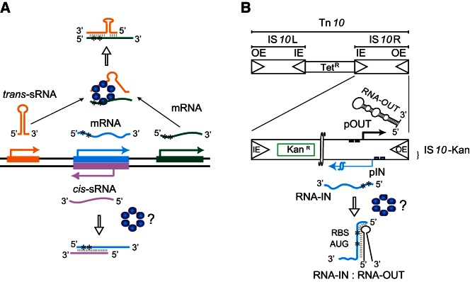
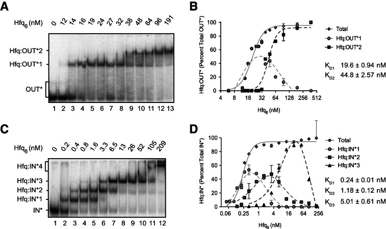
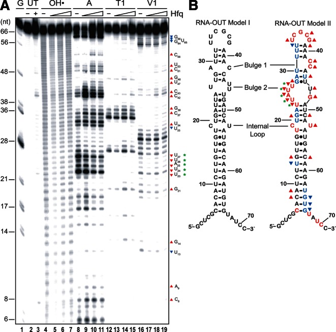
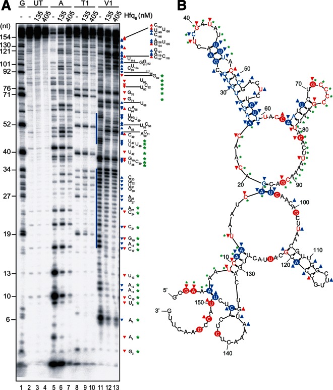
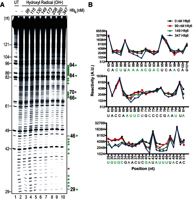
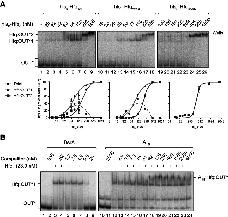
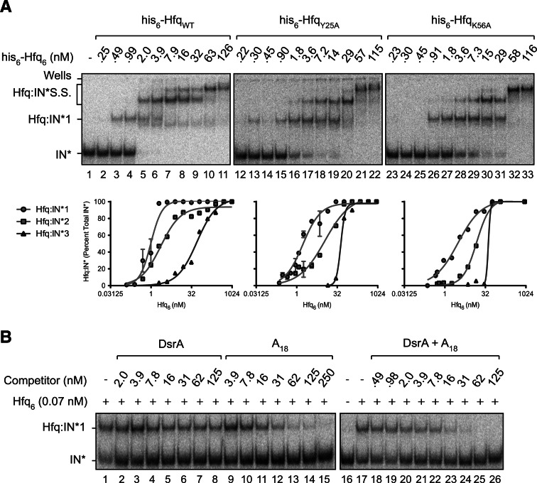
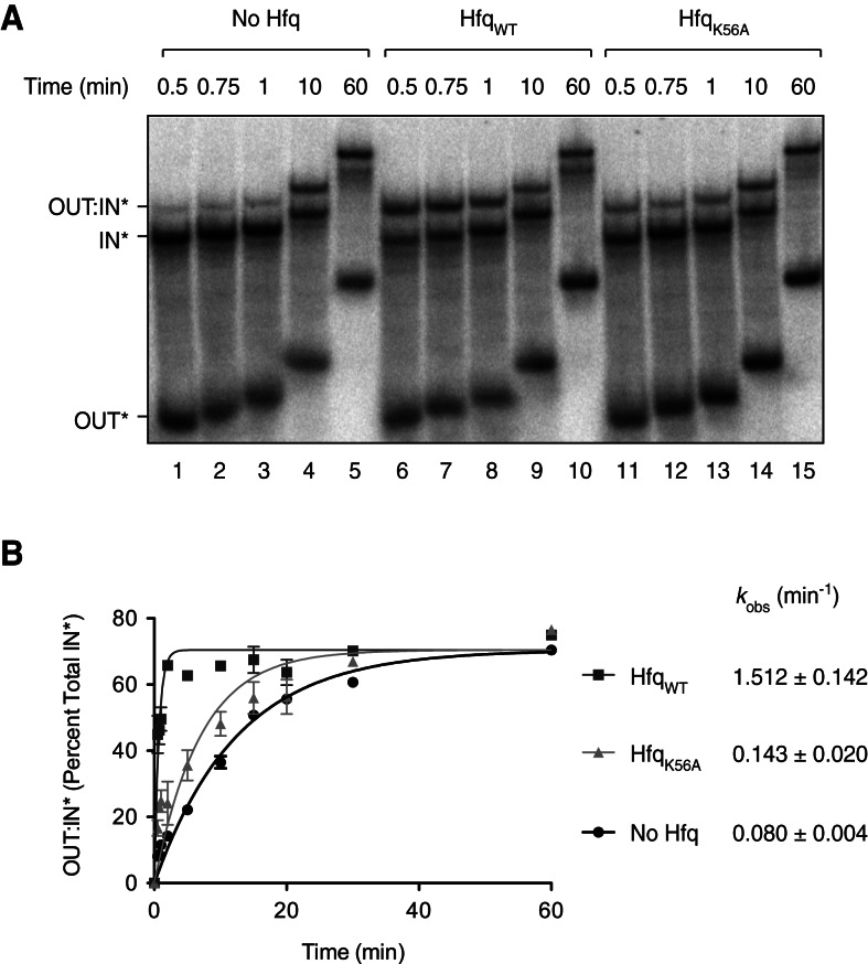
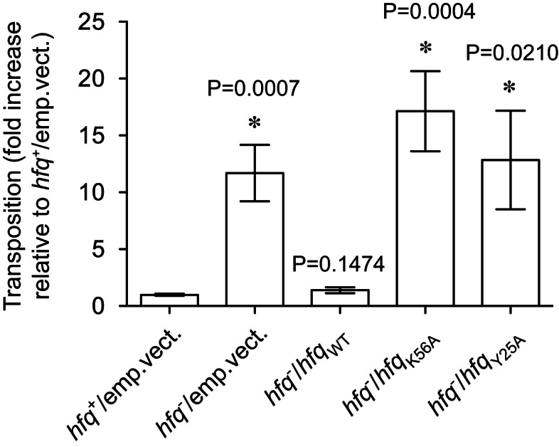
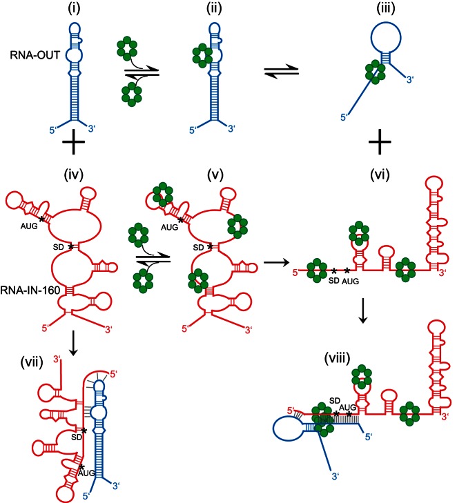
Hfq functions in post-transcriptional gene regulation in a wide range of bacteria, usually by promoting base-pairing of mRNAs and trans-encoded sRNAs that share partial sequence complementarity. It is less clear if Hfq is required for pairing of cis-encoded RNAs (i.e., antisense RNAs) with their target mRNAs. In the current work, we have characterized the interactions between Escherichia coli Hfq and the components of the Tn10/IS10 antisense system, RNA-IN and RNA-OUT. We show that Hfq interacts with RNA-OUT through its proximal RNA-binding surface, as is typical for Hfq and trans-encoded sRNAs. In contrast, RNA-IN binds both proximal and distal RNA-binding surfaces in Hfq with a higher affinity for the latter, as is typical for mRNA interactions in canonical sRNA-mRNA pairs. Importantly, an amino acid substitution in Hfq that interferes with RNA binding to the proximal site negatively impacts RNA-IN:OUT pairing in vitro and suppresses the ability of Hfq to negatively regulate IS10 transposition in vivo. We also show that Hfq binding to RNA-IN and RNA-OUT alters secondary structure elements in both of these RNAs and speculate that this could be important in how Hfq facilitates RNA-IN:OUT pairing. Based on the results presented here, we suggest that Hfq could be involved in regulating RNA pairing in other antisense systems, including systems encoded by other transposable elements.


Figures

FIGURE 1.
FIGURE 1.
Small regulatory RNAs (sRNAs) and the Tn10/IS10 antisense system. (A) Cis- vs. trans-encoded sRNAs. Transcribed strands of three different genes and their corresponding RNAs (color coded) are shown. Pairing of a trans-sRNA (gold) and an mRNA (green) and of a cis-sRNA (pink) and an mRNA (cyan) is shown. Hfq (blue hexamer) catalyzes pairing in the former case where there is partial sequence complementarity between partners, but it is unclear if it also catalyzes pairing in the latter case where there is perfect sequence complementarity between partners. Asterisks (*) define the translation initiation region (TIR) of the mRNAs. (B) Structure of Tn10 and IS10-Kan. Tn10 is a 9147-bp composite transposon that confers tetracycline resistance (TetR). Tn10 is comprised of IS10-Left and IS10-Right, the latter of which encodes a functional transposase protein that catalyzes DNA cleavage and joining events involving the “outside” (OE) and “inside” (IE) ends. The transposase mRNA (RNA-IN) is encoded from the promoter pIN (blue squares). A second promoter (pOUT–black squares) within IS10-Right encodes a cis-sRNA (also referred to as an antisense RNA), RNA-OUT. To follow transposition of IS10-Right in E. coli, a KanR gene cassette was cloned into IS10-Right, creating IS10-Kan. RNA-OUT is depicted as a stable stem–loop structure (black) and RNA-IN is depicted as a blue line with asterisks defining the TIR. RNA-OUT is known to pair with RNA-IN, and this inhibits translation of RNA-IN, thereby down-regulating transposition. Hfq can enhance the rate of RNA-IN:OUT pairing in vitro, but it is not known if Hfq plays a role in this antisense system in vivo.
FIGURE 2.
FIGURE 2.
Hfq binds with high and moderate affinities to RNA-IN and RNA-OUT in vitro. 32P-labeled RNA-OUT (A) or RNA-IN (C) was mixed with varying concentrations (reported per hexamer) of purified Hfq protein, and reactions were subject to EMSA as described in Materials and Methods. Band intensities were quantified (ImageQuant), and the percent of each shifted species (relative to total labeled RNA) was plotted vs. Hfq6 concentration (B,D). RNA-OUT formed two complexes with Hfq, Hfq:OUT*1, and Hfq:OUT*2. RNA-IN formed four complexes with Hfq, Hfq:IN*1, Hfq:IN*2, Hfq:IN*3, and Hfq:IN*4. Apparent dissociation constants (KD) are indicated; see Table 1 for a summary of KD values and Hill coefficients determined in this study. RNA-OUT and RNA-IN were present at a final concentration of ∼0.1 nM. Error bars represent standard error from two experiments. KD is reported ± standard error.
FIGURE 3.
FIGURE 3.
Structure-probe analysis of RNA-OUT and Hfq:RNA-OUT complex. (A) 32P-labeled RNA-OUT (65 nM) was incubated with or without Hfq as indicated before hydroxyl radical (lanes 47) or ribonuclease (A, T1, or V1; lanes 819) treatments. Reactions, including untreated RNA (lanes 2,3) and a G-ladder (lane 1), were analyzed on a 10% denaturing polyacrylamide gel. Nucleotide labeling is relative to the RNA-OUT in vitro transcriptional start site, which includes two extra nucleotides introduced by T7 RNA polymerase at the 5′ end of the RNA. Where Hfq was included, it was present at 1460, 2190, and 4380 nM. (B) A previous model of RNA-OUT (Model I) is compared to the model derived from the current work (Model II). Colored letters represent RNase-sensitive positions in RNA-OUT observed in the absence of Hfq. Red indicates cleavage by either RNase A or T1, while blue indicates cleavage by RNase V1. Symbols (triangles and asterisks) are defined in the text.
FIGURE 4.
FIGURE 4.
RNase footprinting of RNA-IN. (A) 32P-labeled RNA-IN-160 (45 nM) was incubated with or without Hfq as indicated before treatment with ribonuclease A, T1, or V1 (lanes 513). Reactions, including RNA not treated with RNase (lanes 24) and a G-ladder (lane 1), were analyzed on a 10% denaturing polyacrylamide gel. Nucleotide labeling is relative to the RNA-IN in vitro transcriptional start site, which is nucleotide 1. Blue bars highlight clusters of V1 sensitivity observed in the absence of Hfq. (B) A model is shown for the secondary structure of RNA-IN-160. The model was produced using Mfold with hard constraints (circled positions) obtained from two independent RNase structure-probe experiments (part A and Supplemental Fig. S2). RNase A/T1 cleavage is indicated with red letters, while V1 cleavage is indicated with blue letters. Symbols (triangles and asterisks) are defined in the text.
FIGURE 5.
FIGURE 5.
Hydroxyl radical footprinting of RNA-IN. (A) 32P-labeled RNA-IN-160 (45 nM) was incubated with increasing concentrations of Hfq (lanes 310) and then subject to hydroxyl radical treatment (lanes 210). Lane 1 contains RNA not treated with hydroxyl radicals. Samples were analyzed as in Figure 4. Nucleotides are numbered as in Figure 4. Green asterisks identify positions protected from hydroxyl radical cleavage in the presence of Hfq, while purple asterisks identify positions where Hfq induced hypersensitivity to hydroxyl radical cleavage. (B) Quantification of band intensities from selected lanes of the gel image in part A is shown. Reactivity is presented in arbitrary units (AU).
FIGURE 6.
FIGURE 6.
RNA-OUT interacts specifically with the proximal RNA-binding surface of Hfq. (A) EMSAs with 32P-labeled RNA-OUT (∼0.4 nM) and either WT or mutant forms of Hfq. HfqY25A is defective in RNA-binding at the distal site, and HfqK56A is defective in RNA-binding at the proximal site. The corresponding binding curves are presented below each gel image. Error bars represent standard error from two experiments. Note that all forms of Hfq used in this experiment possess a his6 epitope tag at their C termini. Species are labeled as in Figure 2. (B) EMSAs performed in the presence of competitor RNAs. HfqWT (untagged) was first mixed with various concentrations of DsrA or A18 RNA for 5 min, and 32P-labeled RNA-OUT (0.4 nM) was added. After an additional 15 min, reactions were subjected to polyacrylamide gel electrophoresis. A species expected to represent a ternary complex is labeled A18:Hfq:OUT*. IC50 values were calculated from curves shown in Supplemental Figure S4 and are reported in Table 2.
FIGURE 7.
FIGURE 7.
RNA-IN interacts with the distal and proximal RNA-binding surfaces of Hfq. (A) EMSAs with 32P-labeled RNA-IN (0.17 nM) and either WT or mutant forms of his6-tagged Hfq. Species are labeled as in Figure 2. Binding curves are shown below the corresponding EMSA, and apparent KD values are reported in Table 1. Error bars represent standard error from two experiments. (B) EMSAs performed in the presence of competitor RNAs. Competitor experiments were performed as described in Figure 6B except that RNA-IN was present at a concentration of 0.17 nM. For lanes 1826, a 1:1 mix of DsrA and A18 was serially diluted to the indicated concentrations before competition. IC50 values were calculated from curves shown in Supplemental Figure S4 and are reported in Table 2.
FIGURE 8.
FIGURE 8.
RNA-IN:RNA-OUT pairing reactions. (A) 32P-labeled RNA-IN-160 (0.85 nM) was mixed with excess 32P-labeled RNA-OUT (8.5 nM) and, where indicated, untagged WT or K56A Hfq (45 nM). Note that RNA-OUT had a lower specific activity than RNA-IN. At the indicated time points, pairing reactions were stopped by treatment with a phenol/water mix and immediately loaded onto a 6% native polyacrylamide gel. (B) The amount of RNA-OUT:RNA-IN* complex (OUT:IN*) was determined as a percentage of total RNA-IN* for each time point and plotted as a function of time. Error bars represent the standard error from three experiments. The observed rate constant (kobs) is indicated for each reaction. These values were derived from curves corresponding to the equation describing the rate of exponential association, presented in Materials and Methods.
FIGURE 9.
FIGURE 9.
IS10-Kan transposition is derepressed in E. coli encoding Y25A and K56A forms of Hfq. E. coli cells (hfq+ or hfq) were cotransformed with pDH602 (encodes IS10-Kan) and a compatible plasmid encoding untagged Hfq (WT, K56A, or Y25A) or the corresponding “empty vector” control. Relative transposition frequencies were measured using the conjugal mating out assay (see Materials and Methods for details). An average transposition frequency (4.03 × 10⁻³ events per mL of mating mixture) was calculated for the hfq+ strain (hfq+/emp.vect.) from 15 independent “donor” colonies across four independent experiments, and this value was set at 1. All other transposition values are expressed relative to this value where Hfq-directed repression of transposition is at its maximal level. Bars indicate the mean; the error bars indicate standard error on the mean. From left to right, the n value for each treatment group is 15, 15, 14, 16, and 11—these were compiled from at least two (and up to four) independent experiments. An asterisk (*) indicates that means were significantly different from the hfq+ control group; P values are indicated above the corresponding bars.
FIGURE 10.
FIGURE 10.
Model for RNA-IN:OUT antisense pairing in the presence vs. absence of Hfq. The Hfq-independent pairing pathway is shown on the left-hand side (structures i, iv, and vii) and the Hfq-dependent pathway is shown on the right-hand side (structures iii, vi, and viii). In structures (ii) and (v) Hfq is shown bound to RNA-OUT and RNA-IN, respectively, but conformational changes in the RNAs have not yet taken place. Other structures are described in the text. Hfq hexamers are indicated by green circles. The start codon (AUG) and Shine-Dalgarno sequence (SD) of RNA-IN are indicated by asterisks (*) at the first nucleotide of each sequence. Intramolecular base pairs in RNA-OUT/IN are indicated by blue and red, respectively. Intermolecular base pairs between RNA-OUT and RNA-IN are in gray.

References

    1. Altuvia S, Weinstein-Fischer D, Zhang A, Postow L, Storz G 1997. A small, stable RNA induced by oxidative stress: Role as a pleiotropic regulator and antimutator. Cell 90: 43–53 - PubMed
    1. Arini A, Keller MP, Arber W 1997. An antisense RNA in IS30 regulates the translational expression of the transposase. Biol Chem 378: 1421–1431 - PubMed
    1. Bolivar F, Backman K 1979. Plasmids of Escherichia coli as cloning vectors. Methods Enzymol 68: 245–267 - PubMed
    1. Brennan RG, Link TM 2007. Hfq structure, function and ligand binding. Curr Opin Microbiol 10: 125–133 - PubMed
    1. Brescia CC, Mikulecky PJ, Feig AL, Sledjeski DD 2003. Identification of the Hfq-binding site on DsrA RNA: Hfq binds without altering DsrA secondary structure. RNA 9: 33–43 - PMC - PubMed

Publication types

MeSH terms

LinkOut - more resources