

Review
doi: 10.1007/s00401-013-1106-9.
Epub 2013 Mar 20.
What do we know about IDH1/2 mutations so far, and how do we use it?
Affiliations
- PMID: 23512379
- PMCID: PMC3633675
- DOI: 10.1007/s00401-013-1106-9
Item in Clipboard
Review
What do we know about IDH1/2 mutations so far, and how do we use it?
Acta Neuropathol.
2013 May.
Abstract
Whole genome analyses have facilitated the discovery of clinically relevant genetic alterations in a variety of diseases, most notably cancer. A prominent example of this was the discovery of mutations in isocitrate dehydrogenases 1 and 2 (IDH1/2) in a sizeable proportion of gliomas and some other neoplasms. Herein the normal functions of these enzymes, how the mutations alter their catalytic properties, the effects of their D-2-hydroxyglutarate metabolite, technical considerations in diagnostic neuropathology, implications about prognosis and therapeutic considerations, and practical applications and controversies regarding IDH1/2 mutation testing are discussed.
Figures
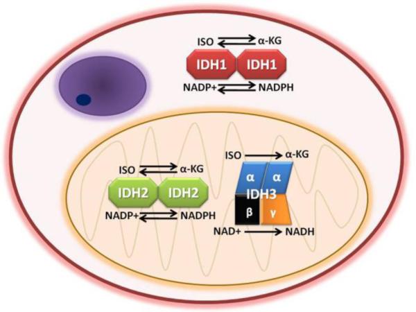
All three enzymes oxidize isocitrate (ISO) to alpha-ketoglutarate (α-KG). IDH1 and IDH2 are homodimers, whereas IDH3 is a heterotetramer. IDH1 and IDH2 utilize nicotinamide adenine dinucleotide phosphate (NADP+) as a cofactor, generating NADPH. IDH3 uses NAD+ and produces NADH. IDH2 and 3 are located in mitochondria while IDH1 is in the cytosol and peroxisomes. In certain circumstances IDH1 and IDH2 can reduce α-KG to isocitrate, whereas IDH3 is unidirectional. (The structure in the upper left of the cell depicts a nucleus.)
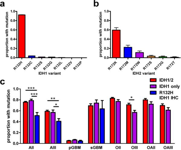
(a) In a pooled analysis of over 3400 gliomas from 37 studies in which mutation subtypes were reported, R132H was the most common IDH1 variant, comprising 92% of all IDH1 mutations (P < 0.0001). The rarest was R132P, occurring in a single case [54]. (b) In contrast, there was less preference for a specific type of IDH2 mutation, though the R172K variant was present in 60% of IDH2-mutant tumors (P < 0.0001, N = 89). (c) Inter-study mutation frequencies significantly differ in grades II-III astrocytomas and grade III oligodendrogliomas, depending on whether the studies tested for both IDH1 and IDH2 mutations (red bars, N = 4324 gliomas from 26 studies), screened for just IDH1 mutations (purple bars, N = 2075 gliomas from 12 studies), or only used the R132H IDH1 antibody (blue bars, N = 794 gliomas from 6 studies). Not enough oligodendroglial tumors have been interrogated with R132H IDH1 antibody to be reliably compared with the other columns. Of note, mean mutation frequencies in IDH1-only studies sometimes barely exceeded the frequencies reported in IDH1/2 papers (purple versus red bars in AII and sGBM subgroups). This apparent incongruity can be explained by inter-cohort variations, especially given how rare IDH2 mutations are in astrocytic tumors (see Figure 4). A list of the studies from which these data are derived is in Supplemental Table 1. All data bars in a-c represent mean ± SEM; statistical analyses were done via Student's t-test or ANOVA with Kruskal-Wallis post hoc test, as appropriate. *P < 0.05; **P < 0.01; ***P < 0.001. AII = grade II diffuse astrocytoma; AIII = grade III anaplastic astrocytoma; pGBM = primary GBM; sGBM = secondary GBM; OII = grade II oligodendroglioma; OIII = grade III anaplastic oligodendroglioma; OAII = grade II oligoastrocytoma; OAIII = grade III anaplastic oligoastrocytoma.
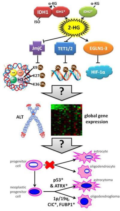
Unlike mutant IDH2, mutant IDH1 is more efficient when it heterodimerizes with a wild-type partner. Both mutations convert α-KG to 2-HG. This 2-HG compound inhibits some enzymes that use α-KG as a cofactor, including JmjC domain-containing histone demethylases and TET DNA demethylases. The result of this inhibition is a net upregulation of histone and DNA methylation, the former occurring at key amino acid residues that are mutated in some non-IDH1/2-driven gliomas (involving the H3F3A gene encoding histone 3.3). Other proteins involved in chaperoning histone H3.3 include ATRX and DAXX, both of which can be mutated in IDH1/2-wt and IDH1/2-mutant gliomas (though ATRX is far more likely to be mutated than DAXX)[149]. EGLN1-3 prolyl hydroxylases may actually be activated by 2-HG, thereby increasing degradation of Hif-1α. The exact results of all these alterations are not yet clear, but they definitely cause global modifications of gene expression and may promote Alternative Lengthening of Telomeres (ALT). EGLN activation in particular appears to slow down the differentiation of glial precursors, perhaps providing a greater opportunity for additional mutations to arise. Mutant TP53 and/or ATRX tend to produce astrocytomas, whereas 1p/19q codeletion withCIC and/or FUBP1 mutations produce oligodendrogliomas. Asterisks (*) denote mutated proteins.
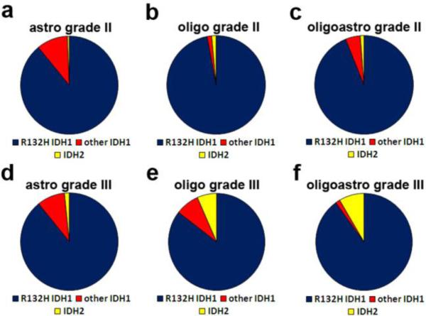
Combining data from multiple studies that included details on mutation subtypes by histologic diagnosis (see Figure 2), about 89% of IDH1/2 mutations in grade II-III astrocytic tumors were R132H IDH1 (a & d), with another 10% being other IDH1 mutations. Only 0.5-1% of those gliomas had an IDH2 mutation. In contrast, 7-8% of grade III oligodendrogliomas and oligoastrocytomas were IDH2-mutant (e & f). But this increased proportion of IDH2 mutations was only seen in grade III tumors; 94-97% of grade II oligodendrogliomas and oligoastrocytomas were R132H IDH1, and only 1% were IDH2. A list of the studies from which these data are derived is in Supplemental Table 1.
References
-
- Abbas S, Lugthart S, Kavelaars FG, et al. Acquired mutations in the genes encoding IDH1 and IDH2 both are recurrent aberrations in acute myeloid leukemia: prevalence and prognostic value. Blood. 2010;116(12):2122–6. - PubMed
-
- Aghili M, Zahedi F, Rafiee E. Hydroxyglutaric aciduria and malignant brain tumor: a case report and literature review. J Neurooncol. 2009;91:233–6. - PubMed
-
- Ahmad K, Henikoff S. The histone variant H3.3 marks active chromatin by replication-independent nucleosome assembly. Mol Cell. 2002;9(6):1191–200. - PubMed
-
- Ahmadi R, Stockhammer F, Becker N, et al. No prognostic value of IDH1 mutations in a series of 100 WHO grade II astrocytomas. J Neurooncol. 2012;109(1):15–22. - PubMed
Publication types
MeSH terms
Substances
Grants and funding
LinkOut - more resources
Full Text Sources
Other Literature Sources
Medical
Miscellaneous
