. 2013 Mar 15;27(6):683-98.
doi: 10.1101/gad.211011.112.

ETV1 directs androgen metabolism and confers aggressive prostate cancer in targeted mice and patients

Affiliations

ETV1 directs androgen metabolism and confers aggressive prostate cancer in targeted mice and patients

Esther Baena et al. Genes Dev. .

Abstract

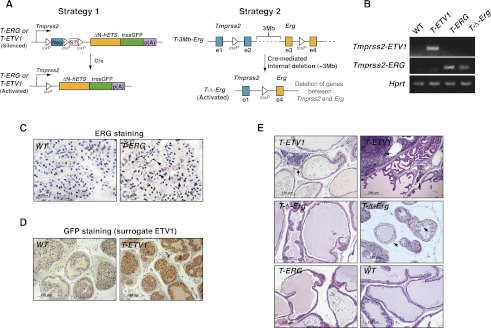
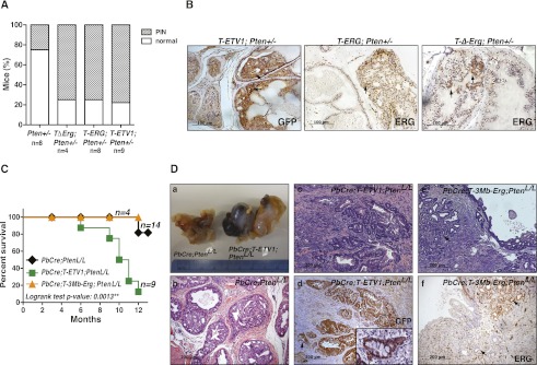
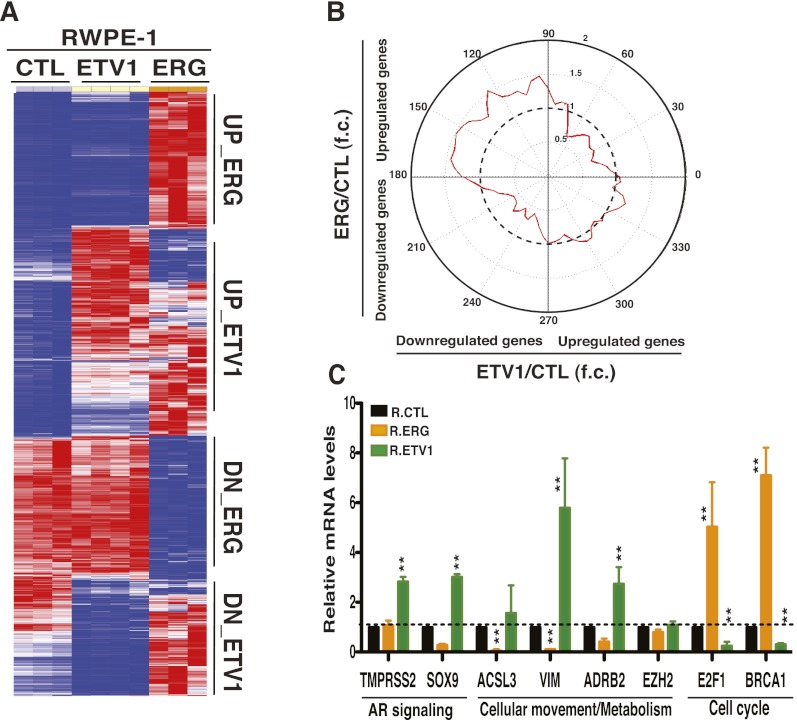
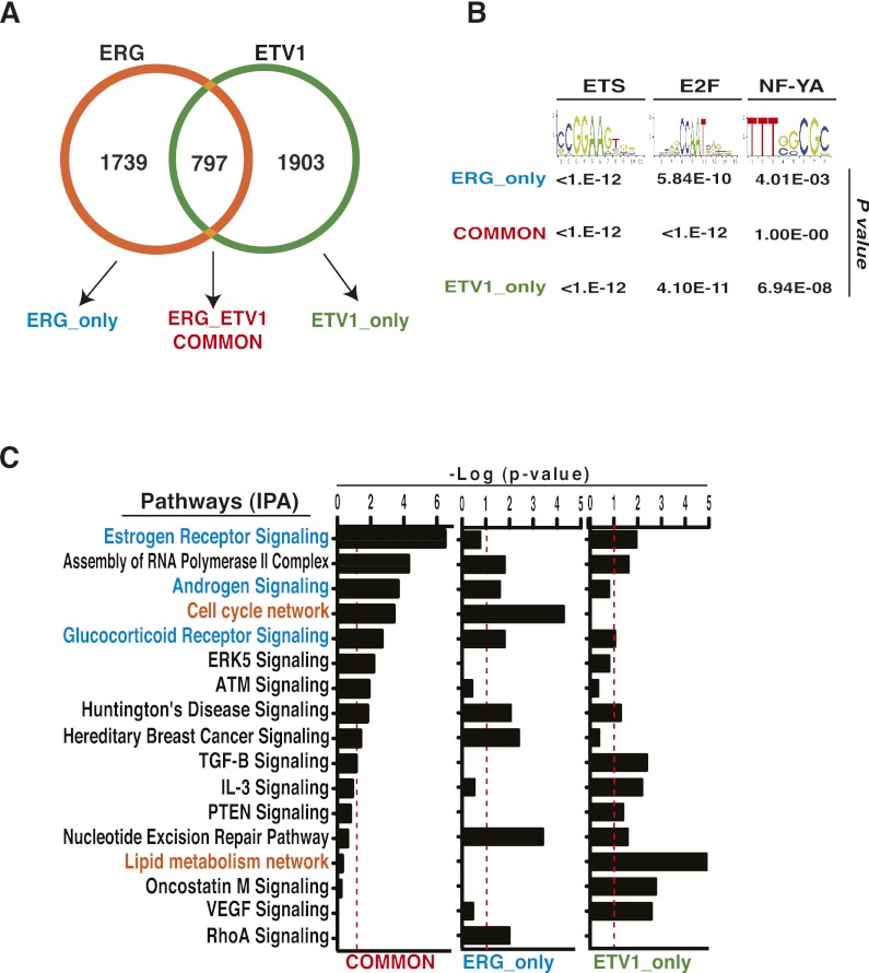
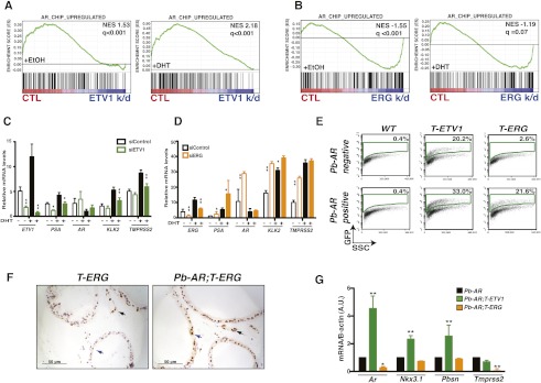
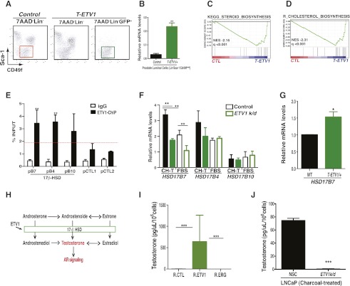
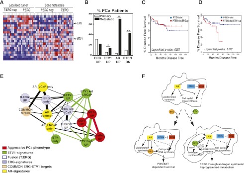
Distinguishing aggressive from indolent disease and developing effective therapy for advanced disease are the major challenges in prostate cancer research. Chromosomal rearrangements involving ETS transcription factors, such as ERG and ETV1, occur frequently in prostate cancer. How they contribute to tumorigenesis and whether they play similar or distinct in vivo roles remain elusive. Here we show that in mice with ERG or ETV1 targeted to the endogenous Tmprss2 locus, either factor cooperated with loss of a single copy of Pten, leading to localized cancer, but only ETV1 appeared to support development of invasive adenocarcinoma under the background of full Pten loss. Mechanistic studies demonstrated that ERG and ETV1 control a common transcriptional network but largely in an opposing fashion. In particular, while ERG negatively regulates the androgen receptor (AR) transcriptional program, ETV1 cooperates with AR signaling by favoring activation of the AR transcriptional program. Furthermore, we found that ETV1 expression, but not that of ERG, promotes autonomous testosterone production. Last, we confirmed the association of an ETV1 expression signature with aggressive disease and poorer outcome in patient data. The distinct biology of ETV1-associated prostate cancer suggests that this disease class may require new therapies directed to underlying programs controlled by ETV1.


Figures

Figure 1.
Figure 1.
Tmprss2-ERG (with or without interstitial deletion) and Tmprss2-ETV1 expression are insufficient to initiate prostate tumorigenesis. (A) Targeting strategies for engineering Tmprss2-ERG and Tmprss2-ETV1 knock-ins. Strategy 1 is based on direct knock-in of N terminus-truncated human ERG or ETV1 cDNA (ΔN-hETS) into the murine Tmprss2 locus. Strategy 2 is based on the introduction of loxP sites to murine Tmprss2 and Erg loci by sequential gene targeting in mouse embryonic stem cells so that the 3-Mb interstitial region can be deleted by Cre-mediated recombination and meanwhile generate the Tmprss2-Erg gene fusion. Details of gene targeting are shown in Supplemental Figures S1 and S2. (B) RT–PCR showing expression of the Tmprss2-ETV1 fusion transcripts in T-ETV1 knock-in prostates and expression of the Tmprss2-ERG fusion transcripts in T-ERG and T-Δ-Erg knock-in prostates but not in wild-type (WT) prostates. (C) IHC staining showing moderate ERG expression (arrows) in the anterior lobes of a T-ERG knock-in male but not in the wild-type male. (D) IHC staining showing homogeneous GFP expression (as surrogate for ETV1) in the anterior lobes of a T-ETV1 knock-in male but not in the wild-type male. (E) Hemotoxylin and eosin (H&E) staining showing normal prostate histology from all three knock-ins (showing ventral lobes except those of T-ETV1). Arrows in T-ETV1 pictures indicate inflammation in T-ETV1 knock-in males ([left] slight inflammation in the lateral lobe of a young knock-in male; [right] extensive inflammation in the anterior lobe of a 30-mo-old knock-in male). (Right) Arrows in the T-Δ-Erg picture indicate abnormal-looking (lightly stained “foamy”-looking cytoplasm, randomly distributed nuclei) prostate cells,observed in four out of 21 of T-Δ-Erg males. Bars, 100 μm (200 μm in top right picture). All animals analyzed in C–E were ∼10 mo of age unless otherwise indicated.
Figure 2.
Figure 2.
Cooperation of Tmprss2-ERG and Tmprss2-ETV1 gene fusions with Pten loss. (A) Bar graph summarizing histology of prostates from Pten+/−, T-Δ-Erg;Pten+/−, T-ERG;Pten+/−, and T-ETV1;Pten+/− males. All males were at 6–15 mo of age when checked. The youngest Pten+/−, T-Δ-Erg;Pten+/−, T-ERG;Pten+/−, and T-ETV1;Pten+/− males in which PIN lesions were detected were at 13, 9.5, 6.5, and 6 mo of age, respectively. (B) IHC staining showing GFP expression (as a surrogate for ETV1 expression) in the PIN lesion of a T-ETV1;Pten+/− male (left) and ERG expression in the PIN lesions of a T-ERG;Pten+/− male (middle) and a T-Δ-Erg;Pten+/− male (right). Note, in all cases, GFP or ERG staining in PIN lesions (arrows) is stronger than that in normal-appearing prostate cells. Bars, 100 μm. (C) Kaplan-Meier survival curve. All males were monitored for survival for at least 1 yr. The four PbCre;T-3Mb-Erg;PtenL/L males all survived to 1 yr and were euthanized for histology. The majority of PbCre;PtenL/L control males were still alive even after 15 mo. Log-rank test: (**) P = 0.0013 for PbCre;T-ETV1;PtenL/L in relation to PbCre;PtenL/L controls. (D) Cancer phenotypes in PbCre;PtenL/L males with or without ETS fusions. (Panel a) Gross appearance of prostates from a 10-mo-old PbCre;T-ETV1;PtenL/L male showing large tumor and prostatic cyst (right) and a 13-mo-old PbCre;PtenL/L control male (left). (Panel b) Typical localized prostate cancer seen in a control PbCre;PtenL/L male. (Panel c) Invasive prostate adenocarcinoma seen in a PbCre;T-ETV1;PtenL/L male. (Panel d) GFP staining (as a surrogate for ETV1 expression) in invasive prostate adenocarcinoma cells (brown) in a PbCre;T-ETV1;PtenL/L male (a magnified view of GFP+ invasive prostate cancer cells is shown in the inset). (Panel e) Invasive prostate adenocarcinoma cells (arrow) detected in an aged PbCre;T-3Mb-Erg;PtenL/L male. (Panel f) IHC staining of Erg (brown) revealing Erg+ and Erg invasive prostate tumor cells (arrows; from the same male as in panel e) within the same section. Bars, 200 μm.
Figure 3.
Figure 3.
ERG and ETV1 regulate a common program in immortalized nontumorigenic RWPE-1 prostate cells but in an opposing fashion. (A) Expression profiling of ERG-overexpressing (R.ERG) and ETV1-overexpressing (R.ETV1) RWPE-1 cells compared with BirA-expressing controls (CTL). Heat map generated by hierarchical clustering and by applying Pearson correlation and the complete linkage rule. The heat map shows differentially expressed genes (fold change, >1.5; false discovery rate [FDR], <0.05). (Red) Highest expression; (blue) lowest expression. (B) Bidimensional plot comparing expression profiles of genes differentially expressed (fold change, >1.5) in R.ERG versus R.CTL and in R.ETV1 versus R.CTL RWPE-1 cells. The red line represents the distribution of genes. The dotted line corresponds to a gene density fold change of 1. (C) RT–PCR analysis of select genes associated with prostate cancer pathways upon ERG or ETV1 overexpression in RWPE-1 cells. n = 3 per group. Error bars, SEM; t-test: (**) P < 0.01. If no P-value is indicated, P > 0.05.
Figure 4.
Figure 4.
ERG and ETV1 drive specific transcriptional programs. (A) Venn diagram of targets occupied by ERG and ETV1. The intensity of binding at each probe was calculated by model-based analysis of tiling array (MAT) (Johnson et al. 2006). MAT scores were then normalized by quantile–quantile normalization (Bolstad et al. 2003) between ETV1 and ERG ChIP-on-chip experiments. Target loci were defined as the peaks associated with P-value < 10−4. (B) Enrichment of ETS-binding motifs and other indicated motifs in all ChIP target subsets. The Fisher exact test was applied. (C) IPA analysis of ChIP-defined target gene sets implicating common target genes in nuclear receptor signaling pathways and ETV1 unique targets in lipid metabolism network. The significance of enrichment of each gene set is shown as −Log (P-value).
Figure 5.
Figure 5.
ERG and ETV1 regulate AR signaling in an opposite manner. (A) Androgen-induced genes are depleted in ETV1-silenced LNCaP cells upon 16-h androgen stimulation ([left] no androgen stimulation; [right] with androgen stimulation). The androgen-induced signature was obtained from the common AR ChIP targets in LNCaP and VCaP cells that were up-regulated in them upon androgen stimulation. (B) Androgen-induced genes are significantly enriched in ERG-silenced VCaP cells upon 16-h androgen stimulation compared with controls ([left] no androgen stimulation; [right] with androgen stimulation). (C,D) ETV1 silencing specifically decreases expression of AR-associated genes (C), whereas ERG silencing increases their expression (D). Mean, n = 3; error bars, SEM; t-test: (*) P < 0.05; (**) P < 0.01. If no P-value is indicated, P > 0.05. (E) Flow cytometry analysis demonstrating robust GFP+ population in the T-ETV1 prostates but not in the T-ERG prostates. However, in the presence of the Pb-AR transgene, GFP expression can be readily detected in Pb-AR;T-ERG prostates; in addition, GFP expression in Pb-AR;T-ETV1 prostates appear to be further elevated. (F) IHC staining showing weak ERG staining in the ventral lobe of a T-ERG knock-in male (blue arrow; compared with strong Erg staining in the endothelial cells [black arrow]) but much stronger ERG staining in the ventral lobe of a Pb-AR;T-ERG male (blue arrow; almost comparable with ERG staining in endothelial cells in the same section [black arrow]). Bars, 50 μm. (G) Real-time PCR quantification showing up-regulation of most AR target genes in Pb-AR;T-ETV1 prostates and slight down-regulation of them in Pb-AR;T-ERG prostates in relation to those of Pb-AR-alone prostates. Mean, n = 3; error bars, SEM; t-test: (*) P < 0.05, (**) P < 0.01. If no P-value is indicated, P > 0.05.
Figure 6.
Figure 6.
ETV1 regulates steroid metabolism in prostate cells. (A, right plot) Flow cytometry profiles and gating strategies showing GFP+ prostate luminal cells (LinSca-1CD49fmed) sorted from T-ETV1 knock-in males used for microarray analysis. (Left plot) The LinSca-1CD49fmed prostate luminal cells sorted from wild-type (WT) control males were used as the control. (B) Real-time PCR quantification confirming ectopic ETV1 expression in sorted GFP+ prostate luminal cells from T-ETV1 knock-in mice (mean, n = 3 samples per group; error bars, SEM). (C,D) Steroid and cholesterol biosynthesis pathways are the top pathways significantly enriched in T-ETV1 knock-in prostate luminal cells compared with controls. Note that a critical enzyme in the steroid biosynthesis pathway, HSD17B7, is also a key enzyme in the steroid hormone biosynthesis pathway, which is enriched in metastatic prostate cancers (Supplemental Fig. S15). (E) ChIP-PCR validation of ETV1 binding to HSD17B7 (pB7), HSD17B4 (pB4), and HSD17B10 (pB10) promoters (mean, n = 5; error bars, SEM). (pCTL1 and pCTL2) Nonspecific promoter control regions. (F) Only HSD17B7 levels significantly decreased upon knockdown of ETV1 (k/d) (mean, n = 3, error bars, SEM) under both the androgen-deprivation condition ([CH-T] charcoal-treated) and the regular condition in the presence of serum (FBS). Conversely, HSD17B7 expression increased upon ETV1 overexpression in RPWE-1 cells (Supplemental Fig. S16A). t-test: (**) P < 0.01. If no P-value is indicated, P > 0.05. (G) T-ETV1 knock-in prostate cells exhibit increased Hsd17b7 expression levels compared with wild-type controls (mean, n = 3; error bars, SEM). t-test: (*) P-value < 0.05. (H) Schematic diagram showing the key role of 17-β HSD enzymes, including HSD17B7, in converting androgen and estrogen from their less active forms to active forms. (I) ETV1 overexpression in RWPE-1 cells promoted the elevation of the endogenous testosterone level, while no changes were observed upon ERG overexpression (mean, n = 4; error bars, SEM). Testosterone levels per 106 cells (R.ETV1 mean = 642.16 pg/μL; R.ERG mean = 0.49 pg/μL; R.CTL mean = 1.89 pg/μL). t-test: (***) P < 0.001. (J) Testosterone levels were reduced in androgen-deprived (charcoal-treated) LNCaP cells upon stable ETV1 silencing (k/d) as compared with controls (mean, n = 3; error bars, SEM). (NSC) Nonsilencing shRNA control. Testosterone levels per 106 cells (NSC mean = 74.69 pg/μL; R.ERG mean = 0.49 pg/μL; ETV1k/d mean = 0.56 pg/μLr). t-test: (***) P < 0.001.
Figure 7.
Figure 7.
ETV1, rather than ERG, expression and the program it drives are associated with advanced prostate cancer in multiple patient cohorts. (A) Heat map showing ERG and ETV1 expression pattern in localized and bone metastatic prostate cancer samples using the Beth-Israel (BI) cohort data set (Stanbrough et al. 2006). Heat map generated by hierarchical clustering and by applying Pearson correlation and the complete linkage rule. Heat map showing differentially expressed select genes (fold change, >2; FDR, <0.05). (B) Graph showing ERG and ETV1 expression along prostate cancer progression from localized to metastatic samples in the MSKCC cohort (Taylor et al. 2010). The graph reveals that the number of patients carrying ETV1 overexpression (fold change, >3), PTEN deletion, and AR alterations (amplification and expression fold change, >3) increased in metastatic samples compared with localized prostate tumors, while patients carrying high levels of ERG (fold change, >3) did not increase over time (also in Supplemental Fig. S18B). t-test: (*) P-value < 0.05; (**) P-value < 0.01. If no P-value is indicated, P > 0.05. (C) Disease-free survival plot showing that among all patients in the MSKCC cohort with PTEN deletion (n = 21), those with ERG overexpression (n = 4) exhibited no survival difference from the rest of patient with PTEN deletion. P = 0.553 by log-rank test. (D) Disease-free survival plot showing that among all patients in the MSKCC cohort with PTEN deletion (n = 21), those with ETV1 overexpression (n = 8) exhibited much worse survival compared with remaining patients with PTEN deletion. (*) P = 0.015 by log-rank test. (E) Correlation between ERG- and ETV1-associated gene sets with patient prognosis. Overlap between genes enriched in patient samples associated with indolent or aggressive prostate cancer from the MSKCC cohort (Taylor et al. 2010) and ETV1 or ERG gene sets defined in Figures 3 and 4. ETV1-associated genes are enriched in patients with a higher Gleason score in the Swedish cohort (also in Supplemental Fig. S18C; Setlur et al. 2008). “UP” represents those genes up-regulated in the shown category with a fold change of >1.5. The significance of overlap of these gene sets was calculated by the Fisher exact test and visualized as connecting line width (cutoff, P = 0.01). (Red) Aggressive prostate cancer-associated; (green) ETV1-associated gene sets; (blue) TMPRSS2-ERG fusion-associated gene sets; (purple) ERG signature-associated gene sets; (yellow) AR-associated gene sets; (orange) common targets of ERG and ETV1. (F) Model illustrating the differential contribution by ERG and ETV1 to prostate tumorigenesis under the PTEN loss background. See the text for details.

References

    1. Attard G, Clark J, Ambroisine L, Fisher G, Kovacs G, Flohr P, Berney D, Foster CS, Fletcher A, Gerald WL, et al. 2008a. Duplication of the fusion of TMPRSS2 to ERG sequences identifies fatal human prostate cancer. Oncogene 27: 253–263 - PMC - PubMed
    1. Attard G, Clark J, Ambroisine L, Mills IG, Fisher G, Flohr P, Reid A, Edwards S, Kovacs G, Berney D, et al. 2008b. Heterogeneity and clinical significance of ETV1 translocations in human prostate cancer. Br J Cancer 99: 314–320 - PMC - PubMed
    1. Bauer DE, Hatzivassiliou G, Zhao F, Andreadis C, Thompson CB 2005. ATP citrate lyase is an important component of cell growth and transformation. Oncogene 24: 6314–6322 - PubMed
    1. Bismar TA, Dolph M, Teng L-H, Liu S, Donnelly B 2012. ERG protein expression reflects hormonal treatment response and is associated with Gleason score and prostate cancer specific mortality. Eur J Cancer 48: 538–546 - PubMed
    1. Bolstad BM, Irizarry RA, Astrand M, Speed TP 2003. A comparison of normalization methods for high density oligonucleotide array data based on variance and bias. Bioinformatics 19: 185–193 - PubMed

Publication types

MeSH terms

Associated data