

Two or more impingement and/or instability deformities are often present in patients with hip pain
- PMID: 23512747
- PMCID: PMC3825892
- DOI: 10.1007/s11999-013-2918-6
Two or more impingement and/or instability deformities are often present in patients with hip pain
Abstract
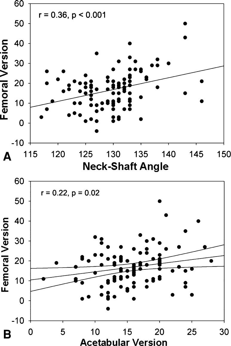
Background: Damage to the hip can occur due to impingement or instability caused by anatomic factors such as femoral and acetabular version, neck-shaft angle, alpha angle, and lateral center-edge angle (CEA). The associations between these anatomic factors and how often they occur in a painful hip are unclear but if unaddressed might explain failed hip preservation surgery.
Questions/purposes: We determined (1) the influence of sex on the expression of impingement-related or instability-related factors, (2) the associations among these factors, and (3) how often both impingement and/or instability factors occur in the same hip.
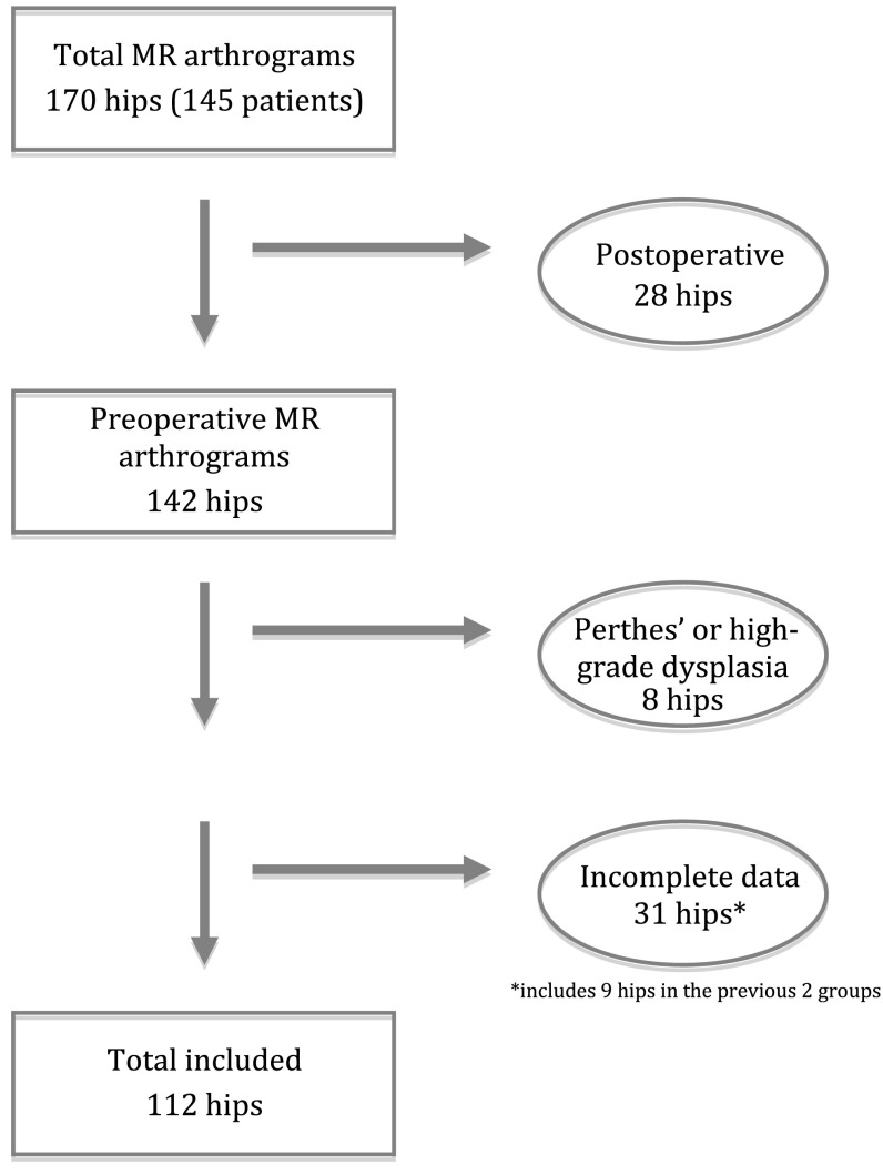
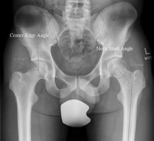
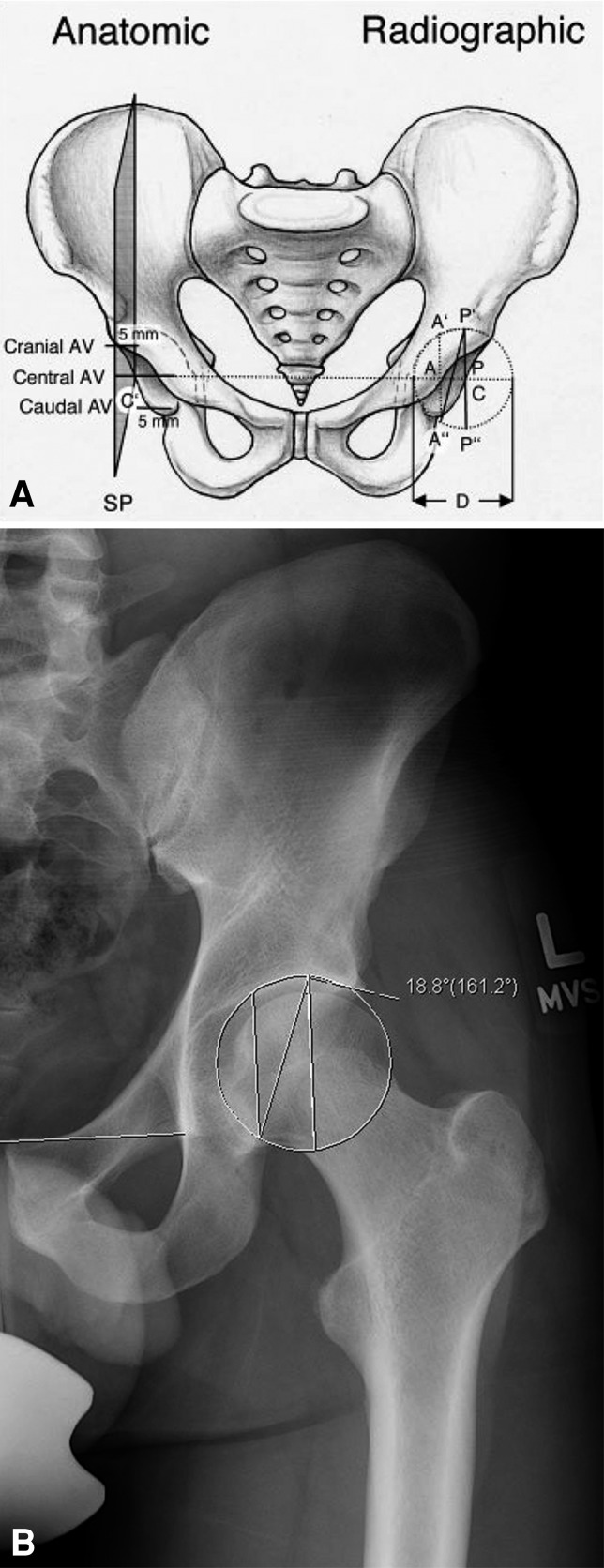
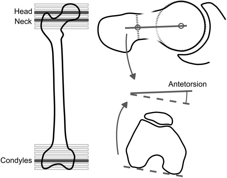
Methods: We retrospectively reviewed a cohort of 170 hips (145 patients) undergoing MR arthrography of the hip for any reason. We excluded 58 hips with high-grade dysplasia, Perthes' sequelae, previous surgery, or incomplete radiographic information, leaving 112 hips (96 patients). We measured femoral version and alpha angles on MR arthrograms. Acetabular anteversion, lateral CEA, and neck-shaft angle were measured on pelvic radiographs.
Results: We observed a correlation between sex and alpha angle. Weak or no correlations were observed between the other five parameters. In 66% of hips, two or more (of five) impingement parameters, and in 51% of hips, two or more (of five) instability parameters were found.
Conclusions: Patients with hip pain frequently have several anatomic factors potentially contributing to chondrolabral damage. To address pathologic hip loading due to impingement and/or instability, all of the anatomic influences should be known. As we found no associations between anatomic factors, we recommend an individualized assessment of each painful hip.
Figures
References
-
- Akiyama M, Nakashima Y, Fujii M, Sato T, Yamamoto T, Mawatari T, Motomura G, Matsuda S, Iwamoto Y. Femoral anteversion is correlated with acetabular version and coverage in Asian women with anterior and global deficient subgroups of hip dysplasia: a CT study. Skeletal Radiol. 2012;41:1411–1418. doi: 10.1007/s00256-012-1368-7. - DOI - PubMed
-
- Argenson JN, Ryembault E, Flecher X, Brassart N, Parratte S, Aubaniac JM. Three-dimensional anatomy of the hip in osteoarthritis after developmental dysplasia. J Bone Joint Surg Br. 2005;87:1192–1196. - PubMed
MeSH terms
LinkOut - more resources
Full Text Sources
Other Literature Sources
Medical
Research Materials
